Bibliographic and Educational Resources in Fetal Medicine

This platform is designed to serve as a comprehensive educational and bibliographic tool for healthcare professionals involved in fetal medicine. Covering a broad range of up-to-date topics within the field, it offers structured access to recent scientific literature and an array of pedagogical resources tailored to clinicians, educators, and trainees.

Each topic is anchored in a selection of current publications, accompanied by in-depth summaries that go far beyond traditional abstracts—offering clear, clinically relevant insights without the time burden of reading full articles. These summaries serve as a gateway to the original literature, allowing users to identify which articles merit deeper exploration.

In addition to these detailed reviews, users will find a rich library of supplementary materials: topic overviews, FAQs, glossaries, synthesis sheets, thematic podcasts, fully structured course outlines adaptable for teaching, and ready-to-use PowerPoint slide decks. All resources are open-access and formatted for easy integration into academic or clinical training programs.

By offering practical, well-structured resources, this platform enables members of the fetal medicine community to quickly update their knowledge on selected topics. It also provides ready-to-use educational materials that can be easily adapted for instructional purposes.

Congenital Cytomegalovirus Infection

Overview

  • Viral Characteristics and Transmission Routes: Cytomegalovirus (CMV) is an enveloped DNA virus belonging to the herpes virus family. It establishes a lifelong latent period after primary infection, residing in monocytes and granulocytes, and can reactivate or lead to reinfection with different strains. Transmission occurs through direct contact with contaminated bodily fluids such as urine, saliva, blood, genital secretions, breast milk, and through organ transplants.
  • Risk Factors: The primary risk factor for maternal infection in women of reproductive age is exposure to the contaminated saliva and urine of young children, particularly those younger than 2 years, who can shed the virus for extended periods. Sexual transmission is also a significant route.
  • Types of Maternal Infection: Vertical transmission to the fetus can occur through:
    • Primary infection (MPI): When a seronegative mother (IgG and IgM negative) seroconverts during pregnancy. This is associated with the highest risk of vertical transmission (30–40%) and greater potential for severe congenital infection.
    • Non-primary maternal infection (NMPI): This includes reactivation of a latent virus or reinfection with a different CMV strain in previously immune women. While the risk of fetal infection is much lower (1–3%) compared to primary infection, it still accounts for about half of cCMV cases in high-income countries due to high seroprevalence in the general population.
  • Timing of Maternal Infection and Transmission Rates: The rate of vertical transmission varies with the gestational age at which the mother acquires primary infection:
    • Periconceptional period (from 4 weeks before to 3–6 weeks after the last menstrual period): 5.5% to 36.8% transmission, with a fetal insult rate (CNS malformation, TOP, or neurological symptoms at birth) of 28.8%.
    • First trimester: 20–40% transmission, with a fetal insult rate of 19.3%.
    • Second trimester: 45% to 72% transmission, with a fetal insult rate of 0.9%.
    • Third trimester: 47% to 78% transmission, with a fetal insult rate of 0.4%.
    • Crucially, severe long-term sequelae in infected fetuses are primarily associated with maternal primary infection occurring in the first trimester of pregnancy.
  • Other Transmission Considerations:
    • Breast milk from CMV-infected mothers can transmit the virus to preterm and low-birthweight newborns, potentially causing symptoms. Pasteurization of milk can reduce this risk.
    • Twin pregnancies show a higher vertical transmission rate (58.7%) compared to singletons, with a 50% discordance rate between twins. Monozygotic twins have higher concordance (84.6% vs. 37.5% in dizygotic), suggesting possible genetic susceptibility.
  • Fetal Impact: CMV can directly damage the fetus or indirectly affect it through placental dysfunction, potentially leading to miscarriage, preterm birth, or fetal growth restriction (FGR). The virus replicates in trophoblast cells, which are vital for implantation and pregnancy maintenance. This can impair placental development, induce inflammation, and contribute to complications like placental insufficiency and FGR.
  • Progression: Fetal susceptibility to infection increases with gestational age, possibly due to cytotrophoblast differentiation. The virus crosses the placenta, infects fetal organs, and has tropism for reticuloendothelial cells and the central nervous system (CNS). Fetal lesions develop continuously and heterogeneously, which can lead to missed diagnoses during routine ultrasounds.
  • Symptoms at Birth: Approximately 10-50% of newborns from mothers with primary CMV infection are symptomatic at birth. These symptoms can include:
    • FGR (50%).
    • Jaundice (67%).
    • Hepatosplenomegaly (60%).
    • Generalized petechiae (76%), purpura, thrombocytopenia (77%).
    • Hydrops, pneumonitis.
    • Microcephaly (53%), abnormal brain imaging (calcifications, periventricular hyperinflammation, ventriculomegaly, subependymal cysts, striated lenticular vasculopathy).
    • Seizures (7%), chorioretinitis, hearing loss.
    • Bone abnormalities, abnormal dentition, anemia, hypotonia/lethargy (27%), arterial hypertension.
  • Long-Term Sequelae: Long-term sequelae affect 40-60% of symptomatic children and around 13.5% of asymptomatic children, with symptomatic infections being more frequent and severe. These include:
    • Sensorineural hearing loss (SNHL): The most common sequela, often progressive, unilateral or bilateral, and can manifest later in childhood. It is attributed to cCMV in approximately 21% of hearing loss at birth and 25% by 4 years of age. Even asymptomatic newborns can develop SNHL (6-23%). Bilateral SNHL and/or neurological sequelae are considered severe long-term sequelae.
    • Neurological deficits: Mental retardation, seizure disorders, cerebral palsy, microcephaly, and delayed neuropsychomotor development. Around 30-35% of newborns infected after primary maternal CMV infection in the first trimester develop significant neurological sequelae.
    • Vision impairment: Chorioretinitis, optic atrophy, cortical visual impairment, and strabismus.
    • Vestibular dysfunction: Can occur in symptomatic and asymptomatic newborns, with or without hearing loss, and may affect early motor development.
    • Children with cCMV may also exhibit poorer outcomes in psychological development, cognitive processes, social communication, and attention control.
  • Maternal Serology: Essential for identifying women at risk of primary infection.
    • IgG and IgM Testing: Diagnosis of primary maternal infection is based on CMV IgM detection, with latest generation assays having high sensitivity (>98%) for detecting recent infection (within the previous month). However, IgM can persist for months and lead to false positives, necessitating further testing.
    • IgG Avidity Testing: Crucial for dating infection. Low or intermediate IgG avidity in the first trimester suggests recent primary infection. High avidity generally excludes recent infection, though intermediate avidity results may require retesting or expert consultation.
    • Timing of Serology: Initial serology should be performed as early as possible in the first trimester, with retesting every 4 weeks until 14–16 weeks for seronegative women. Serology is also indicated if pregnant women show symptoms compatible with primary CMV infection or if abnormal ultrasound features suggest fetal infection.
  • Fetal Diagnosis:
    • Amniocentesis: The gold standard for diagnosing fetal CMV infection. CMV PCR on amniotic fluid (AF) is performed from 17+0 weeks gestation onwards, provided that maternal infection occurred at least 8 weeks prior to allow for viral shedding into the amniotic fluid. It has high specificity (around 100%) and sensitivity (87-95%). A negative amniocentesis generally ensures absence of long-term sequelae.
    • Chorionic Villus Sampling (CVS): A newer approach being explored for first-trimester diagnosis of placental infection. PCR amplification of the CMV genome in chorionic villi has shown high specificity (100%) and negative predictive value (91%) for diagnosis of placental infection, suggesting its potential to exclude CMV-related embryopathy leading to neurological and sensorineural deficits. However, its sensitivity is lower (50%) compared to amniocentesis for definitive fetal infection diagnosis, and long-term follow-up is needed to confirm its prognostic value.
    • Prenatal Ultrasound (US) and Magnetic Resonance Imaging (MRI): Used to identify CMV-associated findings and provide prognostic information.
      • Routine detailed ultrasound is performed at 20–24 and 30–34 weeks. However, it is not an appropriate screening tool for congenital CMV infection leading to long-term sequelae due to its low sensitivity (26% for severe sequelae) and the non-specific nature of CMV features. Non-specific infection-related findings (e.g., small-for-gestational-age (SGA), hyperechogenic bowel, ventriculomegaly, placentomegaly) are often present but may not raise suspicion.
      • Targeted ultrasound of known infected fetuses has high sensitivity (91% for long-term sequelae, 100% for severe sequelae) and negative predictive value (96% and 100% respectively). Awareness of sonologists and knowledge of maternal serological status are key to its diagnostic performance.
      • Fetal MRI is recommended between 28 and 34 weeks in infected fetuses, especially for suspected cerebral abnormalities, as it can reveal significant pathology missed by ultrasound. Severe brain involvement on imaging is a predictor of poor prognosis.
  • Neonatal Diagnosis: To distinguish congenital from postnatal infection, PCR should be performed on a sample collected within the first 3 weeks of birth.
    • Urine or saliva: Considered the gold standard for neonatal diagnosis due to high sensitivity. A positive saliva PCR should be confirmed by a urine PCR.
    • Dried Blood Spots (DBS): Can be used for retrospective diagnosis in older children, but sensitivity is debated due to lower viral loads in blood and methodological differences.
    • Indications for Neonatal Testing: Suspected or confirmed primary maternal CMV infection, abnormalities on fetal imaging, suspected hearing loss at birth, symmetric IUGR, very preterm/low birth weight infants, or unexplained symptoms/laboratory abnormalities consistent with cCMV.
  • Key Prognostic Factors:
    • Timing of Maternal Infection: As noted above, maternal infection in the first trimester is associated with nearly all long-term sequelae.
    • Amniocentesis Results: A negative CMV PCR in amniotic fluid is strongly associated with absence of severe neonatal symptoms, SNHL, or neurodevelopmental delay.
    • Prenatal Imaging: The absence of CNS abnormalities on ultrasound and MRI predicts a good prognosis, with a very low risk of symptomatic newborns, abnormal neurodevelopment, or hearing loss. Severe cerebral abnormalities, especially microcephaly, are strong predictors of poor outcomes.
    • Fetal Blood Markers: Platelet count (<50,000/mm³) at cordocentesis, high fetal viremia (viral load > 30,000 copies/mL), and high fetal β2-microglobulin counts are associated with more severe disease and poor prognosis.
    • Neonatal Viral Load: A high neonatal blood viral load correlates with symptomatic disease and sequelae, especially SNHL.
  • Primary Prevention:
    • Hygiene Measures: Advising hygienic modifications for all pregnant women, particularly seronegative ones, is recommended to reduce the risk of primary infection. Starting these measures prior to conception and throughout the first trimester is key to reducing cCMV-related disabilities. Examples include avoiding intimate contact (kissing, sharing food/cutlery) with young children.
    • Vaccine: No CMV vaccine is currently available, but candidates are progressing through clinical trials.
  • Secondary Prevention (for maternal primary infection):
    • Valaciclovir: Oral valaciclovir at a dose of 8 g/day (2g four times a day to minimize renal side effects) is recommended for maternal primary infection in the periconceptional period or first trimester, administered as early as possible after diagnosis until amniocentesis results are available. Studies show it reduces vertical transmission by 70-71% and symptomatic newborns at birth. Side effects are generally mild, but acute renal failure has been reported and is usually reversible upon cessation.
    • Hyperimmune Globulin (HIG): Routine administration of HIG (100 IU/kg every 4 weeks) is not recommended to prevent vertical transmission based on RCTs. Higher doses (200 IU/kg every 2 weeks) in very recent first-trimester primary infection may be considered, but evidence is less strong.
  • Minimally Invasive Management:
    • Prone positioning: Often the first-line non-invasive treatment, as it can be sufficient in some cases. However, its efficacy is controversial, and prolonged prone positioning raises concerns due to the increased incidence of sudden infant death syndrome (SIDS).
    • Nasopharyngeal Airway (NPA): A simple and effective second-line option if prone positioning is insufficient, bypassing UAO and facilitating feeding.
    • Pre-Epiglottic Baton Plate (PEBP): An orthodontic device that positions the tongue anteriorly to alleviate UAO. This method is highly specialized but has shown promising results in improving airway obstruction and feeding.
    • High-flow nasal cannula (HFNC) and Continuous Positive Airway Pressure (CPAP): These therapies can help manage UAO and can be administered in various settings, sometimes in combination with NPA.
    • Feeding management: Includes temporary nasogastric tube placement for feeding difficulties, with gastrostomy tubes being a valid alternative for prolonged issues. Palatal plates can also improve feeding and sucking activity.
  • Surgical Management: Considered when non-invasive treatments fail or in severe, life-threatening cases.
    • Mandibular Distraction Osteogenesis (MDO): This has become the gold standard surgical technique. MDO involves lengthening the mandible, which expands the oral volume and alleviates glossoptosis-related airway obstruction, often reducing or eliminating the need for tracheostomy. The Fast and Early Mandibular Osteodistraction (FEMOD) protocol, involving immediate and rapid distraction post-osteotomy, has shown good long-term outcomes with minimal dental or facial scarring.
    • Tongue-Lip Adhesion (TLA): An older surgical option that anchors the tongue to the lower lip. While providing immediate benefits, it can lead to complications such as dehiscence and infections and is less frequently used now compared to MDO.
    • Tracheostomy: A life-saving intervention for severe respiratory crises, especially in Pfeiffer syndrome. However, decannulation can be challenging, and it is generally considered when other treatments are unsuccessful.

There is notable variability in treatment approaches between centers, with European centers often preferring less invasive methods, while North American centers show a stronger preference for MDO.

  • Prenatal Treatment (in confirmed fetal infection):
    • Valaciclovir: In cases of confirmed fetal infection, valaciclovir 8 g/day may be considered after discussion with an expert team, with the aim of reducing the risk of sequelae.
  • Neonatal Treatment:
    • Valganciclovir/Ganciclovir: Recommended for newborns with significant CMV-related symptoms at birth, including CNS involvement (like microcephaly, SNHL, chorioretinitis, or white matter abnormalities), or severe disease (hepatitis, pneumonia, thrombocytopenia).
    • Dosage and Duration: Ganciclovir (6 mg/kg/day intravenously for 42 days) or valganciclovir (16 mg/kg/dose orally twice daily for 42 days or 6 months). Longer treatment (6 months) has shown greater efficacy in preserving hearing and improving neurodevelopmental scores compared to shorter courses (6 weeks).
    • Timing: Treatment should be started as soon as possible, ideally before 1 month of age. Treatment initiated between 1 and 3 months may also be beneficial, particularly for SNHL.
    • Isolated symptoms: Infants with isolated persistent hepatitis or thrombocytopenia may be treated for 6 weeks, but treatment is not recommended for isolated IUGR.
    • Monitoring: Full blood count and liver function tests should be checked regularly during antiviral treatment due to potential side effects like neutropenia.
  • Long-Term Follow-up:
    • Children with cCMV and confirmed transmission in the first trimester or unknown timing of transmission should be followed up to at least 6 years of age to ensure specialized management.
    • Children with clinical symptoms at birth and/or evidence of long-term sequelae (neurologic disease, SNHL, chorioretinitis, neurodevelopmental impairment) should be seen annually up to 6 years of age.
    • Asymptomatic children with normal imaging and documented maternal primary infection in the second or third trimester may follow standard pediatric care.
  • Ophthalmologic Follow-up: Recommended only for infants with retinitis at birth.
  • Neurodevelopmental Assessment: Recommended at 24–36 months of age in high-risk children, with further follow-up as needed. A pediatric neurologist should evaluate children with neurological symptoms or significant neuroimaging findings.
  • Hearing Follow-up:
    • For infants with normal hearing at birth, with unknown timing of CMV infection or known first-trimester infection, hearing follow-up is recommended until at least 5 years of age due to the risk of delayed SNHL.
    • Regular hearing testing is required for as long as needed (potentially lifelong) in cases of hearing loss at birth.
    • Hearing follow-up is not recommended for children with proven MPI in the third trimester and normal hearing at birth.
  • Vestibular Testing: Recommended within the first year of life in high-risk children (first trimester maternal infection, unknown timing, hearing loss, or developmental delay).
  • Screening: Routine universal serological screening for CMV in pregnancy is not yet widely implemented in many countries, though there is growing evidence supporting its benefits for early diagnosis and intervention.
  • Treatment Guidelines Heterogeneity: There is heterogeneity among clinical practice guidelines regarding the timing of invasive testing, ultrasound surveillance, and treatment protocols for CMV in pregnancy. However, recent guidelines are increasingly recommending valaciclovir for primary maternal infection.
  • Unmet Needs in Biomarkers: Current serological tests and DNAemia measurements have limitations in accurately timing infection, differentiating primary from non-primary infections, and predicting fetal risk or treatment response.
    • Novel Biomarkers: Research is ongoing for new biomarkers such as HCMV-specific T-cell responses (e.g., ELISpot) and non-invasive prenatal testing (NIPT) for cell-free CMV DNA (cfDNA-HCMV) in maternal blood. Placental exosomes are also being explored as potential diagnostic and prognostic markers.
    • Predictive Biomarkers: A critical unmet need is the development of biomarkers that can assess treatment efficacy, detect early signs of drug resistance, and predict adverse effects to tailor antiviral treatment strategies more effectively.

FAQ

The global prevalence of CMV seroprevalence is estimated at 83% in the general population and 86% in women of childbearing age. In Europe, seroprevalence in pregnant women ranges from 50–85%.

Vertical transmission can occur through primary maternal infection (MPI), reactivation of latent virus, or reinfection with a different strain (the latter two are considered non-primary infections). The risk of transmission is highest during a primary maternal infection. CMV is spread through direct contact with contaminated bodily secretions such as urine, saliva, genital secretions, breast milk, and blood.

Primary CMV infection during pregnancy carries the greatest risk for congenital infection, with approximately 30–40% of these fetuses being infected at birth. In contrast, only 1% to 3% of newborns from mothers with non-primary infection are affected. However, if acquired, the risk of postnatal neurological sequelae from non-primary infection is similar to that of primary infection.

Yes, the risk of long-term sequelae in a fetus is primarily limited to maternal primary infection (MPI) acquired in the first trimester of pregnancy. The rates of clinical sequelae decrease significantly with advancing gestational age: 28.8% for periconceptional infection, 19.3% for first-trimester MPI, 0.9% for second-trimester MPI, and 0.4% for third-trimester MPI. Similarly, sensorineural hearing loss and/or delayed neuropsychomotor development outcomes are 22.8% for first-trimester MPI, 0.1% for second-trimester, and 0% for third-trimester.

Approximately 10–50% of newborns delivered to mothers with primary CMV infection are symptomatic. Clinical findings can include:

    • Fetal growth restriction (FGR) (50%).
    • Jaundice (67%).
    • Hepatosplenomegaly (60%).
    • Generalized petechiae (76%) or purpura.
    • Thrombocytopenia (77%).
    • Microcephaly (53%).
    • Abnormal brain imaging (calcifications, periventricular hyperinflammation, ventriculomegaly, subependymal cysts, striated lenticular vasculopathy).
    • Seizures (7%).
    • Chorioretinitis.
    • Hearing loss.
    • Hydrops, pneumonitis, bone abnormalities, abnormal dentition, anemia, hypotonia/lethargy (27%), arterial hypertension.

Long-term sequelae occur in 40–60% of symptomatic congenital CMV infections and in approximately 13.5% of asymptomatic infections. These include:

    • Sensorineural hearing loss (SNHL), which can be progressive, unilateral or bilateral, and may manifest later in childhood.
    • Vision loss.
    • Mental retardation.
    • Seizure disorders.
    • Cerebral palsy.
    • Visual abnormalities (chorioretinitis, optic atrophy, cortical visual impairment, strabismus).
    • Delayed neuropsychomotor development.
    • Vestibular dysfunction, which can be congenital or delayed.
    • Poorer psychological development, cognitive decline, and attention control.

Diagnosis is based on CMV IgM and IgG antibody testing, followed by IgG avidity tests in IgM-positive cases.

    • Confirmed primary infection is defined by seroconversion (new onset of positive CMV IgG in a previously seronegative woman) or CMV IgG+, with low avidity, and IgM+ in the first trimester. Low avidity suggests infection less than 6 weeks (<15%) or less than 12 weeks (<35%).
    • Presumed non-primary infection is defined by CMV IgG+ before pregnancy or CMV IgG+ and IgM- in the first trimester, or a four-fold increase in IgG titers in paired tests without IgM, or positive IgG and IgM with high IgG avidity.
    • Some recent guidelines suggest testing for CMV PCR in maternal blood in cases of negative IgG and positive IgM to confirm a very recent primary infection.

Knowing the timing of maternal infection is crucial because the risk of severe sequelae in the fetus is primarily limited to infections acquired in the first trimester. An initial serology is recommended as soon as possible, followed by retesting every 4 weeks until 14–16 weeks in seronegative women.

The gold standard for diagnosing fetal CMV infection is CMV PCR in the amniotic fluid (AF) sampled by amniocentesis. Amniocentesis should be performed at or after 17 weeks’ gestation and at least 8 weeks after maternal primary infection (MPI) for optimal sensitivity. Earlier studies suggested after 21 weeks. Rarely, false positive results can occur due to contamination by maternal blood.

Diagnosis of placental infection following maternal primary infection (MPI) in early pregnancy may be achieved by PCR amplification of the viral genome in chorionic villi (CVS). If CMV-PCR in chorionic villi is negative after 12 weeks, it could potentially exclude CMV-related embryopathy leading to neurological and sensorineural deficits, although this needs long-term follow-up confirmation. CVS can be performed as early as 11–14 weeks of gestation.

Routine detailed ultrasound examination in pregnancy is not an appropriate screening tool for congenital CMV infection that leads to long-term sequelae. However, targeted ultrasound of known infected fetuses has a high sensitivity (91%) and negative predictive value (96%) for detection of long-term sequelae, and 100% sensitivity for severe long-term sequelae. The non-specific nature and evolution of CMV ultrasound features, and a lack of caregiver awareness, are likely explanations for the poor performance of routine ultrasound.

Fetal ultrasound findings that could suggest CMV infection include: small-for-gestational-age (<10th percentile), microcephaly, ventriculomegaly, abnormal cerebral midline, abnormal posterior fossa, abnormal cerebellum, hyperechogenic bowel, hepatosplenomegaly, placentomegaly, and oligo- or polyhydramnios. Severe brain involvement on ultrasound or MRI is a predictor of poor prognosis, with microcephaly being the only finding that consistently predicts an unfavorable outcome in up to 95% of cases. MRI can complement ultrasound and detect significant pathology often missed by cranial ultrasound. In some cases, MRI has detected CNS abnormalities when ultrasound was normal.

A negative CMV PCR in amniotic fluid following timely amniocentesis ensures the absence of severe long-term sequelae. Neonates found to be infected after a timely negative amniocentesis (up to 8% of cases) had no sequelae at 2–3 years’ follow-up, indicating that late fetal infection (after amniocentesis) is not associated with long-term sequelae.

Hygienic preventive measures are the only available strategy for primary prevention, as no licensed vaccine is yet available. Exposure to young children (under 2 years old) is the main risk factor for maternal primary infection, as they excrete the virus in urine and saliva for long periods. Measures include washing hands, avoiding intimate contact (kissing lips; sleeping together; or sharing cutlery, food, and drinks) with young children, and are most effective when started prior to conception and throughout the first trimester.

Yes, the administration of oral valacyclovir at a dose of 8 g/day is recommended in cases with maternal primary infection in the periconceptional period or the first trimester of pregnancy, started as early as possible after diagnosis and continued until amniocentesis results. This treatment has been shown to reduce vertical transmission of CMV by 70-71%. The optimal dose regimen to minimize renal side effects is 2g four times per day.

No, intravenous administration of 100 IU/kg hyperimmune globulin (HIG) every 4 weeks was not effective in preventing vertical transmission of CMV during the first and second trimester. However, administering HIG at a dose of 200 IU/kg every 2 weeks in women with very recent primary CMV infection in the first trimester may be considered.

Diagnosis is made by viral detection in body fluids (urine, saliva, and blood) by PCR, culture, or antigen testing (pp65 antigen) up to 3 weeks of life. After this period, it is difficult to differentiate congenital from acquired postnatal infection. CMV PCR on urine or saliva is the recommended method. A positive saliva PCR should be confirmed by a urine sample. Dried Blood Spots (DBS) can be tested retrospectively for CMV DNA, but sensitivity is debated. IgM testing in neonates is not recommended due to low sensitivity.

Newborns with significant CMV-related symptoms at birth should receive 6 months of antiviral treatment. Infants with isolated sensorineural hearing loss (SNHL) should also be treated. For isolated persistent hepatitis or thrombocytopenia, 6 weeks of antiviral treatment is recommended. Treatment should be started as soon as possible and before 1 month of age. Treatment initiated between 1 and 3 months may also be beneficial for SNHL. Valganciclovir is the treatment of choice.

MRI is recommended in all infants with clinical manifestations of CMV at birth, SNHL, chorioretinitis or abnormalities detected on cranial ultrasound (cUS). MRI can also be undertaken in cases of documented maternal primary infection during the first trimester, or where timing of maternal infection is not known. Neuroimaging is the most reliable indicator of CNS involvement. Normal neuroimaging predicts normal or near-normal neurodevelopmental outcome, while major lesions are associated with a poor prognosis.

Children with confirmed transmission in the first trimester or unknown timing of transmission should be followed up to at least 6 years of age to ensure specialized management. For those with documented maternal primary infection in the second and third trimesters, this long-term follow-up may not be necessary. Children with clinical symptoms at birth and/or evidence of long-term sequelae should be seen annually up to at least 6 years of age. Hearing follow-up is recommended until at least 5 years of age for infants with normal hearing at birth, with unknown timing of CMV infection or known first trimester infection. For those with hearing loss at birth, regular testing is required for as long as needed (potentially lifelong). Formal neurodevelopmental assessment at 24–36 months is recommended for high-risk children. Ophthalmological follow-up is only recommended for those with retinitis at birth. Vestibular screening tests should be performed within the first year of life in high-risk children.

The detection of HCMV DNA in maternal blood correlates with a significantly increased risk of vertical transmission, with studies indicating a 5- to 13-fold higher risk depending on the trimester of infection. However, its value as a predictive biomarker for treatment response or neonatal outcomes remains unclear, and it is not a reliable standalone tool for determining the timing of infection.

NIPT is an emerging and promising non-invasive technique for the early detection and monitoring of HCMV infection during pregnancy by analyzing fetal DNA fragments (cfDNA-HCMV) in maternal blood. Studies have shown correlation between cfDNA-HCMV and active maternal infections, and cfDNA-HCMV negative samples correlating with negative qPCR results and serological profiles indicating no recent infections.

Critical gaps remain in biomarker development, therapeutic monitoring, and risk stratification. There is a need for validated predictive biomarkers to assess treatment efficacy, detect early signs of drug resistance, and anticipate adverse effects. Standardization of NIPT for HCMV screening and defining clinical thresholds for intervention are essential. Further research is needed on the role of exosome-derived biomarkers in monitoring immune responses and placental involvement. The management of non-primary infection is not extensively addressed in available guidelines, despite its potential severity.

Bibliography

Leruez-Ville, M., Ren, S., Magny, J.-F., Jacquemard, F., Couderc, S., Garcia, P., Maillotte, A.-M., Benard, M., Pinquier, D., Minodier, P., Astruc, D., Patural, H., Ugolin, M., Parat, S., Guillois, B., Garenne, A., Parodi, M., Bussières, L., Stirnemann, J., Sonigo, P., Millischer, A. E., & Ville, Y. (2021).
Accuracy of prenatal ultrasound screening to identify fetuses infected by cytomegalovirus which will develop severe long-term sequelae. Ultrasound in Obstetrics & Gynecology, 57(1), 97–104.

Leruez-Ville, M., Chatzakis, C., Lilleri, D., Blazquez-Gamero, D., Alarcon, A., Bourgon, N., Foulon, I., Fourgeaud, J., Gonce, A., Jones, C. E., Klapper, P., Krom, A., Lazzarotto, T., Lyall, H., Paixao, P., Papaevangelou, V., Puchhammer, E., Sourvinos, G., Vallely, P., Ville, Y., & Vossen, A. (2024).
Consensus recommendation for prenatal, neonatal and postnatal management of congenital cytomegalovirus infection from the European congenital infection initiative (ECCI). The Lancet Regional Health – Europe, 40, 100892.

Pontes, K. F. M., Nardozza, L. M. M., Peixoto, A. B., Werner, H., Tonni, G., Granese, R., & Araujo Júnior, E. (2024).
Cytomegalovirus and Pregnancy: A Narrative Review. Journal of Clinical Medicine, 13(2), 640.

Sorrenti, S., Elbarbary, N., D’Antonio, F., Di Mascio, D., & Khalil, A. (2025).
Diagnosis and management of congenital Cytomegalovirus: Critical Appraisal of Clinical Practice Guidelines. European Journal of Obstetrics & Gynecology and Reproductive Biology, 306, 172–180.

Rotundo, S., Tassone, M. T., Lionello, R., Fusco, P., Serapide, F., & Russo, A. (2025).
Emerging Prognostic and Predictive Biomarkers for Human Cytomegalovirus Infection During Pregnancy: Unmet Needs and Future Perspectives. Viruses, 17(5), 705.

Faure-Bardon, V., Fourgeaud, J., Guilleminot, T., Magny, J.-F., Salomon, L. J., Bernard, J.-P., Leruez-Ville, M., & Ville, Y. (2021).
First-trimester diagnosis of congenital cytomegalovirus infection after maternal primary infection in early pregnancy: feasibility study of viral genome amplification by PCR on chorionic villi obtained by CVS. Ultrasound in Obstetrics & Gynecology, 57(4), 568–572.

Rincón-Guevara, O., Leung, J., Sugerman, D. E., & Lanzieri, T. M. (2024).
Is valacyclovir being used for cytomegalovirus infection during pregnancy? International Journal of Gynaecology and Obstetrics, 167(1), 468–470.

Faure-Bardon, V., Fourgeaud, J., Stirnemann, J., Leruez-Ville, M., & Ville, Y. (2021).
Secondary prevention of congenital cytomegalovirus infection with valacyclovir following maternal primary infection in early pregnancy. Ultrasound in Obstetrics & Gynecology, 58(4), 576–581.

The study involved a prospective cohort from the Cymepedia study (NCT01923636), which included 255 children with congenital CMV in France between 2013 and 2017. All pregnant women in the cohort received routine detailed fetal ultrasound examinations at 20–24 and 30–34 weeks as part of standard antenatal care. For cases where fetal CMV infection was already known, targeted prenatal ultrasound examinations were also performed. Postnatal follow-up for these children was structured and extended up to 48 months of age, involving comprehensive clinical, audiological, and neurological assessments, including Brunet–Lezine scoring. Long-term sequelae were categorized as mild (isolated unilateral sensorineural hearing loss (SNHL) and/or vestibular disorders) or severe (bilateral SNHL and/or neurological sequelae). The analysis involved retrospectively reviewing all imaging reports, looking for findings related to fetal infection, and correlating them with the timing of CMV diagnosis (in utero or postnatally).

Of the initial 255 children, 237 had complete follow-up data available for a median of 24 months. Key population findings included:

  • Prenatal Diagnosis: 71 (30%) of these 237 cases were diagnosed prenatally, primarily through positive CMV polymerase chain reaction (PCR) from amniotic fluid. In 89% of these prenatally diagnosed cases, maternal primary infection was identified through serology screening, while 11% were detected due to suspicious ultrasound findings.
  • Postnatal Diagnosis: The majority, 166 (70%), were not identified or suspected during the antenatal period and were diagnosed only within three weeks after birth.
  • Long-Term Sequelae: Overall, 17% (40/237) of the children developed long-term sequelae, with 9% having mild sequelae and 8% having severe sequelae. A significant finding was that 72.5% (29/40) of children with long-term sequelae, including 74% (14/19) of those with severe long-term sequelae, were not identified in the prenatal period.

The study then detailed the performance of antenatal imaging in different diagnostic contexts:

  1. Performance in cases diagnosed prenatally (targeted imaging):
  • For these cases, where fetal infection was already known, targeted prenatal imaging demonstrated high accuracy.
  • The sensitivity for predicting all long-term sequelae was 91%, and for severe long-term sequelae, it was 100%.
  • The negative predictive value (NPV) was 96% for all long-term sequelae and 100% for severe sequelae.
  • While the positive predictive value (PPV) and positive likelihood ratio (LR+) were generally low when considering all ultrasound findings, the specificity, PPV, and LR+ for at least one severe cerebral symptom were high for predicting severe outcomes (96%, 80%, and 20, respectively).
  • Severe cerebral abnormalities were identified by targeted ultrasound and MRI in 4 out of 5 (80%) cases that developed severe sequelae.
  1. Performance in cases diagnosed postnatally (routine detailed ultrasound):
  • In this group, routine detailed ultrasound examinations were performed without knowledge of the maternal or fetal CMV status.
  • Retrospective review revealed that non-specific infection-related ultrasound findings had been reported in 48% of cases with long-term sequelae and 64% of those with severe long-term sequelae, but these findings did not raise suspicion of CMV infection at the time.
  • The “could have been” sensitivity of routine ultrasound screening was calculated as 48% for all long-term sequelae and 64% for severe sequelae. The corresponding NPVs were 87% and 95%, respectively.
  • Crucially, no severe cerebral abnormalities were identified by routine ultrasound in any of the 29 children from this group who later developed long-term sequelae.

Comparison of Imaging Results:

  • Small-for-gestational-age (SGA) was the most common ultrasound finding and was observed in similar proportions in both prenatally and postnatally diagnosed groups with long-term sequelae (36% vs. 41%).
  • However, the presence of at least one extracerebral finding suggestive of infection was significantly more frequent in the prenatally diagnosed group (82% vs. 3%, P < 0.001). This difference highlights that subtle signs were often missed in routine settings.
  • Severe cerebral abnormalities were picked up by prenatal imaging (ultrasound and/or MRI) in 80% of severe cases with prenatal diagnoses, whereas they were not detected at all by routine ultrasound in any of the severe cases diagnosed postnatally.

The study concluded that routine detailed ultrasound examination during pregnancy is not an appropriate screening tool for congenital CMV infection that leads to long-term sequelae. This is in stark contrast to the high performance of targeted prenatal imaging when fetal infection is already known. The authors attribute the poor performance of routine ultrasound to several factors:

  • The non-specific nature of CMV ultrasound features and their evolving presentation.
  • A lack of awareness among caregivers about congenital CMV.
  • The fact that fetal lesions are a continuous and heterogeneous process that may go unrecognized if ultrasounds are only performed at two specific time points during pregnancy.

The study emphasizes that awareness of the sonologist regarding congenital CMV and knowledge of the maternal serological status in the first trimester are crucial for improving the diagnostic performance of prenatal ultrasound. The results suggest that implementing maternal serology screening in the first trimester should be considered. The study acknowledges its limitations, such as not being based on a systematic neonatal screening program (which means the true proportion of missed severe cases cannot be estimated) and not including cases of termination of pregnancy, which might affect the assessment of severity. However, its multicenter design across various settings in France is a strength, reflecting real-world medical care diversity.

cCMV poses a significant global health burden, with a prevalence of 0.64% in newborns and a 17–20% risk of serious long-term sequelae in infected children. These updated guidelines, formulated in April 2023 under the patronage of the European Society of Clinical Virology, incorporate major advances since previous 2017 guidelines, notably the demonstrated efficacy of valaciclovir in preventing vertical transmission and the understanding that the risk of major sequelae is largely confined to maternal infections acquired in the first trimester of pregnancy.

To develop these recommendations, a transdisciplinary group of experts from eight European countries convened. They formulated key questions, which were then addressed through a systematic and comprehensive literature search of relevant databases including PubMed, Scopus, Cochrane library, and grey literature up to September 2023. Only English-language papers were reviewed, and studies were selected based on originality and relevance. The quality and validity of the selected studies were meticulously assessed for potential biases using established tools like the Cochrane Risk of Bias tool, ROBINS-I, Newcastle Ottawa Scale, and Quality Assessment of Diagnostic Accuracy Studies-2. The GRADE framework was employed to evaluate the quality of evidence in key domains, with initial scores adjusted for factors such as risk of bias, inconsistency, indirectness, and publication bias, ensuring a transparent and standardized evaluation.

The resulting recommendations cover several critical areas:

Primary Prevention

  • Hygienic Measures: Exposure to young children is the primary risk factor for maternal primary infection (MPI), as infected children excrete the virus in urine and saliva. Hygienic measures have been shown to considerably reduce the risk of contracting MPI during pregnancy. While data on non-primary maternal infection (NMPI) is limited, hygienic modifications are recommended for all pregnant women, ideally starting prior to conception and continuing throughout the first trimester to mitigate cCMV-related disabilities.
  • Awareness and Policy: There is a general lack of public knowledge about CMV in Europe, with only 20–40% of pregnant women having heard of it and even fewer aware of prevention methods. Gaps in knowledge among healthcare professionals have also been identified, underscoring the need for improved education strategies. The guidelines note a lack of uniform EU policy on CMV prevention during pregnancy.

Diagnosis of Maternal Infection

  • Serological Status: Establishing CMV serological status is crucial for identifying women at risk of MPI. Modern IgG assays boast high sensitivity (97–100%) and specificity (96–100%). Ambiguous results should be retested or sent to a reference laboratory.
  • Timing of Serology: Initial serology is recommended as early as possible in pregnancy, followed by retesting every 4 weeks until 14–16 weeks for seronegative women. This is critical because the risk of long-term sequelae is overwhelmingly associated with MPI in the first trimester (23% risk), compared to very low risks in the second (0.1%) or third (0%) trimesters. Serology is also indicated if a pregnant woman presents with symptoms compatible with MPI or if suspicious ultrasound features are observed.
  • Primary Infection Diagnosis: This is primarily based on CMV IgM detection, which offers high sensitivity (>98%) for recent infection but poor specificity, necessitating IgG avidity testing to confirm or exclude MPI. High IgG avidity in the first trimester effectively rules out recent MPI.
  • PCR in Blood/Urine: CMV PCR in blood or urine is not recommended for dating MPI due to the variable persistence of DNAemia. An exception is for cases with isolated positive IgM, where PCR in whole blood can help confirm or exclude ongoing MPI.

Secondary Prevention (Fetal)

  • Valaciclovir Efficacy: Oral valaciclovir at a dose of 8 g/day is strongly recommended for cases of MPI in the periconceptional period or the first trimester of pregnancy. Administering it as early as possible after diagnosis and continuing until amniocentesis significantly reduces vertical transmission by 70%. The recommended dosage regimen is 2g four times per day to minimize renal side effects.
  • Hyperimmune Globulin (HIG): While lower doses (100 IU/kg every 4 weeks) of HIG were found ineffective, HIG at 200 IU/kg every 2 weeks for very recent MPI in the first trimester may be considered, though this is based on limited evidence.

Diagnosis of Fetal Infection and Follow-up of Infected Fetuses

  • Amniotic Fluid PCR: CMV PCR on amniotic fluid (AF) is the gold standard for diagnosing fetal CMV infection. It is recommended from 17+0 weeks gestation, provided that maternal infection occurred at least 8 weeks prior. A negative AF PCR result ensures the absence of long-term sequelae, even if a late fetal infection is subsequently detected.
  • Fetal Imaging: For women with confirmed fetal infection, serial focused fetal ultrasound and magnetic resonance imaging (MRI) in the third trimester are recommended to provide prognostic information. While normal ultrasound and MRI offer a high negative predictive value (close to 100%) for moderate to severe sequelae, there remains a 17% residual risk of isolated unilateral sensorineural hearing loss (SNHL). Severe cerebral abnormalities identified by imaging are associated with a poor prognosis. Fetal treatment with valaciclovir (8 g/day) may be considered for confirmed fetal infection after expert discussion.

Neonatal Diagnosis

  • Timing of Diagnosis: CMV PCR should be performed on a sample collected within 3 weeks of birth to differentiate congenital from postnatal infection effectively.
  • Sample Types: Urine or saliva are preferred due to their high sensitivity (93–100%). Positive saliva results should be confirmed by a urine sample to rule out contamination. Dried blood spots (DBS) are the gold standard for retrospective diagnosis but may miss cases due to lower viral loads. Neonatal IgM testing and maternal/neonatal IgG serology are not recommended for cCMV diagnosis.
  • Indications for Testing: Testing is indicated for infants born to mothers with confirmed MPI during pregnancy, those with abnormalities on prenatal imaging, or neonates presenting with clinical manifestations consistent with cCMV (e.g., petechiae, hepatosplenomegaly, microcephaly, SNHL). CMV testing for symmetric intrauterine growth restriction (IUGR) or very preterm/low birth weight infants may help differentiate congenital versus postnatal infection.

Neonatal Investigation, Neonatal Treatment, and Long-Term Follow-up

  • Neonatal Investigation and Prognosis:
    • A comprehensive assessment including anthropometrics, physical examination, full blood count, liver enzymes, bilirubin, ophthalmologic, and audiologic assessment is crucial.
    • MRI is recommended for all infants with clinical manifestations at birth, SNHL, chorioretinitis, or abnormalities detected on cranial ultrasound (cUS), as it is the most reliable indicator of CNS involvement and provides prognostic information.
    • A high neonatal blood viral load correlates with symptomatic disease and sequelae. Lumbar puncture for CSF testing is not recommended for diagnosis or assessment.
  • Neonatal Treatment:
    • Valganciclovir is the treatment of choice.
    • Six months of antiviral treatment is recommended for newborns with significant CMV-related symptoms at birth.
    • Antiviral treatment is also recommended for infants with isolated hearing loss.
    • Treatment should ideally be initiated as soon as possible and before 1 month of age. Treatment initiated between 1 and 3 months may also be beneficial.
    • Six weeks of antiviral treatment is recommended for infants with isolated persistent hepatitis or thrombocytopenia, but not for isolated IUGR.
    • Regular monitoring of full blood count and liver function tests is required during treatment.
  • Post-natal Follow-up:
    • Ophthalmological follow-up is only recommended for infants with retinitis at birth.
    • Formal neurodevelopmental assessment at 24–36 months is recommended for high-risk children (e.g., first-trimester infection, clinical symptoms at birth, SNHL, neuroimaging abnormalities). These children should be followed up annually until school age (at least 6 years of age). Asymptomatic children with normal imaging and documented MPI in the second or third trimester may follow standard pediatric care.
    • Hearing follow-up is recommended until at least 5 years of age for infants with normal hearing at birth but unknown timing of CMV infection or known first-trimester infection. Those with hearing loss at birth require regular, potentially lifelong, testing.
    • Vestibular screening is advised within the first year of life for high-risk children (first-trimester maternal infection, unknown timing, hearing loss, or developmental delay) if facilities are available.

In conclusion, these guidelines highlight that maternal CMV serology in the first trimester is crucial for early identification of MPI, allowing for effective secondary prevention with valaciclovir. A negative CMV PCR in amniotic fluid following timely amniocentesis provides reassurance regarding long-term sequelae. The recommendations also reinforce the importance of valganciclovir treatment for newborns with CNS-related symptoms and isolated SNHL, emphasizing early initiation. Finally, structured, long-term follow-up is key for high-risk children to ensure specialized management and address potential late-onset sequelae.

Cytomegalovirus (CMV) is recognized as the most prevalent congenital infection worldwide, impacting an estimated 0.7% to 1% of all live births. Despite its substantial global burden, there remains a considerable lack of awareness about CMV and its potentially severe outcomes among both the public and healthcare professionals. Approximately 11% of infected neonates are symptomatic at birth, with a significant risk of 30% to 40% developing long-term neurological sequelae, including conditions like sensorineural hearing loss (SNHL), cerebral palsy, vision impairment, and growth retardation. The economic toll of these sequelae in the United States alone is estimated at $2 billion annually. Historically, the absence of effective treatments meant that universal screening for CMV in pregnant women was not deemed justifiable; however, this perspective is evolving with the advent of promising antiviral therapies.

CMV is an enveloped DNA virus from the herpes family that establishes a lifelong latent infection after primary exposure, residing primarily in monocytes and granulocytes. Vertical transmission can occur through a primary maternal infection (MPI), the reactivation of a latent virus, or even reinfection with a new CMV strain. The virus spreads via direct contact with contaminated bodily secretions, such as urine, saliva, genital fluids, and breast milk. The most significant risk factor for maternal infection is exposure to young children (under two years old), who are known to shed the virus in their saliva and urine for extended periods.

Epidemiological data indicate a high global CMV seroprevalence, with rates of 83% in the general population and 86% among women of childbearing age, even reaching 90% in regions like Brazil. Seroprevalence is notably higher in populations with lower socioeconomic status and in developing countries, which contributes to increased rates of congenital CMV resulting from non-primary infections. While MPI carries a higher intrinsic risk for vertical transmission (approximately 30-40%) and more severe congenital infection, the majority of infected newborns globally are actually born to mothers with pre-existing immunity (non-primary infection), reflecting the high baseline seroprevalence. Vertical transmission rates in MPI vary substantially by gestational trimester, ranging from 20-30% in the first trimester to as high as 72% in the third trimester.

Maternal CMV infection frequently presents with minimal or no symptoms in immunocompetent individuals. However, in immunosuppressed individuals, including the developing fetus, uncontrolled viral replication can lead to viremia and widespread organ involvement, causing conditions such as pneumonitis or hepatitis. The intricate pathophysiology of CMV infection during pregnancy involves direct viral infection of placental cells, which can impair placental development and function, as well as indirect damage mediated by the immune system. These processes can result in adverse outcomes including miscarriage, preterm birth, and fetal growth restriction (FGR). Fetal susceptibility to infection appears to increase with advancing gestational age, possibly due to the ongoing differentiation of cytotrophoblasts.

Fetal disease, when present, is typically progressive. Initial ultrasound findings often reflect systemic infection, manifesting as FGR, abnormal amniotic fluid volume, placentomegaly, or hepatic calcifications. Central nervous system (CNS) abnormalities usually appear later, and severe brain involvement is a critical predictor of poor prognosis. Microcephaly, in particular, is identified as the sole ultrasound finding that consistently predicts an unfavorable outcome, with a high predictive value of up to 95%. Magnetic Resonance Imaging (MRI) serves as a valuable adjunct to ultrasound, capable of revealing subtle abnormalities, especially in CNS, that might be missed by ultrasound, particularly in cases of first-trimester infection. Conversely, the absence of CNS abnormalities on both prenatal ultrasound and MRI is associated with a good prognosis. Infected newborns can present with a spectrum of clinical signs, including jaundice, hepatosplenomegaly, petechiae, thrombocytopenia, microcephaly, and abnormal brain imaging.

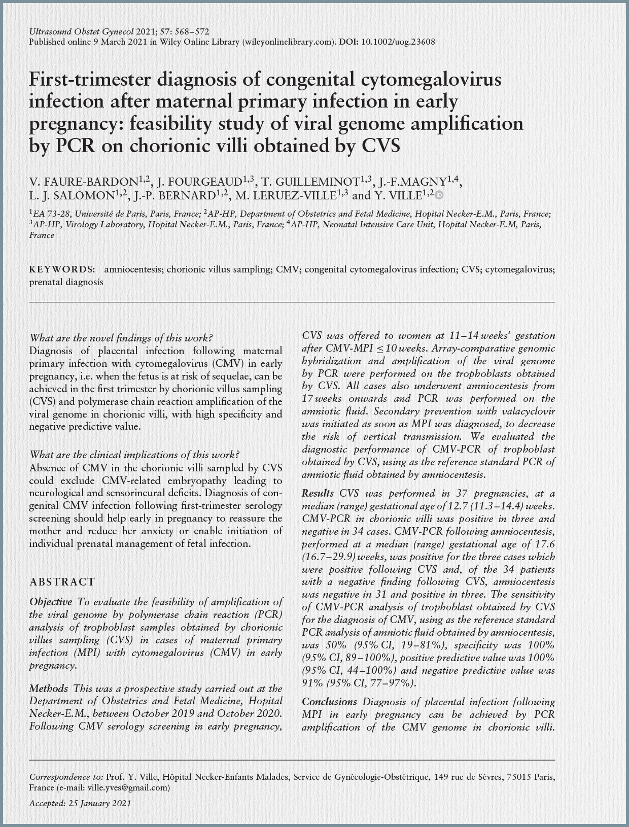
The diagnosis of CMV infection in the fetus is primarily established through CMV polymerase chain reaction (PCR) performed on amniotic fluid, considered the gold standard. Amniocentesis for this purpose is recommended after 21 weeks’ gestation and at least 6 to 8 weeks following documented maternal infection to minimize the risk of false-negative results. For newborns, diagnosis involves viral detection via PCR in urine or saliva samples collected within the first three weeks of life, with saliva noted to contain 10 times higher viral copies than urine.

In terms of prognosis, the review emphasizes that fetal abnormalities and long-term sequelae are predominantly linked to maternal primary infections acquired during the periconceptional period or the first trimester of pregnancy. While vertical transmission rates increase with advancing gestational age at the time of maternal infection, the risk of severe fetal insult significantly declines after the first trimester. Key predictors of poor neonatal prognosis include a high fetal viral load (>30,000 copies/mL) and a low platelet count (<50,000/mm³) in cord blood, alongside the presence of microcephaly on imaging. A negative amniocentesis result is highly reassuring, indicating a very low risk of severe symptoms or neurological sequelae, even if the newborn’s urine later tests positive for CMV. Long-term sequelae can affect both symptomatic (40-60%) and asymptomatic (approximately 13.5%) congenitally infected children, with SNHL being the most common, often progressive and sometimes late in onset. Other potential sequelae include vision loss, intellectual disability, and developmental delays.

Regarding prevention, there is currently no licensed CMV vaccine available, although ongoing research efforts are deeming it a high priority. Behavioral interventions, such as stringent hygiene practices, have shown some promise in reducing infection rates but face challenges related to maternal adherence and a general lack of widespread awareness campaigns.

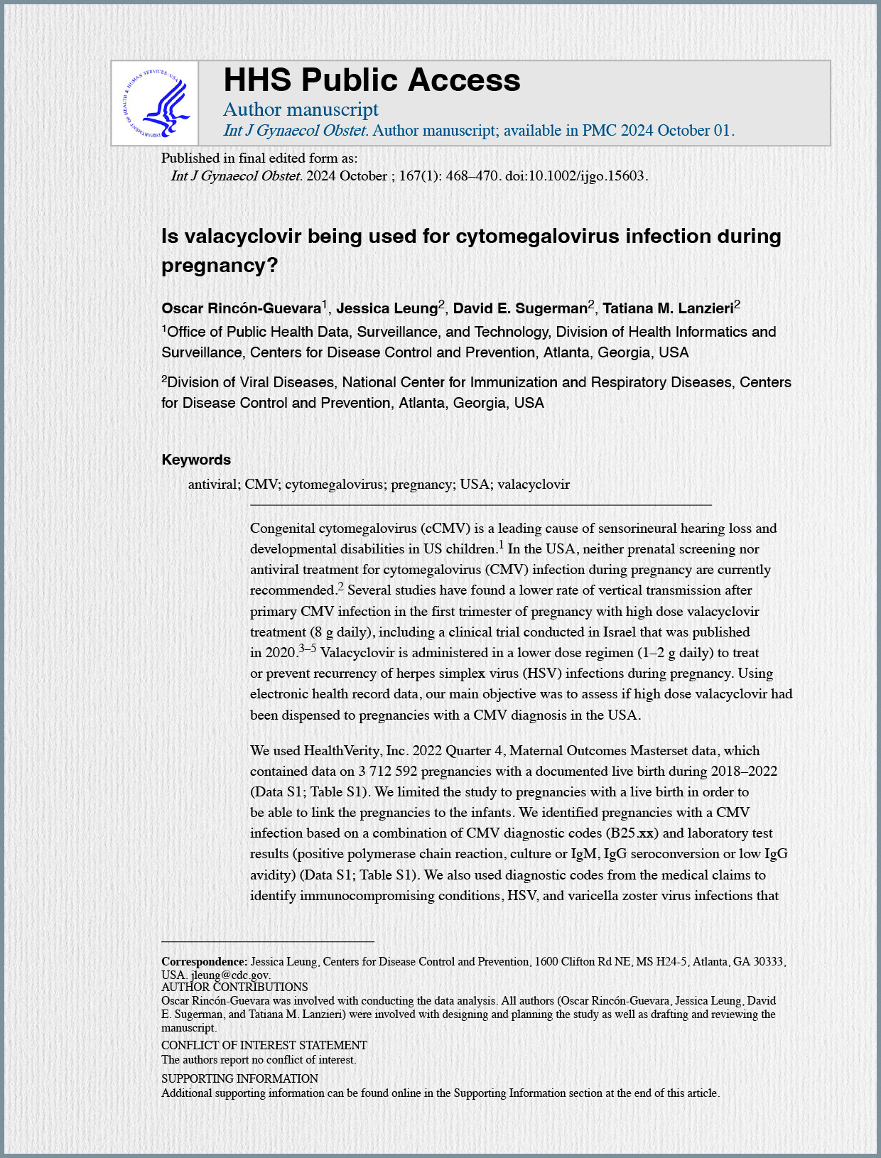
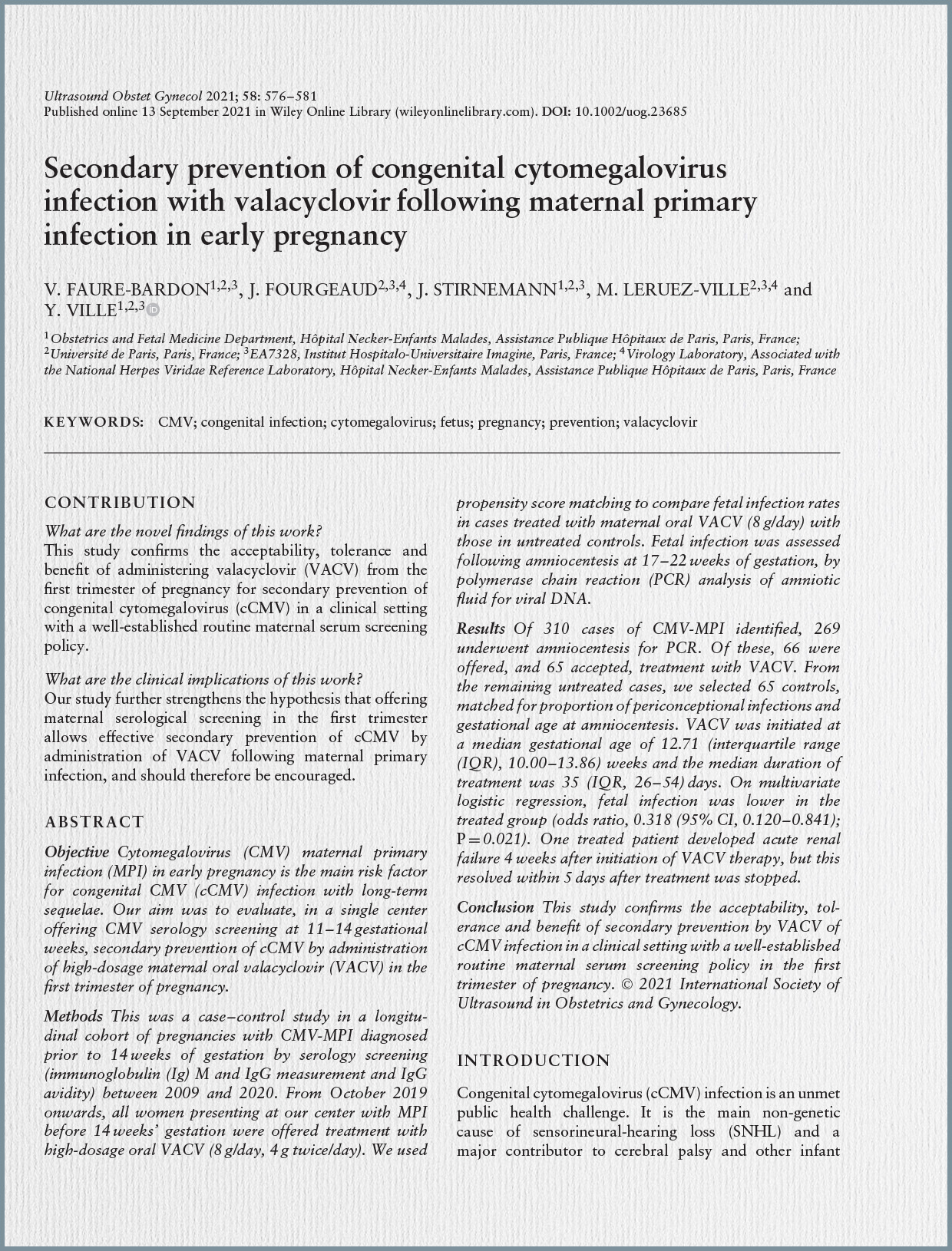
The management of CMV infection during pregnancy has significantly evolved. While potent antivirals like ganciclovir are effective in non-pregnant adults, they are not approved for use in pregnancy due to toxicity concerns. However, valacyclovir, an acyclovir prodrug, stands out as the most promising medication for preventing congenital CMV infection after maternal primary infection in early pregnancy. Clinical studies, including randomized controlled trials, have demonstrated that high-dose oral valacyclovir (8 g/day), administered from the diagnosis of MPI in early pregnancy until amniocentesis, can significantly reduce the rate of vertical transmission by up to 71%. This treatment generally exhibits a favorable safety profile, with rare and reversible side effects such as acute renal failure. Although some studies propose continuing valacyclovir until delivery to prevent late transmission, this approach lacks support from prospective randomized trials. Conversely, the use of human hyperimmune globulin for preventing vertical transmission is largely not supported by current evidence.

For symptomatic newborns, ganciclovir is the drug of choice, administered intravenously, typically for 42 days. It has shown substantial benefits in promoting neuropsychomotor development and reducing the incidence of hearing loss. Valganciclovir, an oral alternative, is also discussed. Treatment is recommended for newborns presenting with CNS involvement, isolated hearing loss, or severe manifestations such as persistent hepatitis or thrombocytopenia.

In conclusion, the review stresses that the historical reluctance to implement universal CMV screening during pregnancy, often attributed to the absence of effective therapeutic options, must be re-evaluated. Given the robust evidence supporting the safety and efficacy of valacyclovir in reducing vertical transmission following early maternal primary infection, the article advocates for amending current guidelines to incorporate CMV screening during the periconceptional period and the first trimester. Early diagnosis is paramount to facilitate timely interventions and mitigate the severe, long-term consequences of congenital CMV infection.

The authors highlight that congenital CMV (cCMV) affects up to 2% of live births globally and stands as the most common acquired cause of neurodevelopmental delay and sensorineural hearing impairment. CMV can be transmitted vertically through primary or non-primary maternal infection, with primary infection posing a higher risk of vertical transmission, ranging from 25–45% in the periconceptional period or first trimester, 45% in the second trimester, and 47–78% in the third trimester. While non-primary infection has a much lower risk of fetal infection (1–2%), if transmission occurs, the risk of postnatal neurological sequelae is similar to that of primary infection, with most symptomatic newborns from CMV-affected mothers actually born from CMV reactivation. The review’s objective was to evaluate the quality and consistency of existing CPGs concerning CMV infection in pregnancy, focusing on diagnosis, treatment, and follow-up.

The methodology involved a systematic literature search across Medline, Embase, and Web of Science databases up to April 1, 2024, in addition to guideline databases and scientific societies’ websites. The search was limited to English-language CPGs and expert consensus statements on CMV diagnosis, treatment, and follow-up in pregnancy. Studies exclusively on screening recommendations were excluded. Quality assessment of the included guidelines was performed using the Appraisal of Guidelines for Research and Evaluation (AGREE) II tool, which assesses six domains and 23 items. A quality score above 60% defined a domain as adequately addressed, with guidelines classified as “high quality” if all six domains scored above 60%. Out of 306 articles identified, 12 CPGs (7 national, 5 international) were ultimately included, with most published in the last 10 years.

Regarding diagnosis, there was a broad consensus among all included CPGs on the criteria for primary maternal CMV infection. All guidelines recommended a combination of positive CMV IgG, IgM, and low IgG avidity as the defining criteria. Seroconversion, marked by the new onset of positive CMV IgG in a previously seronegative woman, was also cited as a criterion for primary infection in 10 out of 12 studies. More recent guidelines also suggested CMV PCR in maternal blood to confirm very recent primary infection in cases with negative IgG and positive IgM. Intermediate CMV IgG avidity was included in the definition in 4 out of 12 studies, and rising CMV IgG titers were mentioned in 2 out of 12. Some guidelines advised investigating earlier antenatal serology when available.

For non-primary maternal infection, diagnostic criteria were less consistently defined, reported by only 3 out of 12 studies. These studies generally defined possible non-primary infection in cases of positive CMV IgG and IgM with high IgG avidity. However, the most recent European expert consensus stated that no valid laboratory test exists to identify women with pre-existing immunity at risk of fetal infection. Other guidelines noted that non-primary maternal infection is diagnosed only when fetal infection is confirmed.

For fetal infection, there was full agreement that CMV PCR in the amniotic fluid sampled by amniocentesis is the gold standard. However, the timing of amniocentesis varied among guidelines: some indicated 17 weeks of gestation, at least 6 to 8 weeks from maternal infection, while others recommended 20 or 21 weeks, with time intervals from infection ranging from 5-6 weeks to at least 8 weeks.

In terms of management, heterogeneity was observed, particularly concerning fetal surveillance in confirmed infections. Serial ultrasound assessment was encouraged by most (9 out of 12) societies. Fetal brain MRI was recommended as a supplementary exam by 7 out of 12 societies, typically in the third trimester (28 to 34 weeks). The possible scenarios of negative amniocentesis or non-confirmed fetal infection were explicitly discussed in only two guidelines, with differing recommendations on continued surveillance. Additional fetal testing, such as fetal blood sampling for platelet count or prognostic assessment, was only suggested by a few societies. Referral to a tertiary unit with expertise in fetal infections was advised by 7 out of 12 guidelines.

Regarding treatment, the guidelines showed evolving trends. Before 2022, most societies did not recommend antiviral use outside research settings, with only one suggesting considering Valacyclovir for primary infection in the first trimester. However, from 2022 onwards, two guidelines explicitly advocated prompt Valacyclovir treatment (8 g/day, administered as 2g/4 times a day) for primary infection to prevent vertical transmission. The European consensus also considered continuing Valacyclovir for confirmed fetal infection after expert discussion. It is notable that only 50% of the included guidelines were published after a key randomized controlled trial in 2020 that highlighted Valacyclovir’s role in preventing vertical transmission. The use of CMV hyperimmune globulins was generally not recommended or recommended against by 9 out of 12 CPGs, although one suggested considering it for fetal infection and another for very recent first-trimester primary infection.

The quality assessment using AGREE II revealed that 8 out of 12 guidelines were deemed “high quality,” 3 “moderate quality,” and 1 “low quality”. The domains with the lowest scores were “Rigor of Development” and “Applicability,” suggesting that guidelines often lack detailed descriptions of their development process and comprehensive advice on implementation.

The review concludes that while there is consensus on diagnostic criteria for primary maternal and fetal CMV infection, significant heterogeneity persists in the timing of invasive fetal testing, fetal ultrasound surveillance, and the use of fetal brain MRI. The recommendations for treatment have shown a progressive inclusion of antiviral therapy, particularly Valacyclovir, over recent years, reflecting new evidence. The authors emphasize that maternal serological screening in pregnancy needs to be reconsidered given the emerging evidence on the benefits of early treatment.

Important clinical practice points not extensively addressed in the current guidelines include the management of non-confirmed fetal infection (e.g., when amniocentesis is declined) and the specific management of non-primary maternal infection, despite evidence that fetal infection in such cases can be as severe as after primary infection. The review notes that the majority of neonates with symptomatic cCMV infection were born to mothers with CMV reactivation, yet only one expert consensus recommended testing neonates in cases of both primary and non-primary maternal infection. This highlights the need for comprehensive prenatal recommendations for managing non-primary infections. The authors acknowledge the study’s strengths, such as extensive literature search and detailed quality assessment, but also its limitations, including the inclusion of lower-quality guidelines and restriction to English-written publications, noting that guidelines will likely be updated as evidence evolves.

The pathogenesis of HCMV during pregnancy is a complex interplay between the virus, the maternal immune system, and the developing fetus. HCMV initially infects epithelial cells via mucosal surfaces, then disseminates systemically, often remaining asymptomatic or with mild maternal symptoms during primary infection. The virus can establish latency in myeloid progenitor cells and reactivate, especially during pregnancy, leading to viral replication and potential fetal transmission. The maternal immune system responds with innate (NK cells, interferons) and adaptive (T-cells, antibodies) mechanisms, though IgG antibodies, which cross the placenta, may not always prevent fetal infection.

Placental infection is a critical step, as HCMV can infect trophoblast cells, disrupting their function and leading to impaired placental development and function. This infection can also induce inflammation, contributing to complications such as placental insufficiency and fetal growth restriction. Primary HCMV infection during pregnancy carries a 30% to 40% risk of intrauterine transmission, with the highest risk occurring in the first trimester. Once infected, the fetus can experience outcomes ranging from asymptomatic infection to severe congenital HCMV (cCMV) disease. The virus can affect various fetal organs, including the brain, liver, and spleen, potentially leading to microcephaly, intracranial calcifications, hearing loss, vision impairment, and developmental delays. Approximately 18% of infants born to women with primary HCMV infection are symptomatic at birth. Crucially, up to 25% of infants asymptomatic at birth developed sequelae within the first two years of life, including sensorineural hearing loss and cognitive deficits. Severe outcomes are more likely when maternal infection occurs in the first half of pregnancy. The frequency of clinical sequelae decreases significantly with advancing gestational age of maternal seroconversion: 28.8% in the periconceptional period, 19.3% in the first trimester, 0.9% in the second trimester, and 0.4% in the third trimester.

The article then delves into the limitations of current diagnostic methods:

  • Serological Diagnostics: While IgM generally indicates recent infection, it is prone to false positives due to cross-reactivity or non-specific factors, and can remain elevated or reappear during reactivations. IgG indicates past exposure but does not pinpoint timing, which is critical in pregnancy. Although a 4-fold increase in IgG has been proposed as a prognostic biomarker, it requires prior serological status, which is not always available. IgG avidity testing helps, with low avidity suggesting recent infection, but results are not always conclusive. These ambiguities often lead to misclassification of infection timing and type, and serology offers limited prognostic power for fetal involvement or severity.
  • HCMV DNAemia (qPCR in blood): Quantitative polymerase chain reaction (qPCR) in blood is highly sensitive for detecting active viral replication, especially in early infection. However, it does not provide information on the precise timing or type of infection (primary, reinfection, or reactivation). HCMV DNA can persist for variable periods, complicating timing inference. While detection of HCMV DNA in maternal blood correlates with a significantly increased risk of vertical transmission (5- to 13-fold higher depending on trimester), its value as a predictive biomarker for treatment response or neonatal outcomes is uncertain.

The review highlights the established role of HCMV DNA in amniotic fluid samples as the gold standard for diagnosing fetal CMV infection. This test is typically performed after approximately 20 weeks + 1 day of gestation, at least 8 weeks after suspected primary maternal infection. Recent studies suggest reliable diagnosis from 17 weeks onwards, provided the 8-week interval from maternal infection is observed, demonstrating a sensitivity of 87.2–95.8% and a specificity close to 100%. Discarding the first 1 mL of fluid reduces maternal cell contamination. The viral load in amniotic fluid has been studied as a possible predictor of symptomatic disease at birth, with one study suggesting >100,000 copies/mL may distinguish symptomatic cases. However, a robust, validated viral load threshold is still needed. CMV DNA in maternal urine samples also shows promise as a valuable marker, with a statistically significant association between its presence and newborn infection, particularly when viral loads are high.

Emerging prognostic and predictive biomarkers are also explored:

  • HCMV T-Cell Immunity: The maternal immune system undergoes delicate modifications during pregnancy. The peripheral HCMV T-cell response offers a promising predictive diagnostic tool for cCMV. While it provides functional insight not offered by serology or PCR, current assays lack specificity to differentiate recent from past infection. Studies evaluating HCMV-specific enzyme-linked immunospot (ELISpot) and interferon gamma-releasing assay (IGRA) have shown inconsistent results, with some suggesting high ELISpot levels correlate with increased fetal transmission risk, while others found no association between T-cell responses and transmission risk, instead emphasizing the importance of viral load. A paradoxically increased risk of cCMV has been linked to strong T-cell responses with low IgG avidity, possibly due to an imbalanced immune response and inflammation. A novel “relative response” (RR) method for IFN-γ responses showed a lower RR (<1.8%) was significantly associated with a reduced transmission risk.
  • Non-Invasive Prenatal Testing (NIPT): NIPT for HCMV DNA in maternal cell-free DNA (cfDNA-HCMV) represents a significant innovation. Large cohort studies have examined its use for early detection. One study detected HCMV DNA in about 0.05% of a general population, indicating its presence even in healthy, asymptomatic individuals. Another multicenter study found that cfDNA-HCMV fragment prevalence correlated with active HCMV infections (recent primary or reactivated), suggesting its utility as a screening method to identify pregnancies at risk for maternal-fetal viral transmission. While cfDNA-HCMV-negative samples largely corresponded to qPCR-negative and serology indicating no recent infection, 67.8% of cfDNA-HCMV-positive samples were qPCR-negative, highlighting qPCR’s limitations for very low viral loads. NIPT is seen as a promising non-invasive method for early detection and understanding virus-host genetic interactions.
  • Exosomes: These small membrane-bound vesicles play roles in viral infections, including immune stimulation and antigen presentation. In pregnancy, placental exosomes are involved in immune regulation and fetal development. HCMV infection significantly alters the composition of exosomes, which could negatively impact placental function and fetal development. While exosomes could serve as non-invasive biomarkers and therapeutic targets, significant research gaps remain.

The article concludes by outlining unmet needs and future perspectives. Despite advancements, critical gaps persist in biomarker development, therapeutic monitoring, and risk stratification. The lack of validated predictive biomarkers hinders the effective tailoring of antiviral treatment strategies, such as valaciclovir therapy. There is a need for biomarkers that can assess treatment efficacy, detect early signs of drug resistance (e.g., resistance-associated mutations in circulating HCMV DNA), and anticipate adverse effects. While maternal DNAemia is a prognostic marker for vertical transmission, its predictive value for treatment response and fetal outcomes needs clarification. Future research should focus on identifying biomarkers that can distinguish pregnancies that would most benefit from antiviral therapy, optimizing timing, and reducing unnecessary drug exposure. Standardization of NIPT for HCMV screening and defining clinical thresholds for intervention are essential. Further exploration of exosome-derived biomarkers is also warranted. The integration of novel predictive biomarkers, personalized treatment approaches, and innovative non-invasive diagnostics is crucial for improving HCMV management in pregnancy, aiming to optimize maternal and fetal outcomes while minimizing unnecessary interventions.

Currently, the definitive diagnosis of fetal CMV infection relies on amplifying the CMV genome in amniotic fluid (AF) using polymerase chain reaction (PCR), which is performed no earlier than 17 weeks of gestation. This waiting period, often several weeks between the detection of maternal primary infection and the possibility of fetal diagnosis, creates considerable psychological burden and anxiety for expectant mothers. Recent research has shown that administering valacyclovir (VACV) following first-trimester MPI can significantly reduce the risk of vertical transmission by 71%. Given these factors, the study hypothesized that early fetal infection could be diagnosed by detecting CMV in the trophoblast as early as 14 weeks of gestation. Furthermore, they posited that the absence of CMV at this early stage in the trophoblast would effectively exclude CMV-related embryopathy leading to neurological and sensorineural deficits. The primary objective of this study was to evaluate the feasibility of performing PCR analysis on trophoblast samples obtained via chorionic villus sampling (CVS) in cases where maternal primary infection occurred at up to 10 weeks of gestation.

The study was a prospective investigation conducted at the Department of Obstetrics and Fetal Medicine, Hopital Necker-E.M., between October 2019 and October 2020. Participating women had a diagnosis of CMV-MPI at or before 10 weeks of gestation and underwent CVS between 11 and 14 weeks. Reasons for offering CVS included the need for early cytogenetic reassurance or cases where women were considering termination of pregnancy due to CMV infection and desired an early risk assessment. All included women were over 18 years old, had a singleton pregnancy with a normal prenatal genetic/cytogenetic diagnosis, and provided consent for CMV-PCR on chorionic villi. Crucially, all participants also agreed to undergo a follow-up amniocentesis for CMV-PCR of amniotic fluid at or after 17 weeks of gestation, ensuring at least an 8-week interval since the estimated date of maternal primary infection.

Maternal primary infection was diagnosed before 14 weeks through serological testing, involving IgG and IgM measurements and IgG avidity tests for IgM-positive cases. A low or intermediate IgG avidity with positive IgG and IgM, or seroconversion, indicated MPI. The reference standard for diagnosing fetal infection was CMV-PCR on amniotic fluid. Amniocentesis was performed as early as 17 weeks, provided an 8-week interval from MPI was observed. This practice was previously validated, demonstrating high diagnostic performance (sensitivity 95.8%, specificity 100%, positive predictive value 100%, negative predictive value 97.7%) for diagnosing cCMV neonatal infection when performed between 17 and 20 weeks with the adequate interval. Both CMV-PCR on trophoblast and amniotic fluid were performed using the CMV R-gene assay, which has a sensitivity of 2.0 log10 copies/mL. All women were offered an 8 g/day regimen of VACV upon biological confirmation of MPI, which continued until amniocentesis results were available. If fetal infection was diagnosed, treatment continued until delivery. Neonatal CMV infection was tested within the first 10 days of life via PCR on urine and/or saliva samples.

The study enrolled 37 pregnancies meeting the criteria. The median gestational age at CVS was 12.7 weeks (range: 11.3–14.4 weeks), and no CVS-related complications were observed. CMV-PCR on chorionic villi was positive in 3 cases and negative in 34. Subsequently, amniocentesis was performed at a median gestational age of 17.6 weeks (range: 16.7–29.9 weeks). All three cases that were positive at CVS were also confirmed as fetal infection by positive CMV-PCR in the amniotic fluid. Of the 34 cases with a negative CVS-PCR result, 31 also had a negative amniotic fluid PCR. However, three cases presented discordant results, with negative CVS-PCR but positive amniotic fluid PCR.

The diagnostic performance of CMV-PCR on trophoblast obtained by CVS, using amniotic fluid PCR as the reference standard, was assessed as follows:

  • Sensitivity: 50% (95% CI, 19–81%).
  • Specificity: 100% (95% CI, 89–100%).
  • Positive Predictive Value (PPV): 100% (95% CI, 44–100%).
  • Negative Predictive Value (NPV): 91% (95% CI, 77–97%).

Postnatal outcome data were available for 24 of the 37 pregnancies at the time of writing, with 13 still ongoing. Among the three cases with concordant CVS-positive/amniotic fluid-positive results, two infants were asymptomatic (one at birth and one at 5 months of age), while one showed unilateral hearing loss. Of the three discordant cases (CVS-negative/amniotic fluid-positive), one pregnancy was terminated (autopsy showed no viral lesions), one infant was asymptomatic at 5 months, and one pregnancy was still ongoing with normal antenatal ultrasound imaging. Maternal CMV viremia before CVS was mostly negative, and in the one case with both positive maternal viremia and trophoblast, the viral DNA load in the trophoblast was 10-fold higher than in maternal blood, suggesting no contamination from the invasive procedure. Discordant CVS-negative/AF-positive cases also had negative maternal blood PCR before CVS, further excluding iatrogenic contamination.

The discussion emphasizes that the findings demonstrate the feasibility of diagnosing placental infection following CMV-MPI in early pregnancy through PCR amplification of the viral genome in trophoblast obtained by CVS. The observed high negative predictive value and specificity are encouraging. This is particularly relevant as there is a growing consensus that first-trimester maternal infection is the sole cause of fetal CMV infection-related neurological impairment and hearing loss, and VACV can significantly decrease vertical transmission. While amniocentesis remains the gold standard, previous studies have shown that 8-10% of negative amniocenteses were followed by the birth of an infected neonate, though these neonates remained asymptomatic. This suggests that later transplacental passage, occurring after the embryonic period, might result in positive neonatal tests but without symptomatic cCMV.

The authors propose that the absence of CMV in the trophoblast at the end of the first trimester should exclude CMV-related embryopathy leading to sequelae, even if later transplacental passage leads to positive findings in amniotic fluid or neonates. The proposed strategy involves performing two invasive tests, CVS and amniocentesis, each carrying a low miscarriage risk of less than 1 in 1000. This dual approach, while novel, aims to reduce maternal anxiety caused by the long waiting period for definitive fetal diagnosis. Early diagnosis would enable informed, shared decision-making, potentially preventing “erratic decisions” like termination based solely on screening results. Although the small sample size and unknown long-term prognosis of discordant cases are limitations, the study concludes that these findings could help establish CVS as the diagnostic test of choice following maternal serology screening in early pregnancy. However, long-term follow-up is necessary to fully confirm whether a negative CVS-PCR after 12 weeks reliably excludes CMV-related embryopathy leading to sequelae.

Despite this significant public health impact, neither prenatal screening for CMV nor antiviral treatment during pregnancy are currently recommended in the USA. This lack of recommendation persists even as several studies, including a notable clinical trial published in 2020 from Israel, have indicated a reduced rate of vertical transmission of CMV following primary infection in the first trimester of pregnancy when high-dose valacyclovir (8 grams daily) is administered. It is also noted that valacyclovir is conventionally used at lower dosages, typically 1–2 grams daily, for the treatment or prevention of recurrent herpes simplex virus (HSV) infections during pregnancy. Against this backdrop, the primary objective of this study was to ascertain whether high-dose valacyclovir has been dispensed to pregnancies with a CMV diagnosis in the USA.

The study utilized a comprehensive dataset from HealthVerity, Inc.’s 2022 Quarter 4 Maternal Outcomes Masterset. This extensive database encompassed 3,712,592 pregnancies with documented live births occurring between 2018 and 2022. The decision to limit the study to pregnancies resulting in live births was made to enable the linkage of data between mothers and their infants. To identify pregnancies affected by CMV infection, the researchers employed a multi-faceted approach, combining CMV diagnostic codes (B25.xx) with documented positive laboratory test results, which included polymerase chain reaction (PCR), culture, IgM positivity, IgG seroconversion, or low IgG avidity. Beyond CMV, the study also leveraged diagnostic codes from medical claims to identify other conditions that might justify antiviral medication use, such as immunocompromising conditions, HSV infections, and varicella zoster virus (VZV) infections. The specific antiviral medications under scrutiny, namely valacyclovir, acyclovir, and famciclovir, were identified through dispensed pharmacy claims. For the diagnosis of cCMV in infants, the study identified relevant diagnostic codes (P35.1 for cCMV infection or B25.xx for CMV disease) within 45 days of the infant’s birth among the linked live births. The ethical considerations for the project were addressed, and it was determined to be exempt from formal review by the Institutional Review Board.

The findings revealed that out of the vast cohort of over 3.7 million pregnancies, 1,884 (0.05%) were identified as having a CMV infection. A breakdown of how these CMV diagnoses were captured showed that 1,023 cases (54%) were identified solely through diagnostic codes, while 654 cases (35%) relied exclusively on positive laboratory test results. A smaller proportion, 207 cases (11%), had both diagnostic codes and positive laboratory tests. The median gestational age at which the first CMV diagnosis was recorded was 25 weeks.

An analysis of co-occurring medical conditions among the CMV-diagnosed pregnancies indicated a higher prevalence of other health issues compared to those without a CMV diagnosis. Specifically, immunocompromising conditions were noted in 114 (6%) of pregnant individuals with CMV infection, a rate significantly higher than the 1% observed in pregnancies without CMV. Similarly, other herpes virus infections, exclusively identified as HSV in this dataset, were present in 261 (14%) of CMV-infected pregnancies, compared to 6% in the non-CMV group.

Regarding the dispensing of antiviral treatments, the study observed that antivirals were dispensed for 185 (10%) of pregnancies that also had a CMV diagnosis. Within this group of 185, 13 (7%) had an immunocompromising condition, and a substantial majority, 114 (62%), had other herpes virus infections, predominantly HSV. Valacyclovir was the most frequently dispensed antiviral medication within this cohort. The median gestational age at which valacyclovir was first dispensed was 34 weeks, with 146 (79%) of these dispensations occurring during the third trimester of pregnancy. The typical daily dose of valacyclovir dispensed was 1000 mg, for a duration of 30 days.

A critical aspect of the study was to identify the use of high-dose valacyclovir (defined as ≥8 grams per day), as this dosage is specifically associated with the prevention of vertical CMV transmission in recent research. The findings showed that a very small proportion, only 17 (9%) of the pregnancies with a CMV diagnosis, received a daily dose of ≥8 grams of valacyclovir. All of these 17 cases had a documented CMV diagnosis during pregnancy. The dispensing of these high doses was primarily observed in the later years of the study period, with 7 cases (41%) in 2020 and 10 cases (59%) in 2021. Consistent with the overall trend, most of these high-dose treatments were initiated during the second or third trimester of pregnancy, and only one of these 17 individuals (6%) was immunocompromised.

Further analysis connected maternal antiviral treatment to cCMV diagnoses in infants. Among 558 pregnancies with a CMV diagnosis that were linked to live births, 71 infants (13%) received a diagnosis of cCMV. Of these 71 cCMV-diagnosed infants, 10 (14%) of their mothers had received documented antiviral treatment during pregnancy. For comparison, among the much larger group of 1,121,663 pregnancies without a recorded CMV diagnosis that were linked to live births, 104 infants (0.01%) were diagnosed with cCMV. Interestingly, among these 104 infants, 50 (48%) of their mothers had received antiviral treatment during pregnancy. The study found no significant difference in the median gestational age at initial treatment (both 34 weeks) or the median daily dose (both 1000 mg) when comparing pregnancies with and without a CMV infection diagnosis.

In their discussion, the authors conclude that the observed characteristics of valacyclovir dispensing, including the typical dosage, duration, and timing, coupled with the high proportion of HSV diagnoses, strongly suggest that the majority of valacyclovir use was intended for the treatment or suppression of recurrent genital herpes, irrespective of whether a CMV diagnosis was also recorded during pregnancy. The study clearly indicates that most pregnant individuals did not receive high-dose valacyclovir, and the limited instances where high doses were dispensed occurred predominantly after the first trimester of pregnancy. The authors acknowledge several limitations of their study, including that it only assessed pregnancies resulting in live birth outcomes, and there is a possibility that diagnostic codes or treatment records might have been missed or inaccurately documented. Furthermore, the study was unable to evaluate the potential side effects of the treatments administered. Despite these limitations, the collected data are considered valuable for future efforts in monitoring trends related to laboratory testing, diagnoses, and treatment practices for CMV infection during pregnancy. The authors emphasize that a comprehensive review of the evidence concerning the efficacy and safety of high-dose valacyclovir administered early in pregnancy for the prevention of vertical CMV transmission would be highly beneficial in shaping future guidelines for prenatal CMV screening and treatment in the USA.

These severe long-term sequelae primarily occur when fetal infection follows MPI in the first trimester of pregnancy. Historically, universal serological screening for CMV in pregnancy was not deemed justifiable due to the absence of an effective therapy to reduce fetal infection rates or improve outcomes for infected fetuses. However, the landscape began to change with a recent randomized controlled trial (RCT) that indicated high-dosage oral VACV could significantly reduce fetal infection rates when administered after first-trimester MPI. The current study aimed to provide further confirmatory evidence regarding the feasibility, acceptability, and impact of VACV treatment, including the absence of significant adverse effects, in a real-world clinical setting that already implements routine maternal serum screening for CMV.

The study employed a case-control design within a longitudinal cohort of pregnancies that had a confirmed CMV-MPI diagnosis before 14 weeks’ gestation through serological screening. The research was conducted at a single center that offered routine serological screening for CMV during the first trimester, specifically between 11 and 14 weeks of gestation, along with combined aneuploidy screening. Maternal primary infection was precisely classified as either periconceptional (<0 weeks of gestation) or occurring during the first trimester (0–14 weeks of gestation). For inclusion in the primary analysis, all patients were required to have undergone amniocentesis for viral DNA PCR analysis, performed at least 8 weeks after the MPI diagnosis and from 17 weeks’ gestation onwards.

A crucial intervention in this study involved the administration of valacyclovir. From October 2019 onwards, all pregnant women presenting at the center with CMV-MPI diagnosed before 14 weeks’ gestation were offered high-dosage oral VACV (8 grams per day, specifically administered as 4 grams twice daily). Treatment commenced as soon as the MPI was biologically confirmed. Its duration depended on the amniocentesis results: if the results were negative for CMV, treatment was discontinued; if positive, it was continued until birth or termination of pregnancy. To monitor for potential side effects, maternal serum transaminase and creatinine levels were measured before treatment initiation and then fortnightly. Control subjects were drawn from women diagnosed with CMV-MPI prior to October 2019, those referred later but already in their second trimester (and thus outside the study’s early-pregnancy treatment window), or those who declined VACV treatment. The primary endpoint for evaluating the treatment’s effectiveness was the rate of fetal infection, determined by PCR analysis of amniotic fluid for CMV-DNA, following amniocentesis performed between 17 and 22 weeks’ gestation.

For the statistical analysis, a propensity score matching technique was utilized to compare the VACV-treated cases with untreated controls. This method allowed for the balancing of key potential confounding factors, specifically the timing of MPI (periconceptional or first-trimester) and the gestational age at amniocentesis. Logistic regression models were then applied to assess the odds ratios (OR) for vertical transmission. The diagnosis of CMV-MPI itself was rigorously established through maternal serology (IgG, IgM, and IgG avidity tests) performed before 14 weeks, using specific commercial assays (LIAISON XL® CMV IgG II and IgM assays, VIDAS® CMV IgG avidity II assay). Avidity index interpretations, such as an index <0.40 indicating MPI in the last 3 months, were strictly followed.

Between 2009 and December 2020, the study identified 310 women with CMV-MPI in early pregnancy. Of these, 269 underwent amniocentesis. When VACV was offered to 66 women from October 2019, a high acceptance rate was observed, with 65 women (98%) opting for the treatment. Treatment was typically initiated at a median gestational age of 12.71 weeks (interquartile range (IQR): 10.00–13.86 weeks), and the median duration of treatment prior to amniocentesis was 35 days (IQR: 26–54 days). A matched control group of 65 untreated women was established.

The study’s results demonstrated a significantly lower rate of vertical transmission in the treatment group compared to the untreated controls. Overall, 12% (8/65) of VACV-treated pregnancies resulted in vertical CMV transmission, in contrast to 29% (19/65) in the untreated control group (P = 0.029). When analyzing subgroups, a similar reduction was observed for cases with first-trimester MPI (P = 0.027), although the difference was not statistically significant for periconceptional MPI (P = 0.60). Multivariate analysis further confirmed that VACV treatment was associated with a significant decrease in vertical CMV transmission (Odds Ratio: 0.318; 95% CI: 0.120–0.841; P = 0.021). Importantly, the study found that the prevention of fetal infection improved with increasing duration of VACV treatment [488, Figure 2b]. Regarding safety, only one out of the 65 treated patients experienced an adverse event: acute renal failure, which resolved within 5 days of discontinuing VACV. This particular patient, despite the confirmed fetal infection, later delivered an asymptomatic newborn.

The authors emphasize that CMV embryopathy can lead to SNHL and neurological deficits in approximately one-third of infected cases, underscoring the importance of prevention studies focusing on MPI occurring before 14 weeks’ gestation. The findings confirm the acceptability and benefit of administering VACV for preventing cCMV infection following early pregnancy MPI. VACV is highlighted as “considerably cheaper” than hyperimmune immunoglobulins, an alternative treatment option. While maternal tolerance to VACV was generally good, the authors stress the need for counseling regarding the potential for renal toxicity and recommend appropriate surveillance of renal function during treatment. They propose that the observed renal toxicity in their study, a reversible acute renal dysfunction, was likely due to acyclovir (ACV) accumulation and crystal precipitation in renal proximal tubule cells, caused by the metabolism of VACV to ACV. To mitigate this, they suggest that administering the 8-gram daily dose in four 2-gram doses throughout the day, rather than two 4-gram doses, might reduce the risk of acute renal failure due to better ACV distribution and lower peak concentrations. The study also suggests that the more pronounced effect of VACV treatment in first-trimester MPI compared to periconceptional infections could be due to the lower baseline transmission risk in periconceptional cases or the delayed initiation of treatment relative to the actual infection time. The use of amniotic fluid PCR at 17–22 weeks gestation as a reliable indicator of transplacental passage is justified, as any false-negative results are typically associated with very late and asymptomatic viral transmission.

A key strength of this study lies in its precise dating of MPI and the rigorous application of propensity score matching to control for potential confounding factors, despite not being a randomized controlled trial itself. However, the authors acknowledge a limitation that their findings might represent a “best-case scenario” due to the well-established screening and care pathways in their reference center.

In conclusion, this study provides strong further evidence supporting the effectiveness of maternal serological screening in the first trimester, followed by secondary prevention with high-dose valacyclovir, to reduce congenital CMV infection following maternal primary infection. The authors “strongly encourage” this practice and call for long-term assessment of these children, potentially through the establishment of an international registry, to comprehensively evaluate the impact of this policy on neonatal short- and long-term cCMV-related morbidity.

Summary sheet

Congenital Cytomegalovirus (cCMV) infection is a significant public health challenge and the most common congenital viral infection worldwide, affecting approximately 0.7% to 1% of all live births. It is the leading non-genetic cause of sensorineural hearing loss (SNHL) and a major contributor to neurodevelopmental delay and cerebral palsy in children.

  1. Transmission & Pathophysiology:
    • CMV, a DNA herpesvirus, can cause lifelong latent infection but can reactivate or lead to reinfection with new strains.
    • Vertical transmission can occur through primary maternal infection (MPI), reactivation, or reinfection, though MPI carries the highest risk of severe congenital infection.
    • The most critical factor for long-term sequelae is maternal infection acquired in the first trimester of pregnancy. The risk of fetal abnormalities from MPI decreases significantly after the first trimester.
    • The virus crosses the placenta, replicates in trophoblast cells, and can directly damage fetal organs, especially the central nervous system (CNS), liver, and spleen.
    • Fetal insult rates (CNS malformation or neurological symptoms at birth) following vertical transmission are significantly higher in periconceptional (28.8%) and first-trimester (19.3%) infections compared to second (0.9%) and third (0.4%) trimesters.
  2. Diagnosis:
    • Maternal Serology:
      • Recommendation: Maternal CMV serology (IgG, IgM, IgG avidity) should be performed in the first trimester of pregnancy, as early as possible, followed by retesting seronegative women every 4 weeks until 14–16 weeks.
      • Diagnosis of Primary Infection: Based on CMV IgG seroconversion (new onset of positive IgG in previously seronegative woman), or positive IgM with positive IgG and low IgG avidity. High avidity generally excludes recent infection (<3 months).
      • Non-primary Infection: More difficult to diagnose serologically; positive IgM with high IgG avidity or rising IgG titers may suggest it.
    • Fetal Diagnosis:
      • Gold Standard: CMV Polymerase Chain Reaction (PCR) on amniotic fluid collected via amniocentesis.
      • Timing: Optimal sensitivity is achieved when amniocentesis is performed at or after 17+0 weeks of gestation and at least 8 weeks after maternal primary infection (MPI).
      • Negative Amniocentesis: A negative CMV PCR in amniotic fluid following timely amniocentesis ensures absence of long-term sequelae. Late fetal infection (after amniocentesis) is generally not associated with long-term sequelae.
      • Emerging Methods: Chorionic villus sampling (CVS) and PCR on chorionic villi in the first trimester (11–14 weeks) can detect placental infection, with high specificity (100%) and negative predictive value (91%) to exclude embryopathy leading to sequelae, but sensitivity is lower (50%). Non-invasive prenatal testing (NIPT) for cell-free CMV DNA (cfDNA-HCMV) shows promise for early detection but requires further validation.
    • Neonatal Diagnosis:
      • Timing: PCR should be performed on a sample collected within 3 weeks of birth (ideally as soon as possible) to distinguish congenital from postnatal infection.
      • Samples: Urine or saliva are preferred over dried blood spots (DBS) due to higher sensitivity, though DBS can be used for retrospective diagnosis. A positive saliva PCR should be confirmed with a urine sample.
      • Indications: Suspected or confirmed MPI during pregnancy, abnormal fetal imaging, clinical manifestations (e.g., SNHL), or symmetric IUGR.
  3. Prognosis & Fetal Imaging:
    • Predictors of Poor Prognosis:
      • Maternal infection timing: Periconceptional period and first trimester.
      • Fetal imaging: Severe brain involvement is a strong predictor of poor prognosis; microcephaly is consistently predictive of unfavorable outcomes (up to 95% of cases).
      • Cordocentesis findings: Platelet count <50,000/mm³ and viral load >30,000 copies/mL.
    • Ultrasound Findings:
      • Routine detailed ultrasound is not an appropriate screening tool for cCMV leading to long-term sequelae without knowledge of maternal serostatus.
      • Targeted ultrasound in known infected fetuses has high sensitivity (91%) and NPV (96%) for detecting long-term sequelae.
      • Extracerebral findings (e.g., FGR, ascites, placentomegaly, hyperechogenic bowel, hepatosplenomegaly) are usually initial symptoms of systemic infection.
      • CNS findings (e.g., ventriculomegaly, periventricular calcifications, microcephaly, posterior fossa abnormalities) indicate more severe disease.
    • MRI:
      • Recommended as a complementary tool in infected fetuses, especially in the third trimester (28–34 weeks), as it can reveal significant CNS pathology missed by ultrasound.
      • Normal ultrasound and MRI findings have a very high negative predictive value (close to 100%) for moderate to severe sequelae, but a residual risk of isolated unilateral SNHL (around 10–17%) remains.
  4. Prevention & Treatment:
    • Primary Prevention (before infection):
      • Hygiene measures (e.g., hand washing, avoiding contact with young children’s urine/saliva) are currently the only available strategy and can significantly reduce risk of MPI. Education for women of childbearing age and healthcare professionals is recommended.
      • No licensed CMV vaccine is currently available, though candidates are in clinical trials.
    • Secondary Prevention (preventing vertical transmission after maternal infection):
      • Valacyclovir: Recommended for maternal primary infection (MPI) in the periconceptional period or first trimester of pregnancy.
        • Dosage: Oral valacyclovir at 8 g/day (preferably 2g four times daily to minimize renal side effects) should be administered as early as possible after diagnosis until amniocentesis.
        • Efficacy: Reduces vertical transmission by ~70–71%.
        • Safety: Generally well-tolerated; rare side effects include reversible acute renal failure.
      • Hyperimmune Globulin (HIG): Not recommended at 100 IU/kg every 4 weeks. Higher doses (200 IU/kg every 2 weeks) for very recent first-trimester MPI may be considered but more evidence is needed.
    • Tertiary Prevention (treating infected neonates):
      • Valganciclovir (or Ganciclovir IV): Recommended for newborns with significant CMV-related symptoms at birth (especially CNS-related symptoms and isolated SNHL).
      • Timing: Should be started as soon as possible and before 1 month of age; treatment between 1 and 3 months may also be beneficial.
      • Benefits: Modest benefits in preserving hearing and improving neurodevelopmental scores.
      • Specific Cases: 6 weeks of treatment for isolated persistent hepatitis or thrombocytopenia; no treatment for isolated IUGR.
  5. Long-term Follow-up:
    • Children with cCMV and confirmed transmission in the first trimester or unknown timing of transmission should be followed up to at least 6 years of age for specialized management.
    • Asymptomatic children with normal imaging and documented MPI in the second or third trimester may follow standard pediatric care.
    • Hearing Follow-up: Recommended until at least 5 years of age for infants with normal hearing at birth but unknown timing of infection or known first-trimester infection. Lifelong regular hearing testing for those with hearing loss at birth.
    • Neurodevelopmental Assessment: At 24–36 months for high-risk children.
    • Ophthalmological Follow-up: Only recommended for infants with retinitis at birth.
    • Vestibular Testing: Recommended within the first year of life for high-risk children (first-trimester maternal infection, unknown timing, hearing loss, developmental delay).

Podcast

Course Outline: Congenital Cytomegalovirus Infection

  • What is Congenital Cytomegalovirus (cCMV)?
    • CMV is an enveloped DNA virus, a member of the herpesvirus family, that establishes lifelong latency after primary infection.
    • It is the most common congenital viral infection worldwide, affecting approximately 0.64% to 1% of all live births.
    • cCMV is the leading non-genetic cause of sensorineural hearing loss (SNHL) and a major contributor to neurodevelopmental delay and cerebral palsy.
    • Around 17–20% of infected children suffer serious long-term effects. Approximately 11% of infected newborns are symptomatic at birth, with 30–40% of these at risk of long-term neurological sequelae.
    • In the US, about 8,000 children annually are diagnosed with neurological sequelae, representing an estimated annual cost of $2 billion.
  • Types of Maternal Infection and Transmission:
    • Vertical transmission can occur through primary maternal infection (MPI), reactivation of latent virus, or reinfection with a different strain.
    • In high-income countries, about half of cCMV cases follow MPI, and the other half follow non-primary maternal infection (reactivation or reinfection).
    • MPI carries the highest risk of severe congenital infection.
    • Risk factors for maternal infection: Direct contact with contaminated body fluids like urine and saliva, particularly from young children (<2 years old). Sexual transmission is also a route.
  • Seroprevalence:
    • Global CMV seroprevalence is estimated at 83% in the general population and 86% in women of childbearing age, reaching 90% in Brazil.
    • In Europe, seroprevalence in pregnant women is 50–85%. Seroprevalence is generally higher in lower socioeconomic groups and developing countries.
  • Viral Mechanism and Placental Invasion:
    • The virus typically enters through mucosal surfaces and replicates in epithelial cells, then disseminates systemically.
    • CMV can infect the placenta directly or by ascending from the cervix.
    • Infection of trophoblast cells, crucial for implantation and pregnancy maintenance, can disrupt placental function, leading to placental insufficiency and fetal growth restriction (FGR).
    • Fetal transmission occurs when the virus crosses the placenta, usually via the hematogenous route.
  • Timing of Maternal Infection and Fetal Consequences:
    • The most critical factor for long-term sequelae is maternal infection acquired in the first trimester of pregnancy.
    • Fetal insult rates (CNS malformation or neurological symptoms at birth) following vertical transmission are significantly higher in periconceptional (28.8%) and first-trimester (19.3%) infections compared to second (0.9%) and third (0.4%) trimesters.
    • Neurological sequelae and SNHL are primarily associated with MPI in the first trimester.
    • The overall risk of vertical transmission increases with gestational age: 20–30% in the first trimester to 72% in the third trimester. However, the risk of severe outcomes is highest with early infection.
    • Brain malformations, like microcephaly, are associated with infection before 18 weeks.
  • Fetal Outcomes:
    • Fetal outcomes range from asymptomatic infection to severe congenital disease.
    • The virus can affect the brain, liver, and spleen, leading to complications like microcephaly, intracranial calcifications, SNHL, vision impairment, and developmental delays.
  • Current Guidelines on Routine Serology Screening:
    • As of June 2022, most international guidelines did not suggest universal screening for CMV during pregnancy, often recommending it only for research purposes or high-risk patients.
    • However, this is evolving, with new evidence supporting routine screening given the efficacy of valacyclovir. Some recent cost-effectiveness studies suggest universal screening combined with treatment could be cost-effective.
    • A significant gap in awareness exists, with only 20-40% of pregnant women having heard of CMV, and few healthcare professionals routinely advising on prevention.
  • Recommended Serology Approach:
    • Maternal CMV serology (IgG, IgM, IgG avidity) should be performed in the first trimester of pregnancy, as early as possible, followed by retesting seronegative women every 4 weeks until 14–16 weeks.
    • Serology is also indicated if maternal symptoms (e.g., prolonged fever, mononucleosis-like syndrome, elevated liver enzymes) or abnormal ultrasound features suggest fetal infection.
  • Interpretation of Serological Results:
    • Primary Infection (MPI) Diagnosis: Based on CMV IgG seroconversion (new onset of positive IgG in previously seronegative woman), or positive IgM with positive IgG and low IgG avidity.
    • IgG Avidity Test: Crucial for dating infection. Low avidity suggests recent infection (<3 months), high avidity excludes recent infection (>3 months). Intermediate avidity requires careful interpretation or retesting.
    • Limitations of IgM/IgG: IgM can remain elevated for months or reappear during reactivation/reinfection, leading to false positives. IgG alone only indicates past exposure.
    • CMV PCR in maternal blood/urine: Not a reliable standalone tool for dating MPI, but can help confirm active infection in cases of isolated positive IgM. A positive CMV PCR in maternal urine may be associated with fetal infection and higher viral load in those who transmit.
  • Diagnosis of Fetal Infection:
    • Gold Standard: CMV Polymerase Chain Reaction (PCR) on amniotic fluid (AF) collected via amniocentesis.
    • Optimal Timing for Amniocentesis: At or after 17+0 weeks of gestation and at least 8 weeks after maternal primary infection (MPI). Earlier amniocentesis (e.g., 17-20 weeks) with an 8-week interval from MPI shows similar diagnostic sensitivity and negative predictive value to later amniocentesis.
    • Specificity of AF PCR is close to 100% and sensitivity around 87-95%.
    • Negative Amniocentesis: A negative CMV PCR in amniotic fluid following timely amniocentesis ensures absence of long-term sequelae. Late fetal infection (after amniocentesis) is not associated with long-term sequelae.
    • Chorionic Villus Sampling (CVS): First-trimester CVS (11–14 weeks) can detect placental infection with high specificity (100%) and negative predictive value (91%) to exclude embryopathy leading to sequelae, but sensitivity is lower (50%). This may help reassure mothers earlier or guide termination decisions. A double invasive test strategy (CVS and amniocentesis) is proposed, with low risk of miscarriage (<1/1000 for each).
  • Prognosis and Fetal Imaging:
    • Predictors of Poor Prognosis: Maternal infection timing (periconceptional period and first trimester), severe brain involvement on imaging (especially microcephaly), high fetal viral load (>30,000 copies/mL) or low platelet count (<50,000/mm³) on cordocentesis.
    • Fetal Ultrasound (US):
      • Routine detailed ultrasound is not an appropriate screening tool for cCMV leading to long-term sequelae without maternal serological status. Routine exams often miss signs or don’t raise suspicion.
      • Targeted ultrasound of known infected fetuses has high sensitivity (91%) and NPV (96%) for long-term sequelae.
      • Findings: Initial systemic findings (FGR, abnormal amniotic fluid, ascites, placentomegaly, hyperechogenic bowel, hepatosplenomegaly, hepatic calcifications) often precede CNS findings. CNS findings include ventriculomegaly, periventricular calcifications, microcephaly, posterior fossa abnormalities.
    • Fetal Magnetic Resonance Imaging (MRI):
      • Recommended as a complementary tool in infected fetuses, especially in the third trimester (28–34 weeks), as it can reveal significant CNS pathology missed by US.
      • Normal US and MRI have a very high NPV (close to 100%) for moderate to severe sequelae, but a residual risk of isolated unilateral SNHL remains (~10–17%).
      • MRI can detect structural anomalies (e.g., cortical malformations, destructive encephalopathy) that US might miss.
    • Categorization of Infection: Mild (no anomalies, normal platelets – good prognosis), Moderate (hyperechogenic bowel, mild ventriculomegaly, isolated calcifications – uncertain prognosis), Severe (ventriculomegaly >15mm, microcephaly, cavitations, hemorrhage, delayed cortical development, thrombocytopenia – poor prognosis).
  • Emerging Prognostic Biomarkers:
    • Non-Invasive Prenatal Testing (NIPT) for cfDNA-HCMV: Promising for early detection, with cfDNA-HCMV correlating with active infection.
    • HCMV T-cell Immunity: T-cell response assays provide functional insight. High HCMV ELISpot levels may correlate with increased fetal transmission risk, suggesting an imbalanced immune response.
    • Exosomes: Placenta-derived exosomes are being explored as non-invasive diagnostic biomarkers, as their composition changes during HCMV infection and may impact placental function.
  • Primary Prevention (Before Maternal Infection):
    • Hygiene measures are the only currently available strategy. These include frequent hand washing (especially after contact with children’s urine/saliva), avoiding sharing food/drinks, and avoiding kissing young children on the mouth. These measures can significantly reduce the risk of MPI.
    • No licensed CMV vaccine is currently available, but candidates are in clinical trials and are a priority.
    • Education campaigns are needed for women of childbearing age and healthcare professionals.
  • Secondary Prevention (Preventing Vertical Transmission After Maternal Infection):
    • Valacyclovir (VACV):
      • Recommendation: Oral valacyclovir at 8 g/day is recommended for maternal primary infection (MPI) in the periconceptional period or first trimester of pregnancy, administered as early as possible after diagnosis until amniocentesis.
      • Dosage: Preferably 2g four times daily to minimize renal side effects, although 4g twice daily has also been used.
      • Efficacy: Reduces vertical transmission by approximately 70–71%. Earlier initiation of treatment is crucial for effectiveness.
      • Safety: Generally well-tolerated. Rare side effects include reversible acute renal failure (2-4%), which resolved upon cessation of treatment. Monitoring of renal function (serum creatinine) is recommended.
      • Continuing VACV in infected fetuses: May be considered after discussion with an expert team, aiming to reduce the risk of sequelae.
    • Hyperimmune Globulin (HIG): Generally not recommended at 100 IU/kg every 4 weeks, as RCTs have shown no efficacy in preventing vertical transmission. Higher doses (200 IU/kg every 2 weeks) for very recent first-trimester MPI may be considered but more evidence is needed.
  • Tertiary Prevention (Neonatal Treatment):
    • Valganciclovir (oral) or Ganciclovir (IV): Recommended for newborns with significant CMV-related symptoms at birth (especially CNS-related symptoms and isolated SNHL).
    • Timing: Should be started as soon as possible and before 1 month of age. Treatment initiated between 1 and 3 months may also be beneficial, but efficacy is reduced.
    • Benefits: Modest benefits in preserving hearing and improving neurodevelopmental scores.
    • Duration: 6 months of antiviral treatment is recommended for newborns with significant symptoms. 6 weeks of treatment may be considered for isolated persistent hepatitis or thrombocytopenia.
    • Non-indications: Treatment is generally not recommended for infants with isolated IUGR without other cCMV manifestations.
  • Neonatal Diagnosis:
    • Timing: PCR should be performed on a sample collected within 3 weeks of birth (ideally as soon as possible) to distinguish congenital from postnatal infection.
    • Samples: Urine or saliva are preferred over dried blood spots (DBS) due to higher sensitivity. A positive saliva PCR should be confirmed with a urine sample. DBS can be used for retrospective diagnosis but may miss cases.
    • Indications for Testing:
      • Mothers with suspected or confirmed primary CMV infection during pregnancy.
      • Infants with abnormalities on fetal imaging potentially associated with CMV.
      • Infants with suspected hearing loss at birth.
      • Infants with symmetric IUGR (weight and head circumference both affected).
      • Very preterm (<32 weeks) or very low birth weight (<1500g) infants to differentiate congenital from postnatal infection.
      • Infants with unexplained symptoms, laboratory abnormalities, or imaging findings consistent with cCMV.
    • Serology in Neonates: IgM testing is not recommended due to low sensitivity. IgG alone cannot confirm or exclude cCMV.
  • Long-term Follow-up:
    • Who needs it? Children with cCMV and confirmed transmission in the first trimester or unknown timing of transmission should be followed up to at least 6 years of age to ensure specialized management. Children with clinical symptoms at birth or evidence of long-term sequelae should also be followed annually up to 6 years.
    • Asymptomatic children with normal imaging and documented MPI in the second or third trimester may follow standard pediatric care.
    • Audiological Assessment:
      • Recommended until at least 5 years of age for infants with normal hearing at birth but unknown timing of infection or known first-trimester infection.
      • Regular hearing testing for as long as required (can be lifelong) in cases of hearing loss at birth. SNHL can be progressive or delayed onset.
      • No hearing follow-up recommended for children with proven MPI in the third trimester and normal hearing at birth.
    • Neurodevelopmental Assessment: Recommended at 24–36 months of age in high-risk children, with further follow-up as needed. cCMV can be associated with cognitive decline and behavioral problems, especially if infected in the first trimester.
    • Ophthalmological Follow-up: Only recommended for infants with retinitis at birth.
    • Vestibular Testing: Recommended within the first year of life in high-risk children (first-trimester maternal infection, unknown timing, hearing loss, or developmental delay). Vestibular problems are common and can impact early motor development.
  • Summary of Key Changes:
    • Growing consensus for first-trimester maternal serology screening.
    • Strong evidence for valacyclovir (8 g/day) in preventing vertical transmission following MPI in early pregnancy.
    • Recognition that long-term sequelae are primarily linked to first-trimester infection.
    • Confirmation that a negative amniotic fluid PCR after timely amniocentesis ensures absence of long-term sequelae.
    • Recommendation for valganciclovir treatment in symptomatic newborns, including isolated SNHL.
    • Refined long-term follow-up recommendations based on timing and severity of infection.
  • Unmet Needs and Future Perspectives:
    • Lack of universal maternal serological screening in most countries.
    • Need for validated predictive biomarkers to assess treatment efficacy and detect drug resistance.
    • Standardization of NIPT for HCMV screening and clear clinical thresholds for intervention.
    • Further research on exosome-derived biomarkers for monitoring immune responses and placental involvement.
    • Management of non-primary maternal infection is not yet extensively addressed, despite its contribution to symptomatic cases.
    • Absence of a licensed CMV vaccine.
    • Need for greater awareness among pregnant women and healthcare providers.
  • Overall Importance:
    • The evolving evidence highlights the shift towards earlier diagnosis and active intervention, emphasizing the critical role of fetal medicine specialists in managing cCMV to improve neonatal outcomes.

Powerpoint Slides

Slide 1: Understanding Congenital Cytomegalovirus (cCMV)

  • Cytomegalovirus (CMV) is an enveloped DNA virus belonging to the herpes virus family, which establishes a lifelong latency period after primary infection.
  • It is the most common congenital infection worldwide, affecting approximately 0.5% to 2% of all live births, with a global prevalence estimated at 0.64%.
  • Despite its high prevalence and serious consequences, congenital CMV infection is poorly understood by the general population compared to other, rarer childhood conditions.
  • cCMV is a leading cause of permanent sequelae in infected children, including sensorineural hearing loss (SNHL), cerebral palsy, severe neurological abnormalities, vision loss, and growth retardation.
  • Globally, approximately 17–20% of children infected with cCMV will suffer from significant long-term effects.

Slide 2: CMV Transmission Pathways

  • CMV is spread through direct contact of mucous membranes with contaminated bodily fluids such as urine, saliva, genital secretions, and breast milk.
  • The primary risk factor for maternal infection is close contact with young children (under 2 years of age), as they can shed the virus in their saliva and urine for up to 24 months.
  • Sexual transmission is another important route of CMV spread. Notably, healthcare workers do not appear to have a higher rate of CMV infection compared to other unexposed groups, suggesting extra care may not be needed for pregnant women in this profession.
  • Vertical transmission to the fetus can occur through three main types of maternal infection: primary infection (first exposure), reactivation of latent virus, or reinfection with a different CMV strain (the latter two are considered non-primary infections).

Slide 3: Maternal CMV Infection Types and Prevalence

  • The global seroprevalence of CMV is high, at 83% in the general population and 86% in women of childbearing age, reaching as high as 90% in some regions like Brazil.
  • In high-income countries, about half of newborns affected by congenital CMV are infected following maternal primary infection (MPI), with the other half occurring after maternal non-primary infection (NPI), which includes reactivation or reinfection.
  • Primary CMV infection during pregnancy poses a significantly higher risk for congenital infection (approximately 30–40% vertical transmission) and has a greater potential for severe congenital disease compared to non-primary infection (1–3% vertical transmission).
  • The incidence of MPI is 1–2% among pregnant women, with an average vertical transmission rate of 32%. Epidemiology of NPI is less documented, but its vertical transmission rate is likely low, estimated to be less than 3.5%.
  • Despite lower transmission rates, the majority of infected newborns globally are born to mothers with pre-existing immunity (NPI), particularly in communities with high CMV seroprevalence.

Slide 4: Timing of Maternal Infection and Fetal Consequences

  • A critical finding is that the risk of severe long-term sequelae in the fetus is predominantly limited to maternal infection acquired in the first trimester of pregnancy.
  • Rates of vertical transmission of CMV vary significantly with the gestational age at which maternal primary infection occurs, ranging from 20–30% in the first trimester to as high as 72% in the third trimester of pregnancy.
  • However, fetal abnormalities, defined as any CNS malformation on ultrasound or neurological symptoms at birth leading to termination of pregnancy, are primarily associated with periconceptional (28.8%) and first-trimester (19.3%) infections, with much lower rates in the second (0.9%) and third (0.4%) trimesters.
  • Similarly, long-term outcomes such as sensorineural hearing loss (SNHL) and/or delayed neuropsychomotor development are reported at 22.8% for first-trimester infections, decreasing to 0.1% for second-trimester, and 0% for third-trimester infections.
  • Maternal viremia typically peaks around 7 weeks after primary infection and can persist for up to 12 weeks, which explains the observed risks associated with pre- and periconceptional infections.

Slide 5: Maternal Serology for CMV Diagnosis

  • Maternal CMV serology should be performed as a key component of prenatal care, ideally in the first trimester of pregnancy.
  • This early screening is crucial because cCMV sequelae are limited to maternal infection acquired in the first trimester.
  • For seronegative women, an initial serology test should be done as early as possible in pregnancy, followed by retesting every 4 weeks until 14–16 weeks of gestation.
  • CMV serology is also indicated in pregnant women who exhibit symptoms compatible with primary CMV infection, such as prolonged moderate fever, mononucleosis syndrome, or elevated liver transaminases.
  • Furthermore, serology may be performed when abnormal ultrasound features suggest potential fetal infection, helping to guide further diagnostic steps.

Slide 6: Interpreting CMV Serology Results

  • The diagnosis of primary maternal infection (MPI) is primarily based on the detection of CMV IgM antibodies, often combined with an IgG avidity test.
  • A high IgG avidity result in the first trimester strongly suggests an infection that occurred more than 3 months prior, effectively excluding a recent MPI in the first trimester or periconceptional period. Low avidity (less than 15%) suggests infection less than 6 weeks prior, and less than 35% suggests less than 12 weeks prior.
  • Confirmed primary infection is typically defined by the presence of both positive CMV IgG and IgM antibodies, alongside a low IgG avidity. Seroconversion, identified by the new onset of positive CMV IgG in a previously seronegative woman, is also a clear criterion for MPI.
  • CMV PCR testing in maternal blood or urine is generally not a reliable standalone tool for precisely determining the timing of MPI. However, in cases with isolated positive IgM, a positive PCR in whole blood can help confirm an ongoing primary infection.
  • If initial CMV IgG results are weakly positive or discordant, it is recommended to repeat the test with a second assay or send the sample to a reference laboratory. Discordant results should be considered equivocal.

Slide 7: Challenges in Maternal Screening Implementation

  • Currently, most European countries do not conduct routine serology screening for CMV during pregnancy.
  • The decision to implement universal screening requires careful evaluation, considering local CMV epidemiology and cost-effectiveness analysis in each country.
  • A recent cost-effectiveness study conducted in France indicated that universal screening combined with valaciclovir treatment would be more cost-effective than existing practices.
  • A significant challenge is the low awareness of CMV among the general public and even healthcare professionals, highlighting a critical need for improved education strategies regarding CMV and its prevention.
  • Presently, there is no uniform European Union policy on the prevention of CMV infection during pregnancy, leading to varying practices across different clinical settings.

Slide 8: Pathophysiology of Fetal Infection

  • The pathogenesis of Human Cytomegalovirus (HCMV) during pregnancy involves a complex interplay of initial infection, subsequent viral reactivation, the impact on both maternal and fetal immune responses, and placental infection.
  • The virus can directly infect the placenta through the bloodstream or by ascending from the cervix, where it replicates within trophoblast cells, which are essential for implantation and maintaining the pregnancy.
  • HCMV infection in the placenta induces inflammation, characterized by increased cytokine production and immune cell infiltration, which can contribute to complications such as placental insufficiency and fetal growth restriction (FGR).
  • While susceptibility to fetal infection tends to increase with advancing gestational age, the frequency of severe clinical sequelae in the fetus or newborn is significantly lower when maternal infection occurs in the second half of pregnancy.
  • Once HCMV crosses the placenta, it can lead to fetal infection, initially affecting fetal organs and subsequently replicating in the tubular epithelium of the fetal kidney, with a strong tropism for reticuloendothelial cells and the central nervous system (CNS).

Slide 9: Ultrasound Findings of Congenital CMV

  • The progression of fetal disease with cCMV is typically gradual, with initial ultrasonographic symptoms often indicative of systemic infection.
  • Common extracerebral findings on ultrasound include fetal growth restriction (FGR), abnormal amniotic fluid volume (oligo- or polyhydramnios), ascites, pleural effusion, skin edema, hydrops, placentomegaly, hyperechogenic bowel, hepatosplenomegaly, and hepatic calcifications.
  • Small-for-gestational-age (SGA) is the most frequently reported ultrasound finding associated with CMV. Although non-specific, it should raise suspicion for CMV infection.
  • Hyperechogenic bowel is another common extracerebral finding, but it is non-specific and can resolve spontaneously, even in normal fetuses or those with other conditions.
  • It is important to note that routine detailed ultrasound examination in pregnancy is not considered an appropriate screening tool for congenital CMV infection that leads to long-term sequelae.

Slide 10: CNS Abnormalities on Ultrasound and MRI

  • Central Nervous System (CNS) findings typically manifest later in the progression of fetal CMV infection, and the presence of severe brain involvement is a strong predictor of poor prognosis.
  • Microcephaly is the most reliable ultrasound finding for predicting an unfavorable outcome, with a poor prognosis in up to 95% of cases.
  • Common CNS abnormalities include ventriculomegaly (mild: <15mm; severe: >15mm), abnormal cerebral midline, abnormal posterior fossa, abnormal cerebellum, periventricular hyperechogenicity, hydrocephalus, porencephaly, lissencephaly, periventricular cysts, and corpus callosum abnormalities.
  • Magnetic Resonance Imaging (MRI) is a valuable complementary tool to ultrasound, often detecting significant pathology that may be missed by cranial ultrasound (cUS), such as white matter abnormalities and cortical malformations.
  • For infected fetuses, fetal brain MRI is recommended in the third trimester (ranging from 28 to 34 weeks), particularly in cases of known maternal primary infection during the first trimester or when the timing of transmission is unknown.

Slide 11: Gold Standard for Fetal Diagnosis

  • CMV PCR on amniotic fluid (AF) is recognized as the gold standard for accurately diagnosing fetal CMV infection.
  • Amniocentesis, the procedure for collecting amniotic fluid, should be performed from 17 weeks and 0 days gestation onwards, ensuring that at least 8 weeks have passed since the suspected maternal primary infection.
  • Under these conditions, CMV PCR on AF demonstrates high sensitivity (87–95%) and nearly 100% specificity for diagnosing fetal infection.
  • False-positive results from amniocentesis are rare and usually occur due to contamination of the sample with maternal fluids. To minimize this risk, the first 1 mL of fluid obtained should be discarded.
  • Chorionic Villus Sampling (CVS) and PCR analysis of trophoblast samples can be used for the diagnosis of placental infection as early as 11–14 weeks in the first trimester. Its high negative predictive value (91%) and specificity (100%) are promising for early exclusion of CMV-related embryopathy.

Slide 12: Prognostic Indicators in Fetal Infection

  • The absence of central nervous system (CNS) abnormalities on both ultrasound and MRI during prenatal care is a crucial indicator for a good prognosis in congenital CMV infection.
  • Conversely, the presence of severe cerebral abnormalities, such as white-matter lesions or microcephaly, identified through targeted ultrasound and MRI, is strongly associated with a poor prognosis.
  • A negative CMV PCR result in amniotic fluid virtually ensures the absence of long-term sequelae, as late fetal infection (occurring after amniocentesis) is not linked to clinically relevant long-term consequences.
  • In cases of positive amniocentesis, a platelet count below 50,000/mm³ obtained via cordocentesis is a significant prognostic factor, carrying an 80% risk of poor outcome (including termination of pregnancy, miscarriage, fetal death, or CNS sequelae).
  • Additionally, high fetal viremia (viral load exceeding 30,000 copies/mL) and elevated fetal ß2-microglobulin counts are associated with more severe disease manifestations.

Slide 13: Fetal Surveillance After Diagnosis

  • For pregnant women with confirmed fetal CMV infection, serial focused fetal ultrasound assessment is strongly encouraged to monitor disease progression.
  • Magnetic Resonance Imaging (MRI) of the fetal brain in the third trimester (typically between 28 and 34 weeks of gestation) provides complementary and prognostic information, often detecting anomalies missed by ultrasound alone.
  • Neuroimaging findings are broadly classified into severe and mild categories, and important extracerebral findings (such as FGR or placentomegaly) are also monitored to assess overall fetal health and potential sequelae.
  • A normal ultrasound and MRI assessment has a high negative predictive value (close to 100%) for moderate to severe sequelae, though a residual risk of isolated unilateral sensorineural hearing loss (SNHL) of about 17% may remain.
  • Even when initial ultrasound findings are normal, follow-up scans or MRI may detect additional brain abnormalities that were not apparent earlier.

Slide 14: Valaciclovir for Secondary Prevention

  • Oral valaciclovir at a dose of 8 g/day is recommended for pregnant women diagnosed with maternal primary infection (MPI) in the periconceptional period or during the first trimester of pregnancy.
  • This antiviral treatment should be initiated as early as possible after the diagnosis of MPI and continued until the results of the amniocentesis are available.
  • Valaciclovir has been shown to significantly reduce vertical CMV transmission rates by 70–71% following maternal primary infection.
  • The effectiveness of valaciclovir in preventing transmission increases with earlier initiation of treatment.
  • The recommended dose regimen of 2g four times per day (total 8g/day) is suggested to minimize the risk of renal side effects, as a regimen of 4g twice a day may be more likely to cause acute renal failure.

Slide 15: Safety and Acceptability of Valaciclovir

  • Valaciclovir is generally considered well-tolerated, even at high doses, and studies have not found an association with fetal malformations when used in pregnancy.
  • Reported mild side effects are infrequent and include nausea, headache, back pain, dyspepsia, and dizziness.
  • Acute renal failure is a rare but documented adverse effect (occurring in 2–4% of cases), typically resolving completely within 5 days to 2 weeks after cessation of treatment.
  • Regular monitoring of serum creatinine levels is recommended during valaciclovir treatment to promptly detect any signs of renal dysfunction.
  • The high acceptability of valaciclovir treatment, demonstrated by a 98% acceptance rate in one study, further supports its feasibility and benefit in clinical settings with established maternal serum screening policies.

Slide 16: Hyperimmune Globulin for Prevention

  • Intravenous administration of 100 IU/kg hyperimmune globulin (HIG) every 4 weeks is not recommended for preventing vertical CMV transmission in pregnant women with primary infection.
  • Multiple randomized controlled trials have concluded that this dosage and regimen of HIG are not effective in preventing vertical transmission during the first and second trimesters.
  • Some studies have even reported adverse effects associated with HIG administration, including an increase in rates of preterm birth and low birthweight.
  • However, in cases of very recent primary CMV infection in the first trimester, the administration of HIG at a higher dose of 200 IU/kg every 2 weeks may be considered, although this is a less common recommendation.

Slide 17: Fetal Treatment with Valaciclovir

  • In pregnancies with confirmed fetal CMV infection, fetal treatment with oral valaciclovir at 8 g/day may be considered, following a comprehensive discussion with an expert medical team.
  • Such treatment has shown potential to decrease the proportion of symptomatic neonates at birth, with reported reductions (e.g., from 66% without treatment to 18% with treatment) and no noticeable maternal or fetal side effects.
  • The primary objective of continuing valaciclovir treatment in fetuses diagnosed with CMV infection is to reduce the risk of long-term sequelae in the infant.
  • It is important to note that more studies are needed to robustly support this specific therapeutic use of valaciclovir for fetal infection.
  • While some authors suggest continuing valaciclovir until the end of pregnancy to prevent late transmission, this approach currently lacks support from prospective randomized trials.

Slide 18: Neonatal Treatment Indications

  • Antiviral treatment is strongly recommended for newborns presenting with significant CMV-related symptoms at birth.
  • This includes infants with central nervous system (CNS) involvement (such as microcephaly, intracranial calcifications, white matter abnormalities, or ventriculomegaly), isolated sensorineural hearing loss (SNHL), chorioretinitis, severe hepatitis, or thrombocytopenia.
  • Treatment should be initiated as soon as possible after birth, ideally before 1 month of age, to maximize efficacy.
  • Even if initiated later, treatment between 1 and 3 months of age may still offer benefit, particularly for infants with SNHL.
  • However, antiviral treatment is generally not recommended for isolated intrauterine growth restriction (IUGR) without other manifestations of cCMV at birth.

Slide 19: Neonatal Treatment: Ganciclovir/Valganciclovir

  • Valganciclovir (oral) is the primary treatment of choice for newborns with congenital CMV infection requiring antiviral therapy.
  • Intravenous ganciclovir may be used in infants who are unable to take oral medication or in very severe cases, with a transition to oral valganciclovir as soon as possible.
  • Typical dosages include valganciclovir at 16 mg/kg/dose every 12 hours for 42 days, or intravenous ganciclovir at 6 mg/kg/day for 42 days.
  • A treatment duration of six months is recommended for newborns with significant symptomatic disease or isolated hearing loss.
  • Regular monitoring of full blood count and liver function tests is essential throughout the antiviral treatment, as neutropenia, which can occur in up to 60% of cases with ganciclovir, is a common side effect.

Slide 20: Outcomes of Neonatal Antiviral Treatment

  • Antiviral treatment with ganciclovir or valganciclovir in symptomatic neonates has shown modest benefits in preserving hearing and improving neurodevelopmental scores when assessed at 24 months of age.
  • Evidence suggests that a longer treatment duration (e.g., 6 months versus 6 weeks) might provide greater efficacy in improving outcomes.
  • The improved outcomes in treated newborns are primarily attributed to the reduction in CMV viral load, which helps to protect the developing brain during its most susceptible period.
  • It is important to note that antiviral treatment does not reverse or improve already-established lesions caused by the infection.
  • Despite treatment, infants may continue to shed the virus in their saliva and urine even after the completion of the antiviral course.

Slide 21: Neonatal Diagnosis and Follow-up Protocol

  • For neonatal diagnosis, CMV PCR on urine or saliva samples collected within the first 3 weeks of life is the gold standard, crucial for distinguishing congenital from postnatal infection.
  • A positive CMV PCR result on a saliva sample should be confirmed with a CMV PCR on a urine sample to rule out false positives due to contamination (e.g., from genital tract or breastfeeding).
  • Children with confirmed congenital CMV (cCMV) and either confirmed first-trimester transmission or unknown timing of transmission should undergo specialized follow-up until at least 6 years of age.
  • Conversely, asymptomatic children with normal imaging and documented maternal primary infection in the second or third trimesters may follow standard pediatric care.
  • At birth, a comprehensive investigation is required, including a complete anthropometric and physical examination, full blood count, liver enzymes, bilirubin, and essential ophthalmologic and audiologic assessments.

Slide 22: Hearing and Vestibular Follow-up

  • For infants with normal hearing at birth but with unknown timing of CMV infection during pregnancy or known first-trimester infection, hearing follow-up is recommended until at least 5 years of age.
  • In cases where hearing loss is present at birth, regular hearing testing is required for as long as needed, which can potentially be lifelong.
  • Progressive sensorineural hearing loss (SNHL) is frequent in cCMV children (affecting over 50%), and those with unilateral SNHL at birth are at risk of developing SNHL in the contralateral ear.
  • The estimated risk of delayed SNHL for asymptomatic children after age 5 years is not significantly different from that of uninfected children.
  • Vestibular screening tests should be performed within the first year of life, especially in high-risk children (those with first-trimester maternal infection, unknown timing of maternal infection, hearing loss, or developmental delay).

Slide 23: Neurodevelopmental and Ophthalmological Follow-up

  • Formal neurodevelopmental assessment at 24–36 months of age is recommended for high-risk children, including those infected during the first trimester or with unknown timing of infection, apparent manifestations at birth, SNHL, chorioretinitis, or neuroimaging abnormalities.
  • Children presenting with clinical symptoms at birth and/or evidence of long-term sequelae (such as neurologic disease, SNHL, chorioretinitis, or neurodevelopmental impairment) should be seen on an annual basis up to at least 6 years of age to ensure specialized management.
  • Ophthalmological follow-up is specifically recommended only for infants diagnosed with retinitis at birth and is not generally required for newborns with a normal retinal examination.
  • All children exhibiting neurological symptoms or significant findings on neuroimaging, as well as those with neurological concerns arising during follow-up, should be evaluated by a pediatric neurologist.
  • There is evidence suggesting a higher incidence of autism, attention-deficit/hyperactivity disorder (ADHD), and behavioral problems among children with cCMV, particularly those infected in the first trimester, underscoring the importance of monitoring until school entry.

Slide 24: Current Gaps in Management and Research

  • There remains significant heterogeneity among clinical practice guidelines regarding the optimal timing for invasive fetal diagnostic testing and the specific protocols for ultrasound surveillance and management of confirmed fetal infection.
  • Despite emerging evidence supporting the benefits of early treatment, many guidelines do not currently recommend routine universal screening for CMV in pregnancy.
  • The management of non-primary maternal CMV infection is not extensively addressed in existing guidelines, despite recent evidence indicating its potential to lead to severe fetal infection similar to primary infection.
  • There is a notable lack of clear guidance for the treatment of fetal signs of infection when amniocentesis is declined by maternal choice or when fetal infection is not definitively confirmed.
  • In the field of antiviral treatment, there is an unmet need for the development of validated predictive biomarkers that can assess treatment efficacy, detect early signs of drug resistance, and anticipate potential adverse effects.

Slide 25: Future Directions and Emerging Biomarkers

  • The future of cCMV management hinges on the integration of novel predictive biomarkers, personalized treatment approaches, and innovative non-invasive diagnostics to optimize outcomes.
  • Non-Invasive Prenatal Testing (NIPT) for cell-free CMV DNA shows significant promise as a non-invasive method for earlier detection and monitoring of fetal infection, although standardization and definition of clinical intervention thresholds require further research.
  • The role of exosomes as diagnostic biomarkers for CMV infection during pregnancy, and their involvement in mother-placenta-fetus communication, is an emerging field that warrants further experimental exploration.
  • Further research into T-cell immunity as a prognostic biomarker for fetal risk and treatment response could provide valuable functional insights beyond what is currently offered by serology and PCR alone.
  • Overall, future research and clinical efforts should concentrate on refining these strategies to optimize maternal and fetal outcomes while simultaneously minimizing unnecessary interventions.