Bibliographic and Educational Resources in Fetal Medicine

This platform is designed to serve as a comprehensive educational and bibliographic resource for healthcare professionals involved in Fetal Medecine. Covering a wide range of up-to-date topics within the field, it offers structured access to recent scientific literature and a variety of pedagogical tools tailored to clinicians, educators, and trainees.

Each topic is grounded in a curated selection of recent publications, accompanied by in-depth summaries that go far beyond traditional abstracts—offering clear, clinically relevant insights without the time burden of reading full articles. These summaries act as gateways to the original literature, helping users identify which articles warrant deeper exploration.

In addition to these detailed reviews, users will find a rich library of supplementary materials: topic overviews, FAQs, glossaries, synthesis sheets, thematic podcasts, fully structured course outlines adaptable for teaching, and ready-to-use PowerPoint slide decks. All resources are open access and formatted for easy integration into academic or clinical training programs.

By providing practical, well-structured content, the platform enables members of the cytogenomics community to efficiently update their knowledge on selected topics. It also offers educational materials that are easily adaptable for instructional use.

Pierre Robin Sequence

Overview

Pierre Robin Sequence (PRS), also known as Robin Sequence, is a congenital anomaly characterized by a classic triad of clinical features: micrognathia, glossoptosis, and upper airway obstruction (UAO). Micrognathia refers to an undersized mandible, which leads to glossoptosis, a posterior displacement or retropositioning of the tongue, thereby obstructing the upper airway. While a U-shaped cleft palate is frequently associated with PRS, occurring in a majority of patients (80–90%), it is not always present and is therefore not considered a mandatory part of the formal diagnostic criteria.

The prevalence of PRS varies considerably across studies, ranging from 1 in 2,000 to 1 in 30,000 live births, or more specifically, from 1:8,500 to 1:20,000 live births. In one study in Northeastern France, the prevalence was reported as 2.29 per 10,000 births. This variability in reported prevalence often stems from discrepancies in the diagnostic criteria used.

PRS can occur in isolation (isolated, idiopathic PRS or non-syndromic PRS – nsPRS) or as part of a broader craniofacial syndrome or in association with other congenital anomalies (syndromic PRS – sPRS). A significant proportion of PRS cases are associated with other conditions; for instance, one study found that 69.7% of PRS cases had associated non-PRS anomalies. The prognosis for patients with isolated PRS is generally good, or with slight handicap, but outcomes may be poor in patients with associated anomalies.

Associated anomalies can include:

  • Chromosomal abnormalities: These are present in a notable percentage of cases, ranging from 0.9% to 16.6% in different series. The most common chromosomal abnormality is 22q11.2 deletion, which has been reported in 10% to 100% of PRS cases with chromosomal anomalies. Other identified chromosomal issues include Trisomy 21 and Trisomy 18, and various deletions and unbalanced translocations (e.g., 4q33-qter, 17q24.3, 2q33.1, 11q23).
  • Recognizable non-chromosomal conditions/syndromes: These were diagnosed in 30.3% of cases in one study. Commonly associated syndromes include Stickler syndrome (the most frequently associated), Treacher Collins syndrome, Larsen syndrome, Nager syndrome, TARP syndrome, Catel-Manzke syndrome, and de Lange syndrome, among others. Over 34 syndromes have been reported in association with PRS.
  • Multiple congenital anomalies (MCA): These affect various organ systems. The most frequently affected systems include the ear, face and neck (35.7% of anomalies in one study), cardiovascular (18.4%), musculoskeletal (11.2%), central nervous (7.1%), urinary (6.1%), and eye (6.1%) systems. For example, the presence of PRS significantly increases the risk of ventricular septal defect, limb deficiency, hypospadias, hydrocephaly, and intestinal malrotation, with high odds ratios.

Genetic mutations play a crucial role, with genetic mutations identified in 30.9% of PRS cases in a systematic review. SOX9 is the most common gene associated with both non-syndromic and syndromic PRS. This gene is essential for cartilage development, and its mutation can lead to skeletal malformations. Other genes implicated include KCNJ2, BMPR1B, COL11A1, COL11A2, and COL2A1. Genetic counseling and testing, including chromosomal microarray and exome sequencing, are recommended for PRS cases, especially those with associated malformations, to aid in diagnosis, tailor investigations, and inform family planning.

Prenatal diagnosis of PRS is of fundamental importance as it allows for psychological preparation of parents and ensures that delivery occurs in a hospital equipped with a pediatric intensive care unit, ready for potential respiratory complications. Early detection helps minimize risks such as severe neonatal hypoxia or the need for emergency tracheostomy. Despite its importance, antenatal diagnosis is currently made in less than a third of reported cases.

Methods for prenatal diagnosis include:

  • Ultrasound (US): This is the first-line examination and can identify features suggestive of PRS, typically during the routine 20-week fetal anomaly scan. Parameters to assess micrognathia include the facial nasomental angle (FNMA), facial-maxillary angle (FMA), and alveolar overjet. Significantly smaller FNMA and FMA and larger overjet are observed in PRS patients. Objective methods also involve comparing fetal lower jaw length (LJL) to gestational age (GA) or femur length (FL). An index relating LJL to GA showed a high sensitivity of 93.75% for micrognathia, while LJL to FL showed 87.5% sensitivity, both at a 95% specificity. Other objective metrics include the inferior facial angle (IFA) and the fronto-naso-mental (FNM) angle. While these angles can be specific for retrognathia, their sensitivity in predicting PRS can be inconsistent. Indirect signs like polyhydramnios (due to impaired swallowing) may also be noted. Challenges for routine US screening include the rarity of the condition, the need for specialist knowledge, and time-consuming manual measurements. Recent advancements in artificial intelligence show potential to automate fetal biometric extraction from ultrasound, which could improve early detection rates.
  • Fetal Magnetic Resonance Imaging (MRI): MRI can be used as an adjuvant in cases with suspicious ultrasound findings or when doubts exist. It offers advantages such as better visualization of glossoptosis and identification of isolated cleft palate, which are difficult to detect on ultrasound. While MRI provides high-resolution images, its high cost and resource demands make it impractical as a first-line screening tool.

Clinical Manifestations and Comorbidities The most critical issues guiding treatment in PRS are upper airway obstruction (UAO) and feeding problems. UAO, if prolonged, can lead to severe consequences such as cerebral anoxia, pulmonary sepsis, malnutrition, and even permanent brain damage due to chronic hypoxia. Feeding difficulties, including sucking and swallowing disorders, choking, and oxygen desaturation during feeding, can severely impact growth and weight gain. Mortality rates associated with PRS are significant, ranging from 1.7% to 65%, with higher rates observed in children with associated syndromes or cardiac/central nervous system comorbidities.

After birth, immediate evaluation for airway obstruction and feeding difficulties is crucial. The severity of PRS is often graded using systems like Cole’s classification, which categorizes newborns based on the signs of respiratory distress and feeding ability into three grades. A multidisciplinary approach involving pediatric intensivists, pulmonologists, neurologists, geneticists, and maxillofacial surgeons is essential for comprehensive evaluation and treatment planning.

Assessment tools include:

  • Clinical assessment combined with pulse oximetry and capillary blood gases.
  • Nasopharyngoscopy helps differentiate tongue-related obstruction from other causes like subglottic anomalies.
  • Polysomnography (PSG): Recommended for all infants with suspected PRS to diagnose and quantify obstructive sleep apnea (OSA). However, interpreting PSG results in neonates can be challenging due to a lack of age-appropriate normative values.
  • Flexible Nasoendoscopy (FNE) and Drug-Induced Sleep Endoscopy (DISE) are also used to evaluate the extent and location of UAO, especially in complex cases where multi-level obstruction might be present.

The management of PRS necessitates a highly specialized, multidisciplinary approach aiming to maintain airway patency, optimize feeding, and prevent complications. Treatment strategies range from conservative to surgical, tailored to the individual patient’s severity:

  • Minimally Invasive Management:
    • Prone positioning: Often the first-line non-invasive treatment, as it can be sufficient in some cases. However, its efficacy is controversial, and prolonged prone positioning raises concerns due to the increased incidence of sudden infant death syndrome (SIDS).
    • Nasopharyngeal Airway (NPA): A simple and effective second-line option if prone positioning is insufficient, bypassing UAO and facilitating feeding.
    • Pre-Epiglottic Baton Plate (PEBP): An orthodontic device that positions the tongue anteriorly to alleviate UAO. This method is highly specialized but has shown promising results in improving airway obstruction and feeding.
    • High-flow nasal cannula (HFNC) and Continuous Positive Airway Pressure (CPAP): These therapies can help manage UAO and can be administered in various settings, sometimes in combination with NPA.
    • Feeding management: Includes temporary nasogastric tube placement for feeding difficulties, with gastrostomy tubes being a valid alternative for prolonged issues. Palatal plates can also improve feeding and sucking activity.
  • Surgical Management: Considered when non-invasive treatments fail or in severe, life-threatening cases.
    • Mandibular Distraction Osteogenesis (MDO): This has become the gold standard surgical technique. MDO involves lengthening the mandible, which expands the oral volume and alleviates glossoptosis-related airway obstruction, often reducing or eliminating the need for tracheostomy. The Fast and Early Mandibular Osteodistraction (FEMOD) protocol, involving immediate and rapid distraction post-osteotomy, has shown good long-term outcomes with minimal dental or facial scarring.
    • Tongue-Lip Adhesion (TLA): An older surgical option that anchors the tongue to the lower lip. While providing immediate benefits, it can lead to complications such as dehiscence and infections and is less frequently used now compared to MDO.
    • Tracheostomy: A life-saving intervention for severe respiratory crises, especially in Pfeiffer syndrome. However, decannulation can be challenging, and it is generally considered when other treatments are unsuccessful.

There is notable variability in treatment approaches between centers, with European centers often preferring less invasive methods, while North American centers show a stronger preference for MDO.

Despite significant progress, several challenges remain in de-escalating axillary surgery:

  • Patient Selection: Determining the appropriate axillary treatment remains a complex decision that must be made by multidisciplinary teams with expertise in personalized breast cancer treatment. Patient selection criteria and SNB evaluation methods can vary significantly across centers.
  • Balancing Efficacy and Morbidity: While reducing surgical morbidity is a key goal, it must be balanced with ensuring effective cancer control and appropriate decisions regarding postoperative systemic treatments, which are still based on axillary status.
  • Defining Nodal Status: The definition of clinically negative nodal status after NST is not yet standardized.
  • Patient Preference: Not all patients may desire de-escalation, with some opting for more aggressive surgery for “peace of mind”.
  • Long-Term Data: More long-term oncological data are still needed, especially for newer procedures like MARI/TAD.

Overall, while de-escalation of axillary surgery has greatly improved patient quality of life without compromising oncologic outcomes in many settings, thorough axillary staging remains crucial in specific cases, and ongoing research aims to further refine and validate these less invasive approaches.

The long-term outcome for PRS patients can be influenced by early intervention and proper management. The risk of cognitive impairment due to prolonged hypoxia is a significant concern. Continuous monitoring of growth and development is essential. Given the complexity, specialized centers with multidisciplinary teams are highly recommended for optimal care. To advance the understanding and improve patient care, there is a call for establishing international observational registries to document epidemiology, treatment, and outcomes for PRS patients, along with further research into antenatal screening (e.g., integrating AI), understanding genotypes, neonatal sleep physiology, and comparing treatment techniques.

FAQ

Pierre Robin Sequence (PRS), also known as Robin Sequence, is a congenital anomaly characterized by a classic triad of clinical features: micrognathia (an undersized mandible), glossoptosis (posterior displacement of the tongue), and upper airway obstruction (UAO). A U-shaped cleft palate is frequently associated with PRS, occurring in 80–90% of patients, but it is not considered a mandatory part of the formal diagnostic criteria. The sequence occurs because micrognathia leads to glossoptosis, which then obstructs the airway.

The reported prevalence of PRS varies, ranging from 1 in 2,000 to 1 in 30,000 live births. More specifically, studies cite prevalences from 1:8,500 to 1:20,000 live births. In a study in Northeastern France, the prevalence was reported as 2.29 per 10,000 births. This variability often stems from discrepancies in the diagnostic criteria used across different studies and the methods of ascertainment.

PRS can occur in isolation (isolated/non-syndromic PRS – nsPRS) or as part of a broader craniofacial syndrome or in association with other congenital anomalies (syndromic PRS – sPRS). A significant proportion of cases, 69.7% in one study, are associated with non-PRS anomalies. The prognosis for patients with isolated PRS is generally good, but outcomes may be poor in patients with associated anomalies.

Associated anomalies can affect various organ systems, with the most frequent being ear, face and neck (35.7%), cardiovascular (18.4%), and musculoskeletal (11.2%) systems in cases with multiple congenital anomalies (MCA). Chromosomal abnormalities are present in 0.9% to 16.6% of cases, with 22q11.2 deletion being the most common. Over 34 syndromes have been reported in association with PRS, with Stickler syndrome being the most frequently associated, followed by Treacher Collins syndrome and Larsen syndrome.

Genetic mutations are identified in 30.9% of PRS cases. SOX9 is the most common gene associated with both non-syndromic and syndromic PRS, crucial for cartilage development. Other implicated genes include KCNJ2, BMPR1B, COL11A1, COL11A2, and COL2A1. Chromosomal issues like Trisomy 21, Trisomy 18, and various deletions (e.g., 4q33-qter, 17q24.3, 2q33.1, 11q23) have also been reported.

Prenatal diagnosis of PRS is of fundamental importance because it allows for psychological preparation of the parents and ensures that delivery occurs in a hospital equipped with a pediatric intensive care unit, ready for potential respiratory complications. This early detection helps minimize risks such as severe neonatal hypoxia or the need for emergency tracheostomy.

Ultrasound (US) is the first-line examination, typically performed during the routine 20-week fetal anomaly scan. Features suggestive of micrognathia include specific measurements like the facial nasomental angle (FNMA), facial-maxillary angle (FMA), and alveolar overjet. A specific index relating fetal lower jaw length (LJL) to gestational age (GA) or femur length (FL) has shown high sensitivity (93.75% for LJL to GA, 87.5% for LJL to FL) for micrognathia.

Fetal Magnetic Resonance Imaging (MRI) can be used as an adjuvant in cases with suspicious ultrasound findings or when doubts exist. MRI offers advantages such as better visualization of glossoptosis and identification of isolated cleft palate, which are difficult to detect on ultrasound. However, its high cost and resource demands make it impractical as a first-line screening tool.

The most critical issues guiding treatment in PRS are upper airway obstruction (UAO) and feeding problems. UAO, if prolonged, can lead to severe consequences such as cerebral anoxia, pulmonary sepsis, malnutrition, and permanent brain damage due to chronic hypoxia. Feeding difficulties, including sucking and swallowing disorders, choking, and oxygen desaturation during feeding, can severely impact growth and weight gain.

Severity is often graded using systems like Cole’s classification, which categorizes newborns based on signs of respiratory distress and feeding ability into three grades. Immediate evaluation for airway obstruction and feeding difficulties is crucial. Assessment tools include clinical evaluation, pulse oximetry, capillary blood gases, nasopharyngoscopy, and polysomnography (PSG) to diagnose and quantify obstructive sleep apnea (OSA).

Management of PRS necessitates a highly specialized, multidisciplinary approach aiming to maintain airway patency, optimize feeding, and prevent complications. Strategies range from conservative (prone positioning, nasopharyngeal airway, pre-epiglottic baton plate, CPAP) to surgical (Mandibular Distraction Osteogenesis, Tongue-Lip Adhesion, Tracheostomy).

  • Omission of ALND after positive SNB: POSNOC (one or two macrometastatic SN, adjuvant therapy alone vs. ALND or axillary RT), and SENOMAC (up to two macrometastases, completion ALND vs. no further axillary surgery, including NAC patients and larger tumors).
  • RT vs. ALND post-NAC: OPBC-03/TAXIS (axillary RT vs. ALND for cN+ patients, including those with residual disease post-NAC, and tailored axillary surgery), Alliance A011202 (ALND vs. axillary RT in cT1–3N1 patients with positive SNB after NAC), and ADARNAT (axillary RT vs. ALND for positive SNB following NST).
  • Omission of RNI: NSABP B-51/RTOG 1304 (necessity of RNI in initially N1 patients who become ypN0 after NAC).
  • Omission of SNB altogether: BOOG 13-08 (omitting SNB in cT1–2N0 patients undergoing BCT), SOAPET (omitting SNB for patients with negative preoperative axillary assessments including LymphPET), and NAUTILUS (omitting axillary surgery in cT1–2N0 with negative axillary US). The AXSANA study continues to compare various axillary surgery techniques. PHERGain evaluates chemotherapy de-escalation in HER2+ EBC based on PET-based pCR.

Initial non-invasive treatments include prone positioning, which repositions the tongue forward. If this is insufficient, a nasopharyngeal airway (NPA) is a simple and effective second-line option. Other options include Pre-Epiglottic Baton Plate (PEBP), an orthodontic device that positions the tongue anteriorly, and respiratory support with high-flow nasal cannula (HFNC) or Continuous Positive Airway Pressure (CPAP).

Surgical management is considered when non-invasive treatments fail or in severe, life-threatening cases. The three primary surgical options are Mandibular Distraction Osteogenesis (MDO), Tongue-Lip Adhesion (TLA), and Tracheostomy.

MDO has become the gold standard surgical technique for PRS. It involves lengthening the mandible, which expands the oral volume and alleviates glossoptosis-related airway obstruction, often reducing or eliminating the need for tracheostomy. Protocols like Fast and Early Mandibular Osteodistraction (FEMOD) have shown good long-term outcomes with minimal dental or facial scarring.

TLA is an older surgical option that anchors the tongue to the lower lip to keep it anteriorly positioned and alleviate upper airway obstruction. While it provides immediate benefits, it can lead to complications such as dehiscence, lacerations, and infections, and often requires secondary interventions. It is less frequently used now compared to MDO.

Tracheostomy is a life-saving intervention for severe respiratory crises. It is generally considered when other non-invasive or less invasive surgical treatments are unsuccessful, or for complex cases with multi-level obstruction (e.g., Pfeiffer syndrome). However, decannulation can be challenging.

Feeding difficulties can be managed with a temporary nasogastric tube as a first intervention. For prolonged issues that impact weight and growth, a gastrostomy tube is a valid alternative. Palatal plates can also be used to restore the separation between the oral cavity and nasal fossae, improving feeding and sucking activity. Optimizing the airway, whether non-invasively or surgically, secondarily helps resolve feeding issues.

The long-term outcome for PRS patients can be significantly influenced by early intervention and proper management. Prolonged hypoxia due to UAO is a significant concern, as it can lead to cognitive impairment and permanent brain damage. Continuous monitoring of growth and development is essential. The prognosis for isolated PRS is generally good, but it can be poor in cases with associated anomalies.

Yes, there is notable variability in treatment approaches between centers. European centers often prefer less invasive methods, while North American centers show a stronger preference for MDO. This is influenced by the philosophy and experience of the operating center.

Future research should prioritize integrating artificial intelligence into antenatal screening to improve diagnosis rates, expanding the understanding of the Robin sequence genotype through genomics studies, advancing the knowledge of neonatal sleep physiology to establish age-appropriate normal values, and comparing minimally invasive and surgical techniques to determine optimal intervention thresholds. Establishing international observational registries to document epidemiology, treatment, and outcomes for PRS patients would also be beneficial.

Bibliography

Stoll, C., Alembick, Y., & Roth, M. P. (2023).
Associated anomalies in Pierre Robin sequence. American Journal of Medical Genetics Part A, 191(9), 2312–2323.

Cascone, P., Quinzi, V., Maffìa, F., Trebbi, E., & Marzo, G. (2023).
The Pierre Robin sequence: a focus on a rare congenital anomaly. European Journal of Paediatric Dentistry, 24(1), 14.

Kruse, T., Neuschulz, J., Wilhelm, L., Ritgen, J., & Braumann, B. (2021).
Prenatal Diagnosis of Robin Sequence: Sensitivity, Specificity, and Clinical Relevance of an Index for Micrognathia. The Cleft Palate-Craniofacial Journal, 58(8), 1012–1019.

Rickart, A. J., Sikdar, O., Jenkinson, A., & Greenough, A. (2024).
Diagnosis and Early Management of Robin Sequence. Children, 11(9), 1094.

Kaufman, M. G., Cassady, C. I., Hyman, C. H., Lee, W., Watcha, M. F., Hippard, H. K., Olutoye, O. A., Khechoyan, D. Y., Monson, L. A., & Buchanan, E. P. (2016).
Prenatal Identification of Pierre Robin Sequence: A Review of the Literature and Look towards the Future. Fetal Diagnosis and Therapy, 39(2), 81–89.

Varadarajan, S., Balaji, T. M., Raj, A. T., Gupta, A. A., Patil, S., Alhazmi, T. H., … & Hedad, I. A. (2021).
Genetic Mutations Associated with Pierre Robin Syndrome/Sequence: A Systematic Review. Molecular Syndromology, 12(2), 69–86.

Pierre Robin Sequence (PRS) Definition and Incidence PRS is characterized by a clinical triad comprising micrognathia, glossoptosis, and concomitant airway obstruction. This sequence of abnormal embryonic development often results in an anatomical configuration that may predispose the fetus to a cleft palate, which develops when the retropositioning of the tongue obstructs the fetal closure of the secondary palatal shelves. Historically, the inclusion of a cleft palate as a pathognomonic feature of PRS has varied among authors, with some considering it optional and others strictly defining PRS by the presence of a typical U-shaped cleft palate. The current study defined PRS by the triad of micrognathia, glossoptosis, and concomitant airway obstruction, with or without cleft palate.

PRS is a relatively common congenital anomaly, with a reported prevalence ranging from 1:8,500 to 1:20,000 live births in various studies. In the specific population studied in northeastern France, covering 387,067 consecutive births from 1979 to 2007, 89 cases of PRS were registered, resulting in a prevalence of 2.29 per 10,000 births. The study noted that 74 of these cases (83.1%) were live births, 2 (2.2%) were stillbirths, and 13 (14.6%) were terminations of pregnancy for fetal anomaly (TOPFA). Cleft palate was present in a significant majority of cases (72 cases, or 80.9%).

High Prevalence of Associated Anomalies A key finding of the study is the high frequency of non-PRS congenital anomalies co-occurring with PRS. Approximately 69.7% of PRS cases in this cohort had associated non-PRS anomalies. This underscores the importance of a thorough screening for other congenital anomalies in individuals diagnosed with PRS.

Classification of Associated Anomalies The associated anomalies were categorized into three main groups:

  1. Chromosomal Abnormalities: These were present in 10 cases (11.2%). The most common chromosomal abnormality identified was the 22q11.2 deletion, found in 3 cases. Other anomalies included one Turner syndrome case and six other autosomal anomalies (four deletions and two unbalanced translocations).
  2. Non-chromosomal Recognizable Conditions (Syndromes): These were diagnosed in 27 cases (30.3%). The most frequently identified syndromes were Stickler syndrome (10 cases) and Treacher Collins syndrome (8 cases). Other conditions included three cases with short stature (one achondroplasia) and one case each of de Lange, fetal alcoholic, Marshall, oculo-auriculo-vertebral, Smith-Lemli-Opitz, and van der Woude syndrome. The diagnoses of these syndromes were made clinically.
  3. Multiple Congenital Anomalies (MCA): This category included 25 cases (28.1%). These were non-syndromic cases where patients had multiple co-occurring anomalies. Across these 25 cases, a total of 98 distinct anomalies were identified. The most common organ systems affected by these MCA were:
    • Ear, face, and neck (35 anomalies, 35.7%).
    • Cardiovascular (18 anomalies, 18.4%), with ventricular septal defects (VSD) being the most common (9 cases, 50%) and atrial septal defects (ASD) (5 cases, 27.8%).
    • Musculoskeletal (11 anomalies, 11.2%), including syndactyly (3 cases, 27.3%), limb deficiency (3 cases, 27.3%), and spine, rib, and sternum anomalies (2 cases, 18.2%).
    • Central nervous system (7 anomalies, 7.1%), with hydrocephaly (2 cases, 28.6%) being specifically noted.
    • Urinary (6 anomalies, 6.1%), including congenital anomaly of the ureter (3 cases, 50%) and other urinary anomalies (3 cases, 50%).
    • Eye (6 anomalies, 6.1%), primarily cataract (3 cases, 50%).
    • Less frequent anomalies included genital (3.1%) and gastrointestinal (3.1%) abnormalities.

In 37 out of 62 cases with associated anomalies (59.7%), the anomalies could be classified into a recognizable pattern or syndrome.

Increased Risk of Specific Anomalies The study also revealed that children with PRS have a significantly higher incidence of other congenital anomalies compared to the general population. The presence of PRS increased the risk for specific defects, with the following odds ratios:

  • Ventricular septal defect: 22.4
  • Limb deficiency: 43.5
  • Hypospadias: 13.0
  • Hydrocephaly: 28.0
  • Intestinal malrotation: 44.0

Methodological Considerations and Challenges in Comparison The authors acknowledge that comparing their findings with other published studies on PRS is challenging due to significant methodological variations. These variations include:

  • Definition of PRS: Different studies use different diagnostic criteria for PRS (e.g., some include cleft palate as mandatory, others do not).
  • Ascertainment Methods: Some registries are passive, while others employ active ascertainment. The current study utilized active registration and examination by a clinical geneticist, ensuring complete ascertainment, including TOPFA and stillbirths.
  • Study Duration and Follow-up: The length of time cases were studied after birth varies, as does the continuation of surveillance for anomalies. This study continued surveillance until 2 years of age.
  • Case Selection and Inclusion/Exclusion Criteria: Differences in how cases are selected (e.g., referred to health facilities vs. all cases born in a geographical area) and specific inclusion/exclusion criteria can impact results. This study was population-based, including all cases of PRS in the defined region.
  • Clinical Expression and Diagnosis: Variations in the clinical expression of associated anomalies and the diagnostic techniques used (e.g., chromosomal microarray was not available during the study period).
  • Sample Size and Population Variability: The size of the sample and true population variations over time also contribute to discrepancies. The authors note that the data used for prevalence estimation in their study were “old” and that the “small number of patients were included” could be a limitation, although complete ascertainment of a homogeneous population was achieved.

Despite these challenges, the study’s strengths include its population-based design, active registration of cases, examination of every case by a clinical geneticist, and the inclusion of terminations of pregnancy for fetal anomalies and stillbirths, ensuring a comprehensive dataset.

Sex Ratio and Mortality The study found a higher male-female ratio of 1.47 in its series (53 males, 36 females), which is consistent with some other reported PRS cohorts. Four children (4.5%) died within the first year of life. The authors note that mortality associated with Robin sequence is reported to be between 1.7% and 65% and that prognosis is generally better for isolated PRS compared to cases with associated anomalies.

Etiology The article briefly mentions that PRS can arise from various causes, including single mutant genes, familial occurrences, known syndromes, chromosome abnormalities, and environmental factors like maternal use of methadone or smoking. However, a detailed discussion of the molecular basis was beyond the scope of this particular paper.

Conclusion and Recommendations The study concludes that PRS is frequently associated with other congenital anomalies, with nearly 70% of cases in this well-defined population exhibiting additional conditions. These associated anomalies are largely responsible for the morbidity and mortality linked to PRS. Given the diverse range of associated conditions, the authors strongly recommend that cases with PRS undergo a careful multidisciplinary checkup. Specifically, a thorough search for associated congenital anomalies is advised, particularly within the ear, face, and neck, cardiovascular, musculoskeletal, urogenital, and central nervous systems. Furthermore, genetic counseling may be indicated for many cases of PRS with associated anomalies. The study emphasizes the need for standardized methods of case classification to allow for more comparable research and a better understanding of the etiology of congenital anomalies.

Definition and Pathogenesis of Pierre Robin Sequence Pierre Robin Sequence is defined by a clinical triad comprising micrognathia, glossoptosis, and upper airway obstruction. The term “sequence” indicates a developmental order: micrognathia (undersized mandible) leads to glossoptosis (posterior displacement or falling back of the tongue). This retropositioning of the tongue obstructs the normal fusion of the two horizontal palatal shelves during fetal development, frequently resulting in a U-shaped cleft palate. While a cleft palate is often associated with PRS, it is important to note that it is not universally present. The incidence of PRS is reported to range from 1:8,500 to 1:20,000 live births. PRS can manifest as an isolated condition or as part of a broader craniofacial syndrome, with Stickler syndrome being the most frequently associated, followed by Larsen syndrome, Treacher Collins syndrome, and Nager syndrome.

Major Comorbidities and Associated Risks The primary comorbidities associated with PRS are upper airway obstruction (UAO) and feeding problems, both stemming from micrognathia and glossoptosis. The tongue falling back into the pharynx creates a significant obstruction. Historically, mortality rates for PRS were very high due to severe respiratory complications such as cerebral anoxia, pulmonary sepsis, and malnutrition. Crucially, prolonged hypoxia has been identified as a high risk factor for permanent brain damage in neonates with PRS.

Severity Classification of PRS To guide clinical management, Cole (2008) proposed a classification system based on the severity of the infant’s symptoms:

  • Grade 1: Infants present with the characteristic features of PRS but show no obvious clinical signs of respiratory distress, and feeding assessment is satisfactory.
  • Grade 2: Infants exhibit PRS features with intermittent signs of respiratory distress, such as inspiratory stridor, and feeding often precipitates respiratory distress.
  • Grade 3: Infants display clear clinical signs of respiratory distress, making oral feeding impossible. The article notes that while the majority of Grade 1 and some Grade 2 cases can be managed with non-invasive treatments, up to 23% of them may still necessitate more invasive interventions.

Importance of Prenatal Diagnosis Prenatal diagnosis of PRS is deemed fundamental as it psychologically prepares parents and allows for delivery in a hospital equipped with a pediatric intensive care unit (PICU), enabling immediate preparedness for potential airway emergencies after birth. Ultrasound (US) is the first-line examination for prenatal diagnosis, with features often visible from the 20th week of gestation. While US has a relatively low sensitivity (72.7%) for detecting micrognathia, the use of a “jaw index”—normalizing mandibular anteroposterior length by biparietal skull width—significantly improves US sensitivity to 100% and specificity to 98.1%. Polyhydramnios, resulting from impaired fetal swallowing, can also serve as a useful indirect ultrasound finding. Magnetic resonance imaging (MRI) is recommended in cases where ultrasound findings are inconclusive. Identifying micrognathia and the subsequent tongue posture during mid-trimester examinations is crucial, as these can indirectly indicate the presence of a cleft palate.

Multidisciplinary Approach to Management The complex nature of PRS necessitates a comprehensive multidisciplinary approach to management. This includes obtaining a correct diagnosis, managing airway obstruction, optimizing feeding, determining the timing for surgical intervention, and addressing any multisystemic abnormalities. Early diagnosis and parental counseling are highlighted as key factors for achieving better outcomes and preventing complications. Post-birth, immediate evaluation for airway obstruction and feeding difficulties is essential. Prolonged obstruction can lead to severe long-term complications including cerebral hypoxia and cognitive impairment. A pediatric intensivist, pulmonologist, neurologist, and geneticist should be involved, particularly for syndromic PRS cases. Nasopharyngoscopy is often employed to differentiate the cause of obstruction.

Airway and Feeding Management Strategies Initial management of airway obstruction typically begins with non-invasive treatments, such as prone positioning and the use of a nasopharyngeal tube. However, about 25% of patients may require more invasive strategies, including intubation or tracheostomy. While these invasive procedures can be life-saving, they often result in longer hospitalizations and increased healthcare costs. Feeding difficulties are managed initially with a temporary nasogastric tube. If difficulties persist and impact growth, a gastrostomy tube may be a necessary alternative. The use of a palatal plate, which restores the separation between the oral cavity and nasal fossae, can improve feeding and partially restore sucking competence. Surgical intervention is considered mandatory and should be performed as early as possible if non-invasive treatments fail, especially in cases with a high Cole grade.

Surgical Treatment Options Historically, tongue-lip adhesion (TLA) was one of the first surgical options, involving anchoring the tongue to the lower lip and mandible to alleviate airway obstruction. However, TLA carries risks such as dehiscence, lacerations, and infections, often requiring secondary interventions. More recently, Mandibular Distraction Osteogenesis (MDO) has become the treatment of choice for micrognathia and subsequent airway obstruction. MDO works on the principle that tension stimulates histogenesis, leading to new bone formation and mandibular lengthening. The primary aim of MDO is to reduce the need for tracheotomy by increasing oral volume and preventing glossoptosis-related events. The surgical protocol involves bilateral jaw osteotomy, placement of a distractor device, and a period of gradual postoperative distraction followed by consolidation. The article highlights the Fast and Early Mandibular Osteodistraction (FEMOD) protocol, where distraction begins intraoperatively and continues daily, aiming for a slight Class III position. Long-term evaluations of the FEMOD protocol have shown favorable outcomes, including no positional change in primary teeth, successful dental bud migration, no dental injuries, and no facial scarring when external fixation is used. This is particularly significant given that tooth agenesis or hypodontia are common in PRS patients.

The article presents a case of a newborn diagnosed prenatally with severe PRS, characterized by micrognathia, glossoptosis, and a U-shaped cleft palate, who required immediate intubation due to respiratory distress. Following stabilization with a nasogastric tube and radiological confirmation of UAO, the infant underwent MDO using the FEMOD protocol. A secondary surgical step involved treating the cleft palate with the pushback technique. Post-operative CT scans confirmed the magnitude of the distraction, and a 2-year follow-up demonstrated symmetrical and adequate mandibular growth with the presence of all deciduous teeth, illustrating the successful outcome of the treatment strategy.

In conclusion, the article strongly advocates for a multidisciplinary approach in managing PRS, emphasizing that it leads to reduced complications and optimal long-term outcomes. For severe manifestations of PRS, surgical intervention, particularly Mandibular Distraction Osteogenesis, is the gold standard, serving as the crucial first step towards resolving respiratory and feeding difficulties and promoting proper mandibular and dental development.

RS is a congenital condition defined by a triad of micrognathia, glossoptosis, and upper airway obstruction (UAO). While a U-shaped cleft palate is prevalent in approximately 90% of patients, it is not considered pathognomonic for diagnosis. The sequence arises from underdeveloped mandible (micrognathia), leading to the tongue falling backward (glossoptosis), which then obstructs the upper airway. This can result in life-threatening respiratory problems immediately after birth, as well as feeding difficulties, developmental abnormalities, mental deficiencies, and even sudden infant death in early life stages. The incidence of RS varies significantly, ranging from 1:3120 in the United States to 1:14000 in Denmark. Approximately 50% of all RS patients present with an additional syndrome, anomaly, or chromosomal abnormality, including conditions like skeletal dysplasia, Treacher Collins, trisomy 18, and trisomy 13.

The Importance and Challenges of Prenatal Diagnosis An accurate prenatal diagnosis of RS is crucial for several reasons: it allows for adequate medical preparation prior to delivery, helps to minimize the risks of severe neonatal hypoxia or emergency tracheostomy, and facilitates the involvement of specialized multidisciplinary teams for optimal post-delivery airway management. However, prenatal diagnosis has historically been challenging. Traditional prenatal ultrasound screening, while highly sensitive for severe fetal malformations, performs less reliably for minor ones, and an isolated cleft palate often remains undetected. Subjective prenatal screening of facial features, including mandibular profile and position, typically yields unsatisfying sensitivity.

Development and Testing of a New Objective Index Objective methods for prenatal diagnosis of micrognathia have been proposed, but they often failed to prevail due to being complicated, time-consuming, or requiring very specific gestational dates. This study aimed to test the sensitivity and specificity of a previously introduced index for objectively assessing fetal mandibular length. This index relies on new biometric parameters that correlate mandibular length with gestational age (GA) or femur length (FL).

For the study, 16 fetuses with subjectively identified suspicious signs of a retrognathic facial profile were selected (from 2015-2020) and clinically confirmed to have RS postnatally (or by autopsy in cases of termination). Their two-dimensional serial ultrasound scans were used to measure the lower jaw lengths (LJL). The LJL was determined by connecting landmarks at the temporomandibular joint and the symphysis menti in a well-defined diagonal sagittal plane. These measurements were then compared to predicted values derived from a control group of 313 healthy fetuses from a prior study. Micrognathia was diagnosed when a prenatal measurement fell outside the 95% prediction interval (i.e., less than two standard deviations from the mean). By setting this diagnostic cutoff, the specificity of the method was designed to be 95%.

Results of the Index Application and Postnatal Findings The results showed a high sensitivity for the index:

  • When based on gestational age (LJL-GA index), 15 out of 16 fetuses (93.75%) with confirmed micrognathia were correctly identified as having a substantially shorter mandibular length than predicted for a healthy fetus at the same GA.
  • When based on femur length (LJL-FL index), 14 out of 16 fetuses (87.5%) were correctly identified. The index based on GA was found to be more sensitive in this sample. The study noted that increasing the specificity beyond 95% would considerably decrease sensitivity.

Postnatal clinical examinations of 14 newborns (excluding two terminated pregnancies) revealed that all had mandibular retrognathia and micrognathia. Respiratory problems requiring initial or continued intervention occurred in 12 of these 14 cases, with some requiring invasive interventions like intubation. The severity of RS was classified using Cole’s grading system, with 12 of the 14 newborns categorized as Grade 2 or 3, indicating definite RS. Two newborns had milder respiratory issues than prenatally expected, classified as Grade 1 (not RS in its strict sense).

A significant finding was the absence of a systematic association between the severity of the anatomical defect identified prenatally and the degree of respiratory impairment at birth. This highlights the variability in clinical manifestation of RS, implying that prenatal anatomical measurements alone may not predict postnatal respiratory severity. Other undetected anomalies, such as laryngomalacia, which are difficult to diagnose prenatally, could also contribute to unforeseen respiratory impairments.

Comparison with Other Diagnostic Approaches The article discusses various prior attempts to objectively diagnose micrognathia, such as the inferior facial angle (IFA) and the fronto-naso-mental (FNM) angle. While these angles show specificity for retrognathia, their sensitivity has been unsatisfying, and they can produce false impressions due to variations in upper jaw or cranial features. The mandibular width to maxilla ratio also showed low sensitivity. The presented LJL index offers advantages over these methods by focusing on sagittal hypoplasia and minimizing false positives associated with retrognathia measures that lack a stable reference.

Glossoptosis, the posterior displacement of the tongue, has received less attention in prenatal diagnosis than micrognathia. While studies have attempted to visualize glossoptosis via ultrasound, these methods are often time-consuming and impractical for routine clinical settings. The study suggests that combining prenatal evaluation of glossoptosis with micrognathia could improve interpretation and differentiate RS from isolated micrognathia. Polyhydramnios, while sometimes associated with RS due to impaired fetal swallowing, is not a specific diagnostic indicator as it can occur in many other conditions.

Role of MRI and Future Clinical Pathway Magnetic Resonance Imaging (MRI) is noted as a potential adjuvant to ultrasound for confirming suggestive findings. MRI can provide reproducible, high-resolution 3D images and is particularly advantageous for outlining posterior palate defects and assessing the tongue’s position relative to the airway due to high contrast resolution. However, its use as a first-line screening modality is considered impractical due to additional cost and resource allocation.

The authors propose a screening algorithm for prenatal identification of RS. This algorithm suggests an initial routine ultrasound screening for retrognathia/micrognathia by measuring the fetal IFA. An abnormal IFA would then prompt a more specific search for glossoptosis, potentially differentiating RS from isolated micrognathia. If findings remain ambiguous, fetal MRI could provide further anatomical evaluation. This diagnostic pathway aims to allow for early risk stratification, enabling appropriate prenatal consultation, planning for delivery in a specialized center equipped for airway emergencies, and reducing perinatal morbidity.

Conclusion The study concludes that mandibular micrognathia can be objectively identified before birth using the proposed index. While the diagnosis of RS is ultimately clinical and confirmed postnatally, objective prenatal findings of micrognathia can prompt early measures to minimize associated risks. The index is sensitive and specific in detecting micrognathia, but it does not provide valid information on the severity of newborns’ respiratory impairment after birth. Therefore, a subjective assessment of the fetal profile, along with observation of cleft palate or abnormal tongue movement, should indicate when to apply the index. When the measured mandibular length falls outside the prediction interval, follow-up examinations for additional developmental abnormalities and karyotyping are recommended. Implementing such a reliable screening protocol is seen as an advance in current clinical practice, requiring multidisciplinary effort and validated prospective clinical trials to decrease perinatal morbidity.

The survey focused on diagnosis, assessment, and management, contacting 211 neonatal units, and received responses from a mix of care levels (8% Level 1, 54% Level 2, 38% Level 3). A significant finding from the survey was the low exposure to RS cases, with 85% of units reporting only one to five births with RS each year. This limited exposure strongly supports the recommendation that care for infants with RS should be centralized in specialist tertiary units, ideally following antenatal diagnosis.

Diagnostic Criteria and Classification

The current diagnostic criteria for RS include micrognathia, glossoptosis, and upper airway obstruction. A U-shaped cleft palate is present in the majority (80-90%) of RS patients, but it is not always present and therefore is not considered a formal diagnostic criterion. The survey revealed significant discrepancies in the diagnostic criteria used by units, as all respondents identified micrognathia as an essential feature, but only two-thirds reported the correct triad of micrognathia, glossoptosis, and upper airway obstruction.

RS can be broadly classified as either isolated or syndromic. Isolated cases present with the core triad, with or without a cleft palate, but no other features or genetic abnormalities. Syndromic cases involve RS with additional clinical features, genetic abnormalities, or a combination of both. Commonly recognized associated conditions include Stickler syndrome, Treacher-Collins syndrome, and 22q.11.2 deletion syndrome. Approximately half of all cases are estimated to be isolated, though this proportion can vary depending on patient numbers and the methods of genetic testing employed.

Antenatal Diagnosis: Challenges and Advancements

Antenatal diagnosis of RS is crucial for proper medical preparation before delivery, allowing for appropriate parental counseling, discussion of prognosis, and preparation for potential airway emergencies. However, the survey mirrored findings from other studies, indicating that less than a third of RS cases are currently diagnosed antenatally. This represents a significant area for improvement.

Ultrasound is the primary modality for prenatal screening. Features suggestive of RS, such as an acute inferior facial angle, frontal nasal mental angle, or facial mental angle, can be identified on a 20-week fetal anomaly scan, though definitive diagnosis is postnatal. Comparison of mandibular length to gestational age can also be used, with reported sensitivities ranging from 84% to 95% and specificities from 81% to 95%. Despite these objective measurements, integration into routine clinical care is hindered by the condition’s rarity, the need for specialist knowledge, and manual post-processing of imaging data. Recent advancements in artificial intelligence show potential to automate fetal biometric extraction from ultrasound, which could greatly improve early antenatal detection of micrognathia in a cost-effective manner.

Fetal MRI is noted as an increasingly available resource that can serve as an adjuvant to ultrasound, especially for suspected micrognathia. MRI offers advantages in assessing the degree of glossoptosis and identifying isolated cleft palate, which can be challenging to detect with ultrasound. This additional imaging can lead to better risk stratification and counseling for parents.

Genetic testing is gaining interest in the antenatal period for cases with potential anomalies. The presence of polyhydramnios may help distinguish between syndromic and isolated cases and influence the decision for further genetic testing. Chromosomal microarray analysis is a useful addition to karyotyping, increasing the detection frequency of abnormalities. While whole genome sequencing (WGS) is considered the gold standard for accurate genetic diagnosis, whole exome sequencing (WES) may be a more practical alternative for antenatal settings due to lower costs and faster results. However, genome-wide sequencing is ethically complex and resource-intensive, often reserved for cases with multiple anomalies after comprehensive counseling.

Parental experiences with counseling highlight that antenatal diagnosis helps understanding and adjustment, though it can also increase anxiety during pregnancy, especially if the child requires prolonged hospitalization post-birth. Early detection and referral to tertiary centers are key to optimizing care.

Delivery and Postnatal Assessment

Given the potential for immediate airway management needs, delivery of infants with RS is prudent in a specialist tertiary center. However, the survey revealed that a low antenatal diagnosis rate meant several infants with RS were born in centers lacking sufficient expertise for emergent neonatal airway issues. While 92% of units had guidelines for difficult neonatal airways, only 13% had a dedicated pediatric difficult airway team, and fewer than half had specialist pediatric anesthetic or ENT surgical cover onsite. This underscores the critical importance of antenatal diagnosis to enable planned delivery in an appropriate facility, potentially via a daytime caesarean section with relevant specialists in attendance.

Postnatal assessment primarily focuses on upper airway obstruction (UAO). Even if early clinical evaluations appear reassuring, problems can emerge later, particularly during sleep or feeding. Polysomnography (PSG) is recommended for all infants suspected of RS to accurately assess UAO, as clinical observations alone are insufficient. The survey showed that while most centers use clinical assessment with pulse oximetry and capillary blood gases, 79% refer suspected RS cases to tertiary services, with only 25% offering PSG directly. Flexible nasoendoscopy (FNE) and drug-induced sleep endoscopy (DISE) are also important methods for evaluating the extent and location of UAO, especially in complex or syndromic cases.

Feeding difficulties are common due to the anatomical factors that contribute to UAO, affecting the suck-swallow-breathe reflex and increasing risks of gastroesophageal reflux and aspiration. Early involvement of speech and language therapists is beneficial, and close monitoring of growth and development is essential. Enteral feeding (often by nasogastric tube) is required in a significant proportion of cases (42-82%), and optimizing the airway ultimately helps address these feeding issues.

Management Approaches

The article emphasizes that despite various proposed treatment algorithms, there remains a lack of consensus and significant variability in the management of RS globally. Care decisions should be made by a multidisciplinary team in high-volume tertiary centers, adapting strategies to individual patient and parent needs.

Minimally invasive management approaches are common:

  • Prone positioning is often the initial trial and may be sufficient in many cases, though its efficacy is controversial and it carries an increased risk of sudden infant death syndrome (SIDS). It should be attempted cautiously with parental communication and potential monitoring.
  • The nasopharyngeal airway (NPA) is a simple, well-tolerated, and effective option if positioning is insufficient, bypassing UAO and allowing for community discharge with proper education and follow-up.
  • The pre-epiglottic baton plate (PEBP) is an orthodontic device designed to bring the tongue base forward and open the airway, showing promising results for UAO and feeding.
  • High-flow nasal cannula (HFNC) and continuous positive airway pressure (CPAP) can also manage UAO, sometimes combined with an NPA.

The survey indicated a strong preference for conservative and minimally invasive management, with 96% of units trialing prone positioning and then progressing to NPA if needed. All respondents agreed that only a small minority of patients require immediate endotracheal intubation.

For severe cases not responding to conservative measures or requiring a secure airway, surgical options include tracheostomy, tongue-lip adhesion (TLA), and mandibular distraction osteogenesis (MDO). MDO, which lengthens the mandible, is gaining favor as it can reduce the need for tracheostomy and has shown positive outcomes in relieving UAO, facilitating decannulation, and improving nutritional intake. However, MDO is not without complications and a substantial burden of care. TLA, which anchors the tongue to the lower lip, is used less frequently due to complications and its efficacy often overlaps with less invasive options like NPA or PEBP.

Conclusion and Future Directions

The article concludes that uncertainty persists regarding the optimal treatment approach for Robin sequence. To advance understanding and improve patient care, the authors recommend the establishment of an international observational registry to document epidemiology, treatment, and outcomes of RS patients.

Key areas for future research include:

  • Integrating artificial intelligence into antenatal screening to improve diagnosis rates.
  • Expanding the understanding of the Robin sequence genotype through genomics studies focused on rare diseases.
  • Advancing knowledge of neonatal sleep physiology to establish age-appropriate normal values and standards for more accurate assessment of interventions.
  • Comparing minimally invasive and surgical techniques to determine optimal thresholds for intervention.

Pierre Robin Sequence: Definition and Clinical Significance Pierre Robin sequence (PRS) is a congenital deformation classically defined by a triad of micrognathia, glossoptosis, and airway obstruction. Micrognathia refers to an undersized mandible, while retrognathia describes a retropositioned mandible. In PRS, the severe micrognathia leads to retrognathia, which causes the tongue to fall backward (glossoptosis), obstructing the airway. A U-shaped cleft palate is also found in approximately 90% of patients with PRS. PRS is considered a relatively rare condition, with reported prevalence rates ranging from 1 in 2,000 to 1 in 30,000 live births. Despite its rarity, the condition can have a high mortality rate, reported to be as high as 30%. The most critical concerns in PRS patients are the combined effects of micrognathia and glossoptosis, which lead to respiratory distress and difficulties with oral feeding. Therefore, prenatal suspicion of PRS is essential to prepare the delivery team for a possible airway emergency. However, a definitive diagnosis of PRS, which is based on clinical findings, can only be confirmed after delivery when airway compromise is demonstrated.

Current Prenatal Diagnostic Modalities: Ultrasound Ultrasound is the most described modality for screening for PRS antenatally and is the standard for prenatal care, serving both screening and diagnostic purposes. It enables the identification of structural abnormalities that may indicate other phenotypic anomalies, syndromes, or genetic conditions. Examination of the fetal face is typically performed using a two-dimensional ultrasound probe in multiple planes:

  • Coronal view: Used to visualize soft tissues, bones, and overall facial symmetry.
  • Sagittal view: Provides a view of the fetal forehead, nasal bridge, and mandible.
  • Transverse view: Allows visualization of the mandible, maxilla, and palate.

The existing medical literature on prenatal findings in PRS is limited, with attempts at prenatal diagnosis largely relying on retrospective reviews of ultrasound findings related to micrognathia, retrognathia, glossoptosis, and polyhydramnios.

Specific Prenatal Findings and Metrics for Diagnosis:

  • Micrognathia and Retrognathia:
    • Subjective Diagnosis: Micrognathia is often diagnosed subjectively, with early detection reported as early as 13 weeks of gestation. However, retrospective analyses have shown low sensitivity, with one study reporting only 55% of confirmed PRS cases being diagnosed prenatally based on subjective micrognathia.
    • Objective Methods: Numerous studies have introduced objective methods for measuring fetal jaw size:
      • Jaw Index: Introduced by Paladini et al., this involves dividing the anteroposterior mandibular distance by the biparietal skull distance. A cutoff of 23 (2 standard deviations below the mean) demonstrated 100% sensitivity and 98.1% specificity. However, it is noted as more difficult and time-consuming to measure accurately compared to newer methods.
      • Mandibular (MD) to Maxillary (MX) Width Ratio: Rotten et al. used 3D reconstructions to measure MD and MX widths. A ratio of <0.785 (2 standard deviations below the mean) was indicative of micrognathia. This method, however, showed only 50% consistency with micrognathia in postnatally confirmed PRS cases.
      • Inferior Facial Angle (IFA): This angle is formed by a line orthogonal to the forehead at the nasofrontal suture and a line from the mentum to the most protrusive lip on a sagittal view. An IFA <49.2° indicated retrognathism. In one study, all 8 PRS patients had an IFA below 2 standard deviations from the mean (35-46°), suggesting it might be a more accurate predictor.
      • Fronto-Naso-Mental (FNM) Angle: This angle is measured from the intersection of a line from the forehead’s most prominent point to the nasal tip and a line from the mentum to the nasal tip in a sagittal view. A measurement below the 5th percentile (142°) indicated retrognathia. It accurately predicted PRS in 4 fetuses with an average angle of 119.25°. While specific, the combination of IFA and FNM angles has shown to be less sensitive than using either alone, with FNM angle associated with more false positives.
      • Mandibular Gap in the Retronasal Triangle View: Sepulveda et al. identified the absence of a mandibular gap in the retronasal triangle view during the first trimester as an early marker for micrognathia. All 12 fetuses with suspected micrognathia (9 measurable) had an absent gap, while all 204 normal fetuses showed a gap. This technique offers the earliest detection but its predictive value for PRS is unknown.
  • Glossoptosis: This is a critical clinical finding but has received limited study for prenatal diagnosis. Bronshtein et al. subjectively evaluated for glossoptosis by observing the echogenic tongue for 20-30 minutes to see if it was posteriorly displaced or failed to reach the anterior mandibular alveolar ridge. This method showed promise in differentiating PRS from isolated micrognathia but is time-consuming and potentially impractical for routine clinical settings.
  • Polyhydramnios: This finding, due to swallowing difficulties, has been associated with micrognathia and PRS. However, the literature is limited and inconsistent; one study found polyhydramnios in only 12 of 20 PRS cases, and it was also present in cases of micrognathia unrelated to PRS. Therefore, it is not currently seen as a beneficial indicator to include in a screening algorithm.

Role of Fetal MRI in Prenatal Diagnosis While ultrasound is the primary screening tool, fetal MRI shows potential as an adjuvant for diagnosing craniofacial abnormalities. MRI offers several advantages:

  • Reproducible, high-resolution three-dimensional images of the face, palate, and jaw.
  • No additional radiation exposure.
  • Better assessment of glossoptosis due to high contrast between low-signal tongue muscle and high-signal amniotic fluid.
  • Ability to outline posterior palate defects.

However, MRI has limitations, including the similar signal intensities of the mandible and adjacent musculature on T2-weighted imaging, making distinction difficult, and the additional cost and resource allocation may render it impractical as a first-line screening modality.

Proposed Screening Algorithm for Prenatal Identification of PRS The paper proposes a screening algorithm to integrate existing measurement techniques into standard prenatal ultrasound schedules to prepare for postnatal care challenges and provide prenatal education to families.

  • Initial Screening: Begin with routine ultrasound screening for retrognathia/micrognathia by measuring the fetal Inferior Facial Angle (IFA) during routine anatomy scans. An IFA <50° would indicate micrognathia.
  • Follow-up for Abnormal IFA: If the IFA is abnormal, the ultrasonographer should then look for glossoptosis. The ability of the tongue to reach the anterior mandibular border during normal movement differentiates findings.
  • Adjunct Imaging: In cases with ambiguous findings or difficulty visualizing the tongue, fetal MRI is recommended for further anatomical evaluation.
  • Outcomes of Screening:
    • Normal IFA (>50°): No intervention needed.
    • Micrognathia without glossoptosis: No further intervention indicated for PRS.
    • Probable Pierre Robin sequence: If micrognathia and glossoptosis are identified, the team should prepare for post-delivery airway intervention.

This algorithm suggests a shift from qualitative to quantitative assessment, potentially improving the accuracy of prenatal diagnosis. The authors advocate for a prospective, multidisciplinary study to validate this proposed algorithm at high-volume institutions or through multi-institutional trials to ensure its consistent and reproducible application. The ultimate goal is to achieve early risk stratification and decrease perinatal morbidity.

Introduction and Definition of Pierre Robin Sequence (PRS)

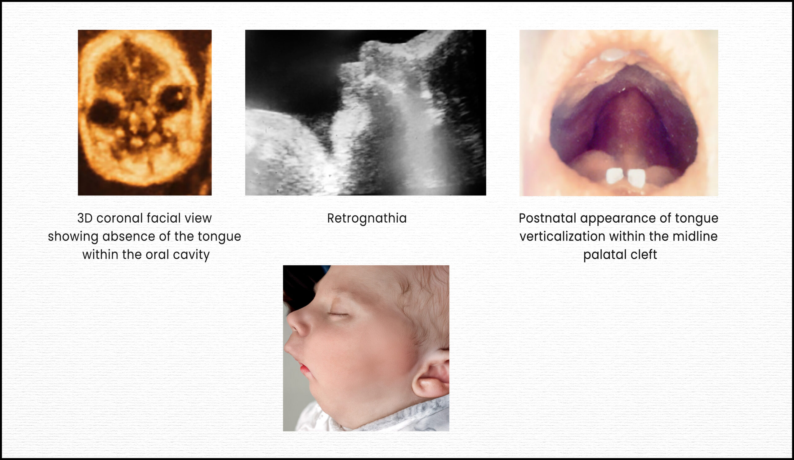
Pierre Robin sequence (PRS) is a rare congenital anomaly characterized by a classic triad of symptoms: micrognathia (undersized jaw), glossoptosis (posterior displacement or falling back of the tongue), and upper airway obstruction. A U-shaped cleft palate is frequently, though not always, associated with this triad. This condition is termed a “sequence” because micrognathia is considered the primary developmental disturbance, which then leads to glossoptosis, and subsequently, airway obstruction. The retropositioning of the tongue can also inhibit the fusion of the palatal shelves, often resulting in the characteristic U-shaped cleft palate.

PRS is a heterogeneous entity, meaning its presentation can vary widely. It can occur as nonsyndromic PRS (nsPRS), where only the core triad (with or without cleft palate) is present, or as syndromic PRS (sPRS), where it is associated with other congenital anomalies or genetic conditions. A distinct classification, PRS Plus, is used for nonsyndromic cases that exhibit additional malformations. The incidence of PRS is reported to range from 1 in 8,500 to 1 in 30,000 live births, with the highest reported incidence in the USA at 1 per 3,120 live births. Syndromic cases account for approximately 50% of all PRS occurrences, and around 34 different syndromes have been linked to PRS.

The clinical significance of PRS lies primarily in the potential for life-threatening respiratory obstruction and feeding difficulties, which contribute to a high mortality rate reported between 1.7% and 65%. Mortality rates are notably higher in children with associated syndromes, particularly those involving cardiac and central nervous system comorbidities. Consequently, the overarching priority in PRS management is to maintain a viable upper respiratory tract.

The Role of Genetic Understanding

Understanding the genetic mutations associated with PRS is crucial for several reasons. Such knowledge can facilitate early confirmatory diagnosis and guide the formulation of appropriate, tailored treatment plans. Furthermore, genetic mutations linked to PRS can be detected prenatally, sometimes as early as the first trimester of pregnancy. This early detection empowers parents to make informed decisions regarding the continuation of the pregnancy, especially given the potential severity of the syndrome. Despite its importance, a comprehensive collection of literature on genetic mutations in PRS has been lacking.

Methodology of the Systematic Review

To address this gap, the authors conducted a systematic review adhering to the Preferred Reporting Items for Systematic Reviews and Meta-Analyses (PRISMA) statement. Their search spanned Web of Science, PubMed, and Scopus databases, utilizing the keywords “Pierre Robin syndrome/sequence AND gene mutation”. The review specifically included original research, case reports, and case series written in English that detailed genetic mutations in PRS, while excluding reviews, conference abstracts, animal models, and articles lacking sufficient genetic information.

Out of an initial 208 articles, 39 met the stringent eligibility criteria for qualitative analysis after a rigorous screening process that involved identifying duplicates and irrelevant studies. The high kappa coefficient values (0.98 and 0.96 for the two screening steps) indicate strong reliability between the reviewers. In terms of bias assessment for the included case series and reports, 34 studies were categorized as having a low risk of bias (meeting 80-100% of quality criteria), while one study had a moderate risk of bias (meeting 60% of criteria).

Key Findings of the Systematic Review

The 39 selected articles collectively reported on 324 cases of PRS. The distribution of these cases highlighted the prevalence of associated conditions:

  • Syndromic PRS (sPRS): 182 cases (56%).
  • Nonsyndromic PRS (nsPRS): 120 cases (37%).
  • PRS Plus: 22 cases (6.8%), referring to nsPRS with additional malformations.

The review identified Stickler syndrome as the most common associated syndrome, appearing in 30 cases (16% of sPRS). Other frequently associated conditions included chromosomal abnormalities (17 cases, 9%), Richieri-Costa-Pereira syndrome (15 cases, 8%), Catel-Manzke Syndrome (10 cases, 5.5%), and TARP Syndrome (6 cases, 3%). It was noted that for 46 sPRS cases (25%), the specific associated syndrome was not mentioned or evaluated.

Various diagnostic techniques were employed to assess genetic mutations, most commonly FISH and G-banding. Other methods included exome sequencing, polymerase chain reaction, microarray, Sanger sequencing, array-CGH, next-generation sequencing, conformation-sensitive gel electrophoresis, and expression profiling.

Crucially, 100 out of the 324 cases (30.9%) had identified genetic mutations. The types of mutations observed varied, with deletions being the most frequent (47%), followed by translocations (20%), duplications (12%), and single nucleotide polymorphisms (9%). Abnormal karyotypes were present in 34 of these cases.

The SOX9 gene was found to be the most commonly mutated gene, identified in 22 cases. SOX9 is critical for cartilage development, and its mutation was linked to isolated PRS, campomelic dysplasia, acampomelic dysplasia, and PRS Plus cases. Specifically, loss of function or haploinsufficiency of SOX9 is associated with lethal skeletal malformations and syndromic PRS. In contrast, disruptions upstream or downstream of the SOX9 gene, which involve highly conserved noncoding cis-regulatory elements, are linked to milder abnormalities and nonsyndromic PRS.

Other genes implicated in isolated PRS included KCNJ2 (12 cases), BMPR1B (5 cases), COL11A1 (6 cases), COL11A2 (1 case), and COL2A1 (4 cases). Chromosome 2 mutations were also found in isolated PRS. For syndromic PRS, the genetic mutations were largely specific to the associated syndrome, such as TGDS in Catel-Manzke syndrome, RBM10 in TARP syndrome, and SNRPB in cerebro-costo-mandibular syndrome. In PRS Plus cases, although SOX9 mutations were common, other mutations like BMP2, NF2, MN1, MAP2K6, BMP4, OTX2, and KCNJ2 were also observed.

Regarding familial inheritance, only 21 of the 39 studies assessed genetic mutations in family members. Several instances of familial inheritance were reported, including autosomal dominant and recessive patterns, as well as maternal inheritance, across various PRS classifications and associated syndromes.

Limitations of the Reviewed Literature and Recommendations

The systematic review highlighted several limitations within the existing literature on PRS genetic mutations. A significant concern was the heterogeneity of the studies; only 5 were cohort studies, with the majority being case reports or case series, often involving very small sample sizes. These small sample sizes hinder robust statistical evaluation of factors like disease progression and treatment response. Furthermore, a notable deficiency was the limited assessment of familial inheritance patterns, with less than half of the studies investigating family members. There remains an ambiguity as to whether the identified genetic mutations are the sole cause of PRS or merely associated findings. The absence of gene ontology and pathway enrichment analysis in most studies (only one study performed this) further limits the understanding of the exact etiopathogenesis and the clinical implications of these mutations.

To advance the understanding and management of PRS, the authors strongly recommend several key actions for future research:

  • Conduct large-scale, multicenter cohort studies to provide sufficient statistical power for evaluating disease progression and treatment outcomes.
  • Implement a standardized protocol for assessing genetic mutations across all cases to minimize bias stemming from variations in diagnostic modalities.
  • Prioritize pedigree analysis to comprehensively assess potential familial inheritance patterns.
  • Integrate gene ontology and pathway enrichment analysis to gain deeper insights into the etiopathogenesis, mutation severity, and clinical manifestations, thereby aiding in more precise treatment planning.

In conclusion, while significant strides have been made in identifying genetic mutations associated with PRS, a more coordinated and large-scale research effort is imperative to fully elucidate its genetic basis and improve patient care.

Summary sheet

  1. Definition and Core Components
  • Pierre Robin Sequence (PRS), also known as Robin Sequence, is a congenital anomaly defined by a classic triad:
    • Micrognathia: an undersized or retropositioned mandible.
    • Glossoptosis: posterior displacement or retropositioning of the tongue.
    • Upper Airway Obstruction (UAO): resulting from the tongue’s position, leading to breathing difficulties.
  • Pathogenesis: The primary developmental disturbance is micrognathia, which leads to glossoptosis, subsequently causing mechanical obstruction of the oropharynx.
  • Associated Cleft Palate: A U-shaped cleft palate is frequently associated with PRS, occurring in 80–90% of patients. However, it is not considered a mandatory part of the formal diagnostic criteria.
  1. Epidemiology
  • Prevalence: Ranges from 1 in 2,000 to 1 in 30,000 live births, with other sources citing 1:8,500 to 1:20,000 live births or a prevalence of 2.29 per 10,000 births in a specific region. Variability in prevalence stems from differences in diagnostic criteria and ascertainment methods.
  • Sex Ratio: A higher male-female ratio (e.g., 1.47 in one study) has been reported, though some studies show no significant difference.
  • Classification: PRS can be:
    • Isolated/Non-syndromic PRS (nsPRS): Only the core triad (with or without cleft palate) is present, without other features or genetic abnormalities.
    • Syndromic PRS (sPRS): PRS occurs as part of a broader craniofacial syndrome or in association with other congenital anomalies. Syndromic cases are more common than isolated PRS (56% vs. 37% in one review).
  1. Associated Anomalies & Genetics
  • Prevalence of Associated Anomalies: A significant proportion of cases (69.7% in one study) are associated with non-PRS anomalies. Overall, 56% of reported PRS cases in a systematic review were syndromic, and 6.8% were PRS Plus (associated with other malformations). These associated anomalies are responsible for most of the morbidity and mortality in PRS.
  • Common Organ Systems Affected (in cases with multiple congenital anomalies):
    • Ear, Face, and Neck (35.7%).
    • Cardiovascular (18.4%).
    • Musculoskeletal (11.2%).
    • Central Nervous System (7.1%).
    • Urinary (6.1%) and Eye (6.1%).
    • PRS increases the risk of ventricular septal defect, limb deficiency, hypospadias, hydrocephaly, and intestinal malrotation.
  • Most Common Syndromes Associated with PRS:
    • Stickler Syndrome (most frequently associated; 16% of sPRS cases).
    • Treacher Collins Syndrome.
    • Larsen Syndrome.
    • Catel-Manzke Syndrome (5.5%).
    • TARP Syndrome (3%).
    • Over 34 syndromes have been associated with PRS.
  • Chromosomal Abnormalities: Present in 0.9% to 16.6% of PRS cases.
    • 22q11.2 deletion is the most common chromosomal abnormality.
    • Other reported abnormalities include Trisomy 21, Trisomy 18, and deletions like 4q33-qter, 17q24.3, 2q33.1, and 11q23.
  • Genetic Mutations: Identified in 30.9% of PRS cases.
    • SOX9 is the most common gene associated with both nsPRS and sPRS, crucial for cartilage development. Mutations upstream or downstream of SOX9 can cause milder abnormalities and nsPRS.
    • Other implicated genes: KCNJ2, BMPR1B, COL11A1, COL11A2, COL2A1.
    • Genetic mutations in sPRS are often specific to the associated syndrome.
    • Molecular testing, including chromosomal microarray, is recommended for PRS cases with associated malformations. Whole exome sequencing (WES) or targeted genetic panels may be preferred over whole genome sequencing (WGS) in the antenatal setting for efficiency and cost.
  1. Prenatal Diagnosis – Key Concepts
  • Fundamental Importance: Prenatal diagnosis allows for psychological preparation of parents and ensures delivery occurs in a hospital equipped with a pediatric intensive care unit, ready for potential respiratory complications. This minimizes risks of severe neonatal hypoxia or emergency tracheostomy.
  • First-Line Modality: Ultrasound (US):
    • Routine 20-week fetal anomaly scan is the typical time for screening.
    • Objective Measurements:
      • Facial Nasomental Angle (FNMA) and Facial-Maxillary Angle (FMA): Significantly smaller in PRS cases (FNMA <136°, FMA <66° often signify micrognathia).
      • Alveolar Overjet: Significantly larger in PRS cases; useful in prenatal diagnosis.
      • Lower Jaw Length (LJL) to Gestational Age (GA) or Femur Length (FL) ratio (Jaw Index): A LJL outside the 95% prediction interval (e.g., LJL < -10.18 + 0.24 * GA in weeks*7+days, or LJL < 3.49 + 0.6 * FL) indicates micrognathia. Sensitivity for LJL to GA is 93.75%, for LJL to FL is 87.5%.
      • Inferior Facial Angle (IFA) and Fronto-Naso-Mental (FNM) Angle: Used to assess retrognathia. IFA <49.2° and FNM angle <142° are indicative, but their sensitivity can be unsatisfying. IFA could serve as an initial screening tool.
    • Indirect Signs: Polyhydramnios (due to impaired swallowing) and abnormal tongue posture (glossoptosis) can be suggestive.
    • Challenges: Ultrasound can have low sensitivity for micrognathia (e.g., 72.7% subjectively) and cleft palate detection is challenging. Integration of objective measurements into routine care is hindered by rarity and need for specialist knowledge.
  • Adjuvant Modality: Fetal MRI:
    • Advantages: Improves understanding of severity, better visualization of glossoptosis, and identification of isolated cleft palate (difficult on US). Offers reproducible, high-resolution 3D images.
    • Role: Used when US findings are suspicious or ambiguous, or for better risk stratification. Not practical as a first-line screening tool due to cost and resources.
  • Future Directions in Prenatal Diagnosis: Integrating artificial intelligence into antenatal screening has potential to improve diagnosis rates.
  1. Postnatal Clinical Assessment & Complications
  • Primary Concerns: Upper airway obstruction (UAO) and feeding problems.
  • Consequences of UAO: Prolonged hypoxia can lead to severe consequences like cerebral anoxia, pulmonary sepsis, malnutrition, and permanent brain damage.
  • Severity Assessment (Cole’s Classification):
    • Grade 1: PRS features, no obvious respiratory distress, satisfactory feeding.
    • Grade 2: PRS features, intermittent respiratory distress (e.g., stridor), feeding precipitates distress.
    • Grade 3: PRS features, clinical signs of respiratory distress, oral feeding impossible.
  • Assessment Tools After Birth:
    • Clinical evaluation, pulse oximetry, capillary blood gases.
    • Nasopharyngoscopy: To distinguish tongue obstruction from other causes (e.g., subglottic anomalies, tracheomalacia).
    • Polysomnography (PSG): Recommended for all suspected PRS infants to diagnose and quantify obstructive sleep apnea (OSA). Interpretation needs to consider age-appropriate normative values, as standard cut-offs are from older children.
  • Feeding Difficulties:
    • Managed initially with a nasogastric tube.
    • For prolonged issues affecting growth, a gastrostomy tube is an alternative.
    • Palatal plates can improve feeding and sucking activity by restoring oral-nasal separation.
    • Optimizing the airway generally resolves feeding issues secondarily.
  1. Management Strategies
  • Multidisciplinary Approach: Essential for correct diagnosis, airway management, feeding optimization, surgical timing, and addressing multisystemic abnormalities.
  • Conservative/Minimally Invasive Management:
    • Prone Positioning: Initial trial, effective in some cases by repositioning the tongue forward. Efficacy is debated, and prolonged use increases SIDS risk.
    • Nasopharyngeal Airway (NPA): Simple, well-tolerated, and effective second-line option to bypass UAO if prone positioning fails.
    • Pre-Epiglottic Baton Plate (PEBP): An orthodontic device positioning the tongue anteriorly. Requires significant expertise.
    • High-Flow Nasal Cannula (HFNC) / Continuous Positive Airway Pressure (CPAP): Can manage UAO and be bridging measures. CPAP can be challenging with a nasogastric tube and carries pressure injury risks.
  • Surgical Management: Considered when non-invasive treatments fail or in severe, life-threatening cases.
    • Mandibular Distraction Osteogenesis (MDO): Gold standard surgical technique. It lengthens the mandible, expanding oral volume and alleviating glossoptosis, often reducing the need for tracheostomy. Protocols like Fast and Early Mandibular Osteodistraction (FEMOD) have shown good long-term outcomes with minimal scarring and preservation of dental development.
    • Tongue-Lip Adhesion (TLA): Older surgical option, anchors tongue to lower lip. Less commonly used now due to complications (dehiscence, lacerations, infections) and often requiring secondary interventions.
    • Tracheostomy: Life-saving for severe respiratory crises, considered when other treatments fail or for complex multi-level obstruction (e.g., Pfeiffer syndrome). Decannulation can be challenging.
  1. Counseling and Delivery
  • Counseling: Antenatal diagnosis aids parental understanding and preparation, though it can increase anxiety. Neonatal genetic counseling can be overwhelming.
  • Delivery: Due to potential for difficult airways, delivery in a specialist tertiary center with a prepared team (pediatric intensivists, pediatric pulmonologist, pediatric neurologist, geneticist) is prudent, ideally via planned caesarean section if concerns are identified.

Podcast

Course Outline: Pierre Robin Sequence

  • Definition and Core Triad (5 minutes)
    • Pierre Robin Sequence (PRS) is classically defined by a triad of micrognathia, glossoptosis, and upper airway obstruction (UAO).
    • Micrognathia refers to an undersized mandible, while retrognathia describes a retropositioned mandible; in severe PRS, micrognathia leads to retrognathia.
    • Glossoptosis is the posterior rotation/displacement of the tongue into the pharynx, which obstructs the airway.
    • Cleft palate is commonly associated, found in roughly 80-90% of PRS patients, often U-shaped. However, it is not always present and therefore not part of the formal diagnostic criteria.
    • The sequence describes a developmental order: micrognathia leads to glossoptosis, which then limits palatal shelf fusion, potentially causing a cleft palate and resulting in neonatal respiratory problems.
  • Prevalence and Clinical Significance (5 minutes)
    • PRS is a relatively rare congenital anomaly, with reported prevalence ranging from 1:8,500 to 1:20,000 live births, though this varies depending on diagnostic criteria. One study reported 2.29 per 10,000 births.
    • The major comorbidities are upper airway obstruction and feeding problems due to micrognathia and glossoptosis.
    • Prenatal suspicion and diagnosis are essential for preparing the delivery team for a possible airway emergency, minimizing hypoxia at birth, and allowing multidisciplinary coordination. This also helps in parental counseling and preparing for the potentially challenging postpartum period.
  • Isolated vs. Syndromic PRS (5 minutes)
    • PRS can occur isolated (core triad with or without cleft palate, no other features or genetic abnormalities) or as syndromic (PRS with additional clinical features or genetic abnormalities).
    • Syndromic cases are more common, making up approximately 50-69.7% of all PRS cases.
    • Commonly recognized associations include Stickler syndrome, Treacher Collins syndrome, and 22q11.2 deletion syndrome. Stickler syndrome is the most frequently associated.
  • Genetic Basis and Associated Mutations (5 minutes)
    • Genetic mutations are noted in about 30.9% of cases.
    • The SOX9 gene is the most common gene associated with both isolated and syndromic PRS, playing a crucial role in cartilage development. Disruptions upstream or downstream of SOX9 can cause milder abnormalities or nonsyndromic PRS.
    • Other genes associated with isolated PRS include KCNJ2, BMPR1B, COL11A1, COL11A2, and COL2A1. Chromosomal abnormalities were present in 11.2% of cases in one study, including 22q11.2 deletion.
    • Genetic mutations in syndromic PRS are specific to the associated syndromes (e.g., TGDS for Catel-Manzke, RBM10 for TARP syndrome, SNRPB for cerebro-costo-mandibular syndrome).
    • Genetic counseling is indicated in many cases with associated anomalies. Chromosomal microarray is a first-tier diagnostic test for individuals with congenital anomalies, and further testing like FISH, qPCR, MLPA, and single gene tests may be advised.
  • Associated Anomalies by Organ System and Environmental Factors (5 minutes)
    • The high prevalence of associated anomalies justifies thorough screening.
    • Most frequent anomalies are in the ear, face, and neck (35.7%), cardiovascular (18.4%), musculoskeletal (11.2%), central nervous (7.1%), urinary (6.1%), and eye (6.1%) systems.
    • Children with PRS have a higher incidence of other congenital anomalies than the general population, increasing the risk of ventricular septal defect, limb deficiency, hypospadias, hydrocephaly, and intestinal malrotation.
    • Some studies suggest an association between maternal use of methadone and increased PRS risk, and maternal smoking has been associated with cleft palate with PRS.
  • Standard Ultrasound Screening and its Purpose (5 minutes)
    • Ultrasound (US) is the first-line examination of choice for prenatal diagnosis of PRS, visible from the 20th week of gestation.
    • Routine 20-week ultrasounds screen facial features and can characterize mandibular morphology and the maxillomandibular relationship.
    • Prenatal diagnosis allows for delivery team preparation for a possible airway emergency and prepares parents psychologically.
  • Key Ultrasound Measurements and Indicators for Micrognathia (15 minutes)
    • Facial Nasomental Angle (FNMA): Measured from mid-sagittal profile images. Significantly smaller in PRS patients (mean 129.6 ± 9 vs. 137.9 ± 2.8 in controls, p<.001), with FNMA <136° signifying micrognathia.
    • Facial-Maxillary Angle (FMA): Also from mid-sagittal profile images. Significantly smaller in PRS patients (mean 64.1 ± 9.3 vs. 75.3 ± 6.5, p<.001), with FMA <66° signifying micrognathia. A smaller median FMA was correlated with increased respiratory support needs.
    • Alveolar Overjet: Significantly larger in the PRS group (mean 3.7 ± 1.3 vs. 2.3 ± 0.8, p<.001). Its utility in prenatal diagnosis is highlighted.
    • Lower Jaw Length (LJL): Can be objectively assessed and related to gestational age (GA) or femur length (FL).
      • An index based on LJL and GA or FL provides means for predicting jaw growth.
      • Sensitivity for micrognathia using LJL-GA index is 93.75% (15 of 16 cases correctly identified) at a 95% specificity.
      • Sensitivity using LJL-FL index is 87.5%.
      • A micrognathia is diagnosed when LJL is substantially shorter than the predicted value of a healthy fetus at the same GA or FL.
    • Jaw Index: Calculated by dividing anteroposterior mandibular distance by biparietal skull distance. A cutoff of 23 (2 standard deviations below the mean) showed a sensitivity of 100.0% and specificity of 98.1%. However, it can be difficult and time-consuming to measure.
    • Inferior Facial Angle (IFA): Measures the angle formed by a line orthogonal to the forehead and a line from the mentum to the protrusive lip. An IFA <49.2° indicated retrognathism. While specific, it may not be as sensitive as other combined measures and can be biased by upper jaw/cranium dysmorphia.
    • Fronto-Naso-Mental (FNM) Angle: Angle from forehead to nose tip to mentum. Retrognathia defined as <142°. Can be accurate, but 3D imaging reduces measurement error.
    • Mandibular (MD) to Maxillary (MX) Width Ratio: MD/MX ratio <0.785 indicated micrognathia in a 3D ultrasound study, but only 50% sensitivity for PRS cases.
    • Absent Mandibular Gap: In the retronasal triangle view (coronal plane) in first-trimester fetuses, the absence of a “mandibular gap” can indicate micrognathia. This is an early objective measure but its utility for PRS prediction is unknown.
  • Assessment of Glossoptosis and Indirect Signs (5 minutes)
    • Glossoptosis: Can be observed by viewing the echogenic tongue to see if it is posteriorly displaced or does not reach the anterior mandibular alveolar ridge during movements. This is a potentially strong predictor to differentiate PRS from isolated micrognathia. However, it requires considerable time (20-30 minutes) and may be impractical.
    • Polyhydramnios: Often an indirect finding due to impaired fetal swallowing secondary to glossoptosis.
    • Cleft Palate: Challenging to detect prenatally; 2D US has sensitivity up to 65%, with 3D US yielding better results.
  • Challenges in Ultrasound Diagnosis (5 minutes)
    • Overall, prenatal screening often relies on subjective standards, yielding low sensitivity.
    • The rarity of PRS and the need for specialist knowledge hinder routine integration of objective measurements.
    • There is no strict association between the anatomical defect severity and the degree of respiratory impairment at birth.
    • Artificial intelligence advancements show potential for automating extraction of fetal biometrics from ultrasound, which could improve early detection.
  • As an Adjuvant Imaging Modality (5 minutes)
    • Fetal Magnetic Resonance Imaging (MRI) is not a first-line screening tool but can be a useful adjuvant in cases with suggestive ultrasound findings or when ultrasound findings are ambiguous.
    • It improves understanding of the condition’s severity.
  • Advantages of Fetal MRI (5 minutes)
    • Improved visualization of glossoptosis: Sagittal MRI sequences easily show tongue position with high contrast conspicuity between tongue muscle and amniotic fluid, indicating advantages over ultrasound.
    • Ability to identify instances of isolated cleft palate.
    • Allows for better risk stratification and counseling of parents.
    • Provides reproducible, high-resolution three-dimensional images of the face, palate, and jaw without additional radiation.
  • Indications for Genetic Testing (5 minutes)
    • Growing interest in more detailed genetic testing, especially when routine screening highlights a possible anomaly.
    • Presence of polyhydramnios may help distinguish between syndromic and isolated cases, influencing the decision for further genetic testing.
  • Methods and Benefits (5 minutes)
    • Chromosomal microarray analysis can detect pathogenic copy number variations and is a useful addition to karyotyping, increasing the frequency of abnormality detection.
    • Whole exome sequencing (WES) may be a more practical alternative to whole genome sequencing (WGS) antenatally, offering lower costs and faster results. WES identifies diagnostic genetic variants in about 8.5% of cases with structural anomalies detected on ultrasound.
    • Accurate genetic diagnosis aids in tailoring further investigations and treatments, and facilitates discussions about risks in future pregnancies.
    • WGS is resource-intensive and often reserved for cases with multiple anomalies, following appropriate counseling and genetic review.
  • Importance of Early Detection (5 minutes)
    • Early diagnosis allows for appropriate prenatal consultation and prepares the treating team for postnatal care challenges, as well as the family for prenatal education.
    • Analysis of parental experiences highlights that antenatal diagnosis aids understanding of the condition and adjustment to potential difficulties.
  • Delivery Considerations (5 minutes)
    • Given the potential for immediate, difficult airway management, cases of suspected PRS should be delivered in a specialist tertiary center.
    • This ensures that children receive optimal care in the delivery room, during assessment, and if intervention is required.
    • A significant minority of units (58%) reported that patients did not have an antenatal diagnosis, leading to births in centers lacking sufficient expertise.
    • Discussion of daytime delivery via planned caesarean section with relevant specialties in attendance is appropriate where concerning features are identified.
  • Transition to Postnatal Care (3 minutes)
    • Postnatal evaluation for airway obstruction and feeding difficulties is crucial.
    • Management of airway obstruction typically starts with minimally invasive treatments like prone positioning and nasopharyngeal tube. Other options include nasopharyngeal airways (NPAs), continuous positive airway pressure (CPAP), and pre-epiglottic baton plates (PEBPs).
    • More severe cases may require surgical intervention like mandibular distraction osteogenesis (MDO) or, less commonly, tongue-lip adhesion (TLA) or tracheostomy.
    • Feeding difficulties are managed with strategies like nasogastric tubes or gastrostomy tubes.
    • Assessment includes clinical observation, oximetry, capillary blood gases, flexible nasoendoscopy (FNE), and polysomnography (PSG).
  • Conclusion and Future Directions (2 minutes)
    • Prenatal diagnosis of PRS is a significant advance that allows for appropriate consultation and care coordination.
    • A multidisciplinary approach is essential for managing PRS, ensuring comprehensive care from prenatal diagnosis through postnatal management.
    • Future research should focus on integrating AI into antenatal screening, expanding genetic understanding, advancing neonatal sleep physiology knowledge, and comparing treatment techniques to improve outcomes.

Powerpoint Slides

Slide 1: Introduction to Pierre Robin Sequence (PRS)

  • Pierre Robin Sequence (PRS) is a congenital anomaly first described in 1923, characterized by a clinical triad.
  • The three core features defining PRS are micrognathia (an undersized lower jaw), glossoptosis (posterior displacement of the tongue), and upper airway obstruction (UAO).
  • A U-shaped cleft palate is frequently associated with PRS, occurring in a majority of patients, although it is not always present and therefore not considered part of the formal diagnostic criteria.
  • The incidence of PRS varies, with reported prevalence rates ranging from 1 in 8,000 to 1 in 20,000 live births, depending on the diagnostic criteria used by different studies.

Slide 2: Pathogenesis of Pierre Robin Sequence

  • PRS is described as a “sequence” because its characteristic triad develops in a specific order.
  • The primary developmental disturbance is micrognathia, or the underdevelopment of the mandible.
  • This underdeveloped lower jaw leads to glossoptosis, where the tongue falls backward into the pharynx, reducing the oral cavity volume and obstructing the fetal closure of the secondary palatal shelves.
  • The retropositioning of the tongue can mechanically obstruct the oropharynx, leading to neonatal respiratory problems.

Slide 3: Classification of PRS: Isolated vs. Syndromic

  • PRS can occur in two main forms: isolated (nonsyndromic PRS or nsPRS), where only the core triad is present, with or without a cleft palate, and no other associated features or genetic abnormalities.
  • Syndromic PRS (sPRS) involves PRS alongside additional clinical features or genetic abnormalities, or a combination of both.
  • It is estimated that nearly half of all PRS cases are syndromic.
  • Commonly recognized syndromes associated with PRS include Stickler syndrome, Treacher Collins syndrome, and 22q11.2 deletion syndrome.

Slide 4: Importance of Prenatal Diagnosis

  • Prenatal suspicion of PRS is essential for preparing the delivery team for a possible airway emergency.
  • An early diagnosis facilitates multidisciplinary team coordination and the initiation of selective treatment steps, which can minimize the risk of severe neonatal hypoxia and avoid emergency interventions like tracheostomy.
  • It also allows for psychological preparation and counseling of parents, enabling them to make informed decisions about the pregnancy and prepare for the potentially challenging postpartum period.
  • Despite its importance, antenatal diagnosis is currently made in less than a third of reported cases, indicating a significant area for improvement.

Slide 5: Prenatal Ultrasound for PRS (Part 1/2)

  • Ultrasound (US) is the first-line examination for prenatal diagnosis of PRS, with features suggestive of the condition visible from the 20th week of gestation during routine anatomy scans.
  • Specific facial measurements can be used to assess micrognathia, including the facial nasomental angle (FNMA), facial-maxillary angle (FMA), and alveolar overjet.
  • FNMA <136° and FMA <66° are indicative of micrognathia. Patients with PRS demonstrate significantly smaller FNMA and FMA compared to controls, and significantly larger alveolar overjet.
  • Alveolar overjet, previously not described in prenatal ultrasound literature, has utility in prenatal diagnosis alongside FMA and FNMA.

Slide 6: Prenatal Ultrasound for PRS (Part 2/2)

  • Other objective methods for diagnosing micrognathia prenatally include the inferior facial angle (IFA) and the fronto-naso-mental (FNM) angle.
  • The IFA, which measures the angle of the facial profile, has been shown to be a specific predictor for retrognathia, with values below 49.2° suggesting micrognathia.
  • Mandibular length (LJL) compared to gestational age (GA) or femur length (FL) is also used; LJL measures below the 95% prediction interval (e.g., LJL < –10.18 + 0.24 * GA in weeks) indicate micrognathia.
  • While these methods show high sensitivity (e.g., 93.75% for LJL based on GA and 87.5% for LJL based on FL) and specificity (95% by design for LJL), their integration into routine care is challenged by the rarity of PRS and the need for specialist knowledge.

Slide 7: Fetal Magnetic Resonance Imaging (MRI) in Diagnosis

  • Fetal MRI can be used as a complement to ultrasound in cases where PRS is suspected or when ultrasound findings are ambiguous.
  • MRI offers advantages such as high-resolution three-dimensional images of the face, palate, and jaw, with no additional radiation exposure.
  • It is particularly useful for assessing the degree of glossoptosis due to the high contrast between the tongue muscle and amniotic fluid, making tongue positioning straightforward to evaluate.
  • MRI can also help identify isolated cleft palate, which can be challenging to detect using ultrasound, and provides better risk stratification and counseling for parents.

Slide 8: Genetic Testing in Prenatal Diagnosis

  • For pregnancies where routine screening highlights a possible anomaly, more detailed genetic testing can be conducted antenatally.
  • The presence of polyhydramnios may help distinguish between syndromic and isolated cases, potentially influencing the decision for further genetic testing.
  • Chromosomal microarray analysis is a useful addition to karyotyping, as it can detect pathogenic copy number variations and increase the frequency of abnormality detection.
  • Whole exome sequencing (WES) and targeted genetic panels are more practical alternatives to whole genome sequencing (WGS) for antenatal settings, offering lower costs and faster results, particularly for cases with multiple anomalies.

Slide 9: Counseling and Delivery Planning

  • Providing parents with detailed information and options for further imaging and comprehensive genetic testing represents the current best standard of care.
  • Prompt evaluation enables parents to make informed decisions about continuing the pregnancy and helps them prepare for the challenging postpartum period.
  • Given the potential for immediate management of a difficult airway, it is prudent that PRS cases be delivered in a specialist tertiary center.
  • A survey revealed that many units lack the expertise for emergent neonatal airway issues, underscoring the importance of antenatal diagnosis to allow for planned delivery with relevant specialties present.

Slide 10: Postnatal Assessment: Upper Airway Obstruction

  • Immediately after birth, newborns with PRS require evaluation for airway obstruction and feeding difficulties.
  • Prolonged airway obstruction can lead to acute and chronic hypoxia, significantly increasing the risk of pulmonary infections, cor pulmonale, heart failure, and cerebral hypoxia with cognitive impairment.
  • Even if early clinical evaluations do not immediately reveal signs of UAO, repeated assessments over the first weeks and months of life are essential, as breathing difficulties can fluctuate and become evident during sleep or feeding.
  • Clinical observations alone are often insufficient; for instance, only about 55% of infants with PRS and obstructive sleep apnea (OSA) snore.

Slide 11: Postnatal Assessment: Feeding Difficulties

  • The same anatomical factors (micrognathia, glossoptosis) that cause airway obstruction also disrupt the suck-swallow-breathe reflex, leading to significant feeding difficulties.
  • Infants with PRS frequently experience gastroesophageal reflux, altered esophageal tone, and an increased risk of aspiration.
  • Early involvement of speech and language therapists is crucial to assist parents and children with feeding, particularly when a cleft palate is present.
  • Close monitoring of growth and weight gain is important, as dysfunctional feeding and increased work of breathing can lead to failure to thrive, necessitating additional support like enteral feeding.

Slide 12: Grading Severity of PRS

  • The severity of PRS needs to be defined to guide treatment decisions.
  • Cole’s classification, proposed in 2008, categorizes the severity of symptoms into three grades based on clinical examination.
  • Grade 1: Newborns show features of PRS but no obvious clinical signs of respiratory distress, and feeding assessment is satisfactory.
  • Grade 2: Newborns exhibit intermittent signs of respiratory distress, such as inspiratory stridor, and feeding precipitates respiratory distress.
  • Grade 3: Newborns present with clinical signs of severe respiratory distress, making oral feeding impossible.

Slide 13: Postnatal Diagnostic Tools for Airway Obstruction

  • A methodical approach to assessment is taken after stabilization, often combining clinical evaluation with objective measures.
  • The majority of centers use clinical assessment along with pulse oximetry and capillary blood gases to evaluate infants with PRS.
  • Flexible nasoendoscopy (FNE) is often necessary to distinguish obstruction due to tongue position from other causes like subglottic anomalies or tracheomalacia.
  • Polysomnography (PSG) is recommended for all infants where obstructive sleep apnea (OSA) is suspected, as clinical signs alone may not be sufficient to capture nocturnal breathing difficulties.

Slide 14: Polysomnography (PSG) in PRS Assessment

  • PSG is widely used to aid in diagnosing OSA in PRS infants, although interpretation in the neonatal population can be challenging.
  • Changes in oxygen saturation and the apnea-hypopnea index (AHI) are commonly used, but normative values are often taken from older children, leading to potential discrepancies.
  • There is a lack of consistency in how UAO severity is assessed and thresholds are set for intervention, highlighting the need for a holistic interpretation of PSG results.
  • Future research should prioritize obtaining a clearer understanding of normal breathing patterns during sleep in the first year of life and implementing standardized reporting tailored to the PRS population.

Slide 15: Flexible Nasoendoscopy (FNE) & Drug-Induced Sleep Endoscopy (DISE)

  • Flexible Nasoendoscopy (FNE) is a crucial tool for assessing the extent and location of upper airway obstruction (UAO).
  • It helps differentiate obstruction caused by glossoptosis from other potential causes like laryngomalacia, cysts, or stenosis, which may not be identifiable prenatally.
  • Drug-Induced Sleep Endoscopy (DISE) is another technique used to evaluate UAO, particularly in complex or syndromic PRS cases where multilevel obstruction may be present.
  • These endoscopic techniques should be integrated into clinical care to provide a comprehensive understanding of the airway anatomy and guide treatment strategies.

Slide 16: Non-Invasive Management Strategies

  • Initial non-invasive treatments are often the first approach for airway obstruction in PRS, starting with prone positioning. While pragmatic, its efficacy is controversial, and prolonged use carries an increased incidence of sudden infant death syndrome (SIDS).
  • If positioning is insufficient, nasopharyngeal airway (NPA) insertion is a simple, well-tolerated, and effective option to bypass UAO, with reported success rates between 60% and 100%.
  • The pre-epiglottic baton plate (PEBP) is an orthodontic device that lies along the palate with an extension to bring the tongue base anteriorly, showing promising results in relieving UAO and improving feeding, though it requires significant expertise.
  • High-flow nasal cannula (HFNC) and continuous positive airway pressure (CPAP) can also manage UAO and may be used as bridging measures or safely administered in community settings, depending on local expertise.

Slide 17: Surgical Management: Overview

  • When non-invasive treatments fail, or for severe cases, surgical intervention becomes necessary and should be performed as soon as possible.
  • The three primary surgical options for managing severe airway obstruction in PRS are tracheostomy, tongue-lip adhesion (TLA), and mandibular distraction osteogenesis (MDO).
  • Historically, tracheostomy was a common option, but it can present challenges with decannulation and home care.
  • TLA involves anchoring the tongue to the lower lip to alleviate obstruction, but it is now less frequently used as its benefits often overlap with less invasive methods like NPA or PEBP.

Slide 18: Mandibular Distraction Osteogenesis (MDO)

  • Mandibular distraction osteogenesis (MDO) has become the treatment of choice for severe cases of micrognathia and subsequent airway obstruction.
  • This technique is based on the principle that tension stimulates histogenesis with bone formation, allowing for the lengthening of the mandible and a more functional airway.
  • The primary aim of MDO in PRS patients is to reduce the need for tracheostomy and improve oral volume.
  • The Fast and Early Mandibular Osteodistraction (FEMOD) protocol is an effective approach, with distraction beginning intraoperatively and continuing daily, aiming for a slight Class III position. Long-term evaluations of FEMOD show preservation of primary teeth development and symmetrical mandibular growth without facial scarring.

Slide 19: Genetic Mutations Associated with PRS

  • Genetic mutations are reported in a significant portion of PRS cases, with 100 out of 324 cases (30.9%) having identified genetic mutations in a systematic review of the literature.
  • SOX9 is the most common gene associated with PRS, found mutated in both isolated PRS and syndromic forms like campomelic dysplasia and acampomelic dysplasia. This gene is crucial for cartilage development.
  • Other genes found to be associated with isolated PRS include KCNJ2, BMPR1B, COL11A1, COL11A2, and COL2A1.
  • The genetic mutations found in syndromic PRS cases are often specific to the associated syndromes, such as TGDS in Catel-Manzke syndrome and RBM10 in TARP syndrome.

Slide 20: Associated Chromosomal Abnormalities

  • Chromosomal abnormalities are present in a notable percentage of PRS cases; in one study, they were found in 10 out of 89 cases (11.2%).
  • The most common chromosomal abnormality is 22q11.2 deletion, which has been reported in varying percentages (e.g., 10% to 100% in different series).
  • Other chromosomal anomalies include Trisomy 21 and Trisomy 18, although they are less frequent.
  • More detailed genetic tests beyond karyotype, such as chromosomal microarray, are advisable for individuals with PRS and any associated malformations, as they can reveal less common but associated chromosomal loci.

Slide 21: Prognosis and Mortality in PRS

  • The prognosis for patients with idiopathic PRS (without associated anomalies) is usually good, or with only slight handicap.
  • However, the outcome can be poor in patients with associated anomalies, as these are responsible for most of the morbidity and mortality.
  • Mortality associated with Robin sequence is reported to range widely, between 1.7% and 65%, with higher rates in children with associated syndromes, cardiac, and central nervous system-related comorbidities.
  • Prolonged hypoxia in PRS neonates has also been reported as a high risk for permanent brain damage.

Slide 22: The Importance of a Multidisciplinary Approach

  • The complexity of Pierre Robin Sequence necessitates a multidisciplinary approach for optimal care.
  • This approach involves a team of specialists to ensure correct diagnosis, effective airway management, optimized feeding, appropriate timing for surgical interventions, and addressing any multisystemic abnormalities.
  • Early diagnosis and parental counseling play a key role in managing PRS, as early activation of the treatment path leads to better outcomes and prevents complications.
  • A multidisciplinary team should discuss and adapt treatment strategies to the specific needs of the patients and their parents, emphasizing patient-centered care.

Slide 23: Future Directions and Research Priorities

  • There remains uncertainty regarding the best approach to treat Robin sequence, emphasizing the need for ongoing research and standardized practices.
  • Integrating artificial intelligence into antenatal screening holds significant potential to improve the rate of early diagnosis of micrognathia.
  • Further research is needed to expand the understanding of the Robin sequence genotype by enrolling patients in genomics studies focusing on rare diseases.
  • Priorities also include advancing the knowledge of neonatal sleep physiology to establish age-appropriate normal values for objective breathing assessments and comparing minimally invasive and surgical techniques to determine optimal intervention thresholds.
  • The establishment of an international observational registry is recommended to document epidemiology, treatment, and outcomes for PRS patients, aiming to improve patient care globally.

Imaging