Bibliographic and Educational Resources in Fetal Medecine

This platform is designed to serve as a comprehensive educational and bibliographic resource for healthcare professionals involved in Fetal Medecine. Covering a wide range of up-to-date topics within the field, it offers structured access to recent scientific literature and a variety of pedagogical tools tailored to clinicians, educators, and trainees.

Each topic is grounded in a curated selection of recent publications, accompanied by in-depth summaries that go far beyond traditional abstracts—offering clear, clinically relevant insights without the time burden of reading full articles. These summaries act as gateways to the original literature, helping users identify which articles warrant deeper exploration.

In addition to these detailed reviews, users will find a rich library of supplementary materials: topic overviews, FAQs, glossaries, synthesis sheets, thematic podcasts, fully structured course outlines adaptable for teaching, and ready-to-use PowerPoint slide decks. All resources are open access and formatted for easy integration into academic or clinical training programs.

By providing practical, well-structured content, the platform enables members of the cytogenomics community to efficiently update their knowledge on selected topics. It also offers educational materials that are easily adaptable for instructional use.

Management of congenital cytomegalovirus infection

Dr Marianne Leruez-Ville

Pr Yves Ville

Overview

Congenital cytomegalovirus (cCMV) infection poses a significant public health challenge, with a global prevalence of 0.64%. It is the most common congenital infection and the leading non-genetic cause of sensorineural hearing loss (SNHL) and neurological damage in children. An estimated 17–20% of infected children experience serious long-term effects.

Recent advances have significantly improved the understanding and management of cCMV, particularly concerning primary maternal infections. Expert consensus guidelines, such as those from the European Congenital Cytomegalovirus Initiative (ECCI), provide updated recommendations for its management, spanning from prevention to postnatal care.

Key Advances in Understanding cCMV
Two major advances have reshaped the approach to cCMV following a primary maternal infection (MPI):

  • Efficacy of Antiviral Treatment: Antiviral treatment, specifically with valaciclovir, has shown efficacy in preventing vertical transmission in pregnant women with MPI.
  • Timing of Risk: Evidence now indicates that the risk of major sequelae is limited to maternal infection acquired in the first trimester of pregnancy.

Management of Congenital Cytomegalovirus Infection
Management of cCMV encompasses prevention, accurate diagnosis, and personalized care throughout prenatal, neonatal, and postnatal periods.

Primary prevention aims to reduce the incidence of maternal CMV infection, especially in seronegative women, thereby preventing cCMV.

  • Hygienic Modifications: Advising on hygienic measures is recommended for all pregnant women, despite a lack of specific data for seropositive women. This includes avoiding contact with body fluids (saliva, tears, urine) from infected individuals, particularly toddlers, from before conception until 14 weeks of gestation. Educational interventions like films, brochures, and calendar reminders have shown to improve hygiene measures and are well-received by pregnant women.
  • Education and Policy: There is a recognized need to improve education strategies for women about CMV prevention, ideally before and during the first trimester. Knowledge gaps among perinatal healthcare professionals have also been identified, highlighting the need for improved professional education. The development of a uniform EU policy for the prevention of primary CMV infection is recommended.

Early and accurate maternal diagnosis through serological tests is crucial for enhancing risk management and prevention strategies.

  • CMV Serology in First Trimester: Maternal CMV serology should be performed in the first trimester of pregnancy, as cCMV sequelae are primarily linked to infections acquired during this period. Routine serology is not recommended beyond 16 weeks, except in cases of suspected infection.
  • Equivocal Results: If CMV IgG results are equivocal, it is recommended to repeat weakly positive IgG results with a second assay or send sera to a reference laboratory. Discordant results should be considered equivocal and declared negative.
  • Valaciclovir for Secondary Prevention: For maternal primary infection in the periconceptional period or first trimester, oral valaciclovir at a dose of 8 g/day is recommended. Treatment should be administered as early as possible after diagnosis and continued until amniocentesis. This regimen has been shown to reduce vertical CMV transmission by 70–71%. A dose regimen of 2g four times per day is recommended to minimize the risk of renal side effects.
  • Hyperimmune Globulin (HIG): Administration of 100 IU/kg HIG every 4 weeks in pregnant women with primary CMV infection is not recommended as it has been found ineffective in preventing vertical transmission. However, for very recent MPI in the first trimester, HIG at 200 IU/kg every 2 weeks may be considered, but with Grade C evidence.
  • Diagnosis of Fetal Infection: CMV PCR on amniotic fluid (AF) is the gold standard for diagnosing fetal CMV infection. It should be collected from 17+0 weeks gestation, provided maternal infection occurred at least 6 weeks earlier. Under these conditions, the specificity of CMV PCR on AF is near 100%, and sensitivity is around 87–95%.
  • Prognostic Markers:
    • Ultrasound and MRI: Serial focused fetal ultrasound assessment and MRI in the third trimester are recommended for infected fetuses, as they provide prognostic information on CMV-associated findings. Severe cerebral abnormalities are associated with a poor prognosis. While normal ultrasound and MRI have a high negative predictive value for moderate to severe sequelae (nearly 100%), there remains a 17% residual risk of unilateral SNHL. Isolated extracerebral features carry a 30% risk of sequelae.
    • Amniotic Fluid Cytokines: Fetal infection and its severity are associated with changes in intra-amniotic cytokines. Increased levels of pro-inflammatory cytokines like IP-10, IL-18, ITAC, and TRAIL (both soluble and extracellular vesicle-associated) have been linked to cCMV infection and its severity. A pattern of specific increase in six proteins (IL-18soluble, TRAILsoluble, CRPsoluble, TRAILsurface, MIGinternal, and RANTESinternal) fitted severely symptomatic infection. These suggest a Th1 immune response polarization. Cytokine profiling shows strong potential for predicting both infection and severity.
    • Fetal Blood Markers: Fetal thrombocytopenia is associated with severe fetal infection. In cases without severe brain ultrasound abnormalities, a fetal blood profile with platelet count >114,000/mm³ and CMV DNA load <4.93 log10 IU/mL, or a low adjusted AF viral load, can further reduce the uncertainty of symptoms at birth to 0-5%. Higher adjusted CMV DNA loads in AF were significantly associated with symptomatic status at birth.
  • Fetal Treatment: In women with confirmed fetal infection, fetal treatment with valaciclovir 8 g/day may be considered after discussion with an expert team. A Phase II study suggested that high-dosage valaciclovir improved the outcome of moderately symptomatic infected fetuses, increasing asymptomatic neonates from 43% (untreated historical cohort) to 82% (treated). It was well-tolerated with high adherence and led to decreased fetal blood viral loads and increased platelet counts.
  • Negative Amniocentesis: Reassurance is recommended for women with a negative CMV PCR in amniotic fluid, as late fetal infection (after amniocentesis) is not associated with long-term sequelae.

To distinguish cCMV from postnatal infection, PCR should be performed on a sample collected within 3 weeks of birth, ideally as soon as possible.

  • Sample Types: Saliva or urine samples are preferred due to high sensitivity. A positive CMV PCR on a saliva sample should be confirmed with a urine sample due to potential contamination. Dried blood spots (DBS) can be used retrospectively but may miss cases due to lower viral loads.
  • Serology Limitations: Neonatal IgM testing has low sensitivity and is not recommended for diagnosis. A negative CMV IgG test at birth in the mother or neonate rules out cCMV, but a positive test cannot confirm or exclude it.
  • Indications for Testing: Testing is indicated for evidence of MPI during pregnancy, indicative features on prenatal ultrasound or MRI, neonatal clinical manifestations (petechiae, hepatosplenomegaly, jaundice, microcephaly, thrombocytopenia), or isolated SNHL. For IUGR, testing should be limited to symmetric IUGR. For very preterm (<32 weeks) or very low birth weight (<1500g) infants, testing may help differentiate infection timing.

The HAS conducted a comprehensive, multidimensional analysis to inform its decision, reviewing the HCSP report, extensive scientific literature (including randomized controlled trials, observational studies, and meta-analyses), modeling test performance, assessing economic implications, analyzing ethical considerations, and integrating data from the SNDS, alongside feedback from experts, learned societies, and patient associations.

The HAS ultimately recommended the implementation of a national systematic screening program for CMV in all pregnant women whose serological status is negative or unknown. This marks a direct reversal of previous French health authority recommendations.

The HAS’s decision was based on several key considerations:

  • Significant Potential Burden: The acknowledged importance of CMV as a cause of severe neurosensory sequelae in newborns, coupled with the prevalence and vertical transmission rates, underscored the public health concern.
  • Established Valacyclovir Efficacy (Hypothesis now “Founded”): Based on the Shahar-Nissan trial and subsequent observational studies and meta-analyses, the hypothesis of valacyclovir’s efficacy in reducing vertical CMV transmission is now considered “founded” by HAS. This efficacy could potentially lead to a reduction of 586 to 2,054 cCMV cases and 511 to 617 cases of severe sequelae annually if screening were universal.
  • Reassuring Safety Profile of Valacyclovir: The available data, including from the CRAT, indicated no particular teratogenicity signals for high-dose valacyclovir during pregnancy, although long-term effects still warrant continued vigilance.
  • Benefits of Serological Status Knowledge: Knowing one’s serological status, especially if seronegative, has been shown to encourage pregnant women to adopt more stringent hygiene measures, which can reduce the risk of primary infection.
  • Consensus on Serological Test Interpretation Algorithms: Standardized algorithms for interpreting IgG, IgM, and IgG avidity tests exist and are recognized by European groups (ECCI) and the French National Reference Center for Herpesviruses (CNR Herpèsvirus).
  • Addressing Current Practice Inequities: The observed widespread, yet heterogeneous, practice of CMV screening and off-label valacyclovir prescription in France highlighted significant disparities in care. Implementing a systematic national program aims to harmonize practices and ensure equitable access to prevention and management across the territory.
  • Positive Economic Evaluations: Several cost-effectiveness analyses, including a French study by Périllaud-Dubois et al. (2023), suggest that systematic screening followed by valacyclovir treatment can be cost-effective, potentially leading to long-term savings by reducing expenses associated with severe cCMV complications.
  • Ethical Acceptability: While ethical dilemmas exist regarding balancing beneficence/non-maleficence with patient autonomy, expert and user association feedback indicated that with clear, informed communication and proper professional training, these concerns could be managed. Patient associations, in particular, largely supported systematic screening on condition of robust information provision and professional education.

The HAS also stipulated that this new systematic screening measure would be re-evaluated after three years to assess its relevance and consider its continuation. Furthermore, HAS recommended continued research to address remaining uncertainties, including updating national epidemiological data, studying valacyclovir’s long-term safety on a larger scale, assessing its effect on fetal/neonatal complications, and evaluating the overall performance of the testing sequence. The importance of developing tailored and comprehensive information for pregnant women regarding the benefits, risks, and persistent uncertainties of screening was also highlighted.

Neonatal investigations assess organ involvement to predict outcome and guide treatment.

  • Initial Assessment: A complete anthropometric and physical examination, full blood count, liver enzymes, bilirubin, ophthalmologic, and audiologic assessments are recommended at birth. MRI is recommended for infants with clinical manifestations, SNHL, chorioretinitis, or abnormalities detected on cranial ultrasound (cUS), or when maternal MPI was in the first trimester or timing of transmission is unknown.
  • Antiviral Treatment:
    • Valganciclovir is the treatment of choice. Ganciclovir may be used if enteral medication is not possible, switching to oral as soon as possible.
    • Duration: Six months of antiviral treatment is recommended for newborns with significant CMV-related symptoms at birth. Comparison of 6 weeks vs. 6 months suggests greater efficacy with longer treatment.
    • Timing: Treatment should be started as soon as possible and before 1 month of age, though initiation between 1 and 3 months may also be beneficial, especially for SNHL.
    • Indications: Treatment is recommended for infants with cCMV and:
      • CNS-related symptoms.
      • Isolated SNHL (sensorineural hearing loss).
      • Chorioretinitis.
      • Validated neuroimaging scores of 2 or 3.
    • Non-recommendations: Treatment is not recommended for infants with isolated IUGR (intrauterine growth restriction) or isolated persistent thrombocytopenia without other manifestations of cCMV at birth. Lumbar puncture for diagnosis or assessment of cCMV is also not recommended, even in symptomatic infants.
  • Discussion with Experts: As valganciclovir/ganciclovir are not licensed for cCMV treatment, all cases should be discussed with a Pediatric Infectious Diseases expert.

Follow-up is crucial for identifying and managing long-term sequelae.

  • Duration: Children with cCMV and confirmed transmission in the first trimester or unknown timing of transmission should be followed up from birth, through treatment, at 6 and 12 months, then annually up to at least 6 years of age. This specialized management may not be necessary for those with documented MPI in the second and third trimesters.
  • Ophthalmological Follow-up: Only recommended for infants with retinitis at birth; not required for newborns with normal retinal examination.
  • Neurodevelopmental Assessment: Formal assessment at 24–36 months is recommended for children at risk for long-term sequelae (first trimester or unknown timing infection, apparent manifestations at birth, SNHL, chorioretinitis, or neuroimaging abnormalities). There is evidence suggesting a higher incidence of autism, attention-deficit/hyperactivity, and behavioral problems in children with cCMV, especially those infected in the first trimester, emphasizing the importance of monitoring until school entry. Asymptomatic infants generally perform as well as healthy controls on neurodevelopmental assessments.
  • Hearing Loss (SNHL): Hearing loss is a common sequela, affecting 7.2-15% of infected children, including those initially asymptomatic. It can be progressive or fluctuating. Regular audiologic assessment (BERA, TEOAE) is essential and should be repeated periodically, often until 3 years of age or more frequently if needed.

Overall, the management of cCMV infection is complex and requires a multidisciplinary approach, with a strong emphasis on early diagnosis, targeted interventions, and long-term follow-up to mitigate the significant burden of this congenital infection.

FAQ

cCMV infection is the most common congenital infection, with a global prevalence of 0.64%. It is recognized as the leading non-genetic cause of sensorineural hearing loss (SNHL) and neurological damage in children. An estimated 17–20% of infected children experience serious long-term effects. The burden is substantial, with approximately 8,000 children affected annually in the United States with neurological sequelae, exceeding the incidence of better-known childhood disorders like Down syndrome, fetal alcohol syndrome, or spina bifida. The annual healthcare cost attributed to cCMV in the United States was estimated at $1.86 billion, and in the United Kingdom at £495–942 million.

Two significant advances have reshaped the approach to cCMV following a primary maternal infection:

  • Demonstration of the efficacy of antiviral treatment, specifically with valaciclovir, in preventing vertical transmission in pregnant women with primary infection.
  • Evidence indicating that the risk of major sequelae is largely limited to maternal infection acquired in the first trimester of pregnancy.

Primary prevention aims to reduce the incidence of maternal CMV infection, particularly in seronegative women. Key recommendations include:

  • Advising on hygienic modifications for all pregnant women, such as avoiding contact with body fluids (saliva, tears, urine) from infected individuals, especially toddlers, from before conception until 14 weeks of gestation.
  • Implementing strategies to improve the education of women about CMV and CMV prevention, ideally before and during the first trimester.
  • Improving knowledge among perinatal healthcare professionals caring for pregnant women.
  • Developing a uniform EU policy for the prevention of primary CMV infection.

Maternal CMV serology should be performed in the first trimester of pregnancy, as cCMV sequelae are primarily linked to infections acquired during this period. Routine serology is not recommended in pregnant women beyond 16 weeks, except in cases of suspected infection.

If CMV IgG results are equivocal (e.g., weakly positive), it is recommended to repeat the test with a second assay or send the sera to a reference laboratory. Sera with discordant results between assays should be considered equivocal and declared negative

Yes, for maternal primary infection in the periconceptional period or first trimester, oral valaciclovir at a dose of 8 g/day is recommended. Treatment should be administered as early as possible after diagnosis and continued until amniocentesis. This regimen has been shown to reduce vertical CMV transmission by 70–71%. To minimize the risk of renal side effects, a dose regimen of 2g four times per day is recommended.

Administration of 100 IU/kg HIG every 4 weeks in pregnant women with primary CMV infection is not recommended as two randomized controlled trials found it ineffective in preventing vertical transmission. However, for very recent MPI in the first trimester, HIG at 200 IU/kg every 2 weeks may be considered, but with Grade C evidence, based on a case-control study.

CMV PCR on amniotic fluid (AF) is the gold standard for diagnosing fetal CMV infection. It should be collected from 17+0 weeks gestation, provided that maternal infection occurred at least 8 weeks earlier. Under these conditions, the specificity of CMV PCR on AF is near 100%, and sensitivity is around 87–95%.

A negative CMV PCR in amniotic fluid following timely amniocentesis ensures absence of long-term sequelae. This is because late fetal infection (after amniocentesis) is not associated with long-term sequelae.

Serial focused fetal ultrasound assessment and MRI in the third trimester are recommended for infected fetuses as they provide prognostic information on CMV-associated findings. Severe cerebral abnormalities are associated with a poor prognosis. While normal ultrasound and MRI have a high negative predictive value for moderate to severe sequelae (nearly 100%), there remains a 17% residual risk of unilateral SNHL. Isolated extracerebral features carry a 30% risk of sequelae. MRI is particularly better for detecting white matter abnormalities compared to ultrasound.

Yes, fetal infection and its severity are associated with changes in the immunological signature in the amniotic fluid. Increased levels of soluble pro-inflammatory cytokines like IP-10, IL-18, ITAC, and TRAIL, as well as extracellular vesicle-associated IP-10, have been linked to cCMV infection. Severity of fetal infection is associated with an increase in twelve cytokines, including five also associated with fetal infection, and a specific pattern of six proteins (IL-18soluble, TRAILsoluble, CRPsoluble, TRAILsurface, MIGinternal, and RANTESinternal) fitted severely symptomatic infection, suggesting a Th1 immune response polarization. This cytokine profiling shows strong potential for predicting both infection and severity.

In women with confirmed fetal infection, fetal treatment with valaciclovir 8 g/day may be considered after discussion with an expert team. A Phase II study suggested that high-dosage valaciclovir improved the outcome of moderately symptomatic infected fetuses, increasing the proportion of asymptomatic neonates from 43% (untreated historical cohort) to 82% (treated). It was well-tolerated with high adherence and led to decreased fetal blood viral loads and increased platelet counts.

To distinguish congenital from postnatal infection, PCR should be performed on a sample collected within 3 weeks of birth, ideally as soon as possible after birth. Saliva or urine samples are preferred due to their high sensitivity (93–100%). A positive CMV PCR on a saliva sample should be confirmed with a urine sample due to potential contamination. Dried blood spots (DBS) can be used for retrospective diagnosis but have variable sensitivity (30-100%) and may miss cases due to lower viral loads. Neonatal IgM testing has low sensitivity and is not recommended for diagnosis.

Testing is indicated for newborns when there is:

  • Evidence of primary maternal infection during pregnancy.
  • Indicative features on prenatal ultrasonography or MRI.
  • Neonatal clinical manifestations consistent with cCMV, including petechiae, hepatosplenomegaly, jaundice, microcephaly, and thrombocytopenia.
  • Isolated sensorineural hearing loss (SNHL), bilateral or unilateral.
  • For intrauterine growth restriction (IUGR), testing should be limited to infants with symmetric IUGR (both weight and head circumference affected).
  • For very preterm (<32 weeks) or very low birth weight (<1500g) infants, testing may help differentiate between congenital and postnatal CMV infection.

Upon virological diagnosis of cCMV, initial investigations should assess organ involvement to predict outcome and guide treatment decisions. These include:

  • A complete anthropometric and physical examination at birth.
  • Full blood count, liver enzymes, and bilirubin (total and conjugated) at birth.
  • Ophthalmologic assessment at birth.
  • Audiologic assessment at birth.
  • MRI in all infants with clinical manifestations at birth, SNHL, chorioretinitis, or abnormalities detected on cranial ultrasound (cUS), or when maternal CMV primary infection occurred during the first trimester, or timing of transmission is not known. Lumbar puncture for diagnosis or assessment of cCMV is not recommended.

Valganciclovir is the treatment of choice. Ganciclovir may be used if enteral medication is not possible, switching to oral as soon as possible. Six months of antiviral treatment is recommended for newborns with significant CMV-related symptoms at birth. Treatment should be started as soon as possible and before 1 month of age. However, treatment initiated between 1 and 3 months may also be beneficial, especially for SNHL. Treatment is specifically recommended for infants with CNS-related symptoms, isolated SNHL, chorioretinitis, or validated neuroimaging scores of 2 or 3. All cases should be discussed with a Pediatric Infectious Diseases expert as valganciclovir/ganciclovir are not licensed for cCMV treatment.

Yes, antiviral treatment is not recommended for infants with isolated intrauterine growth restriction (IUGR) without other manifestations of cCMV at birth. Similarly, it is not recommended for infants with isolated persistent thrombocytopenia without other manifestations of cCMV at birth. Lumbar puncture for the diagnosis or assessment of cCMV is also not recommended, even in infants with symptomatic infection.

Children with cCMV and confirmed transmission in the first trimester or unknown timing of transmission should be followed up from birth, through treatment, at 6 and 12 months of age, then annually up to at least 6 years of age to ensure specialized management. For those with documented maternal primary infection in the second and third trimesters of pregnancy, this specialized follow-up may not be necessary.

Neurodevelopmental Assessment: Formal neurodevelopmental assessment at 24–36 months is recommended for children at risk for long-term sequelae (i.e., infection during first trimester or of unknown timing, apparent manifestations at birth, SNHL, chorioretinitis, or presence of neuroimaging abnormalities). A recent study suggests a higher incidence of autism, attention-deficit/hyperactivity, and behavioral problems among children with cCMV, particularly those infected in the first trimester, emphasizing the importance of monitoring until school entry. Asymptomatic infants generally perform as well as healthy controls on neurodevelopmental assessments.

  • Hearing Loss (SNHL): Hearing loss is a common sequela, affecting 7.2-15% of infected children, including those initially asymptomatic. It can be progressive or fluctuating. Regular audiologic assessment (e.g., BERA, TEOAE) is essential and should be repeated periodically, often until 3 years of age or more frequently if needed.
  • Ophthalmological Follow-up: This is only recommended for those infants with retinitis at birth and not required for newborns with normal retinal examination.

The pathophysiology of fetal injury is complex and incompletely understood. CMV infection of the fetus may alter the “normal blueprint” of the developing brain.

  • Neural Stem Cells: Human CMV replicates efficiently in undifferentiated human neural precursor cells in vitro and can inhibit their differentiation into both neurons and astrocytes and inhibit cell proliferation, potentially accounting for structural and migratory abnormalities in the brain.
  • Immune Response: The infection is associated with an increase in pro-inflammatory cytokines in the amniotic fluid, suggesting a Th1 immune response polarization, which may contribute to fetal injury.
  • Placental Involvement: CMV infection of placental cytotrophoblasts perturbs their cellular gene expression, including reducing the expression of integrin molecules essential for cell adhesion and invasion. This can lead to placental insufficiency, potentially resulting in intrauterine growth reduction and contributing to brain abnormalities like polymicrogyria due to fetal hypoxia.

Do’s and Don’ts

  • Do emphasize early and accurate maternal diagnosis through serological tests to enhance risk management and prevention strategies.
  • Do refine personalized postnatal care based on risk assessments.
  • Do implement strategies to improve education for women about CMV and its prevention, ideally before pregnancy and during the first trimester. Recent educational interventions using films, brochures, and calendar reminders have proven acceptable and effective in improving hygiene measures.
  • Do strive to improve knowledge among healthcare professionals caring for women with cCMV.
  • Do recommend an EU uniform policy for the prevention of primary CMV infection.
  • Do uphold the principle of horizontal equity by ensuring all pregnant women have the opportunity to detect primary maternal CMV infection, enabling informed decisions and access to information about severe fetal CMV.
  • Do acknowledge that systematic CMV screening for first-trimester infection, when combined with timely valaciclovir treatment, is cost-effective and cost-saving.
  • Do advise both parents to avoid contact with body fluids (e.g., saliva, tears, urine) from infected individuals, especially toddlers, from before conception until 14 weeks of gestation. This measure has been shown to decrease primary infection rates.
  • Do consider women with equivocal CMV IgG results as seronegative.
  • Do perform maternal CMV serology in the first trimester of pregnancy, as cCMV sequelae are primarily limited to maternal infections acquired during this period.
  • Do use IgG and IgM testing to diagnose a maternal primary infection.
  • Do use IgG avidity testing to exclude recent infection (less than 90 days).
  • Do use a second avidity test for sera with positive IgM, positive IgG, and equivocal or low avidity.
  • Do consider repeating weakly positive IgG results with a second assay or sending the sera to a reference laboratory, declaring sera positive if both assays are positive, and considering discordant results as equivocal and declaring them negative.
  • Do perform a CMV PCR test in whole blood for isolated positive IgM results to help exclude false positives.
  • Do recommend an initial serology as soon as possible in the first trimester, followed by a retest every 4 weeks until 14–16 weeks for seronegative women.
  • Don’t routinely perform CMV serology in pregnant women beyond 16 weeks, unless there is a specific suspicion.
  • Don’t recommend testing for CMV PCR in blood or urine to diagnose primary infection, as it is not diagnostic.
  • Don’t recommend testing CMV serology or CMV PCR in blood or urine in women with pre-existing immunity, as it is not useful.
  • Do perform CMV PCR on amniotic fluid (AF) for the diagnosis of fetal CMV infection, as it is considered the gold standard.
  • Do collect AF samples from 17+0 weeks gestation, provided that the maternal infection occurred at least 8 weeks earlier. This ensures optimal sensitivity and specificity.
  • Do note that a negative CMV PCR in amniotic fluid following timely amniocentesis ensures absence of long-term sequelae.
  • Do evoke cCMV on prenatal ultrasound when the fetus is small for gestation and shows echogenic bowel, effusions, or any cerebral anomaly.
  • Do recommend fetal ultrasound assessment and MRI assessment in the third trimester for infected fetuses, as it provides crucial prognostic information. Severe cerebral abnormalities detected by these imaging modalities are associated with a poor prognosis.
  • Do use serial targeted ultrasound and MRI for known infected fetuses, as they show greater than 95% sensitivity for brain anomalies. MRI is particularly effective for detecting white matter abnormalities, polymicrogyria, lissencephaly, hippocampal dysplasia, and cerebellar hypoplasia.
  • Do consider fetal blood sampling by cordocentesis to check for fetal platelets and viremia in cases with a positive CMV PCR in AF, as fetal blood parameters (platelet count and viral load) are better predictors of outcome than AF viral load alone.
  • Do recognize that nonsevere ultrasound features, higher DNA load in AF, fetal platelet count ≤114,000/mm3, and DNA load ≥4.93 log IU/mL in fetal blood are associated with a symptomatic status at birth.
  • Do understand that even with normal prenatal imaging, fetuses infected with CMV during the first trimester are at risk for neurodevelopmental childhood sequelae, including deafness and mild developmental delay. Approximately half of cCMV-associated sequelae cases occur without abnormal prenatal imaging findings.
  • Do administer oral valaciclovir at a dose of 8 g/day as early as possible after diagnosis in cases of maternal primary infection in the periconceptional period or the first trimester of pregnancy, and continue until the result of the CMV PCR in amniocentesis. This regimen can reduce vertical transmission rates by 70-71%. Earlier initiation of treatment is crucial for greater efficacy.
  • Do administer valaciclovir using a 2 g four times per day regimen to minimize the risk of renal side effects.
  • Do discuss fetal treatment with valaciclovir 8 g/day (from amniocentesis until birth) with an expert team in women with confirmed fetal infection. Studies suggest it can increase the proportion of asymptomatic neonates. Other treatment regimens are being investigated including valganciclovir and letermovir
  • Do discontinue preventive therapy for women with a negative amniocentesis and provide usual antenatal care.
  • Don’t recommend hyperimmune globulin (HIG) at doses of 100 IU/kg every 4 weeks to prevent vertical transmission.
  • Do perform PCR on a sample collected within 3 weeks of birth (ideally as soon as possible) to distinguish congenital from postnatal infection.
  • Do use urine or saliva for PCR testing, noting that saliva collection has high acceptability and is easier.
  • Do confirm a positive CMV PCR result on a saliva sample with a CMV PCR on a urine sample.
  • Do limit CMV testing to infants with symmetric intrauterine growth restriction (IUGR), where both weight and head circumference are affected.
  • Do consider CMV testing at birth in very preterm (<32 weeks) and very low birth weight infants (<1500 g) to differentiate between congenital and postnatal CMV infection.
  • Don’t routinely test for CMV in all IUGR or preterm infants, as it has a low diagnostic yield.
  • Do perform the same investigations for neonates with cCMV, regardless of whether maternal infection was primary, non-primary, or unknown.
  • Do perform a complete anthropometric and physical examination at birth.
  • Do perform a full blood count, liver enzymes, and bilirubin (total and conjugated) at birth.
  • Do perform an ophthalmologic assessment at birth.
  • Do perform an audiologic assessment at birth.
  • Do perform an MRI in all infants with clinical manifestations at birth, SNHL, chorioretinitis, or abnormalities detected on cranial ultrasound (cUS). MRI could also be undertaken in cases of known maternal CMV primary infection during the first trimester or where the timing of transmission is not known.
  • Do use fetal and neonatal neuroimaging scoring systems to evaluate the individual risk of long-term sequelae.
  • Do perform serology retrospectively on a stored serum from the first trimester if the type and timing of maternal infection are unknown.
  • Do have serology reviewed by a clinical virologist for accurate timing of maternal primary infection.
  • Don’t recommend lumbar puncture for the diagnosis or assessment of cCMV, even in infants with symptomatic infection, due to low diagnostic yield in CSF.
  • Do treat newborns with significant CMV-related symptoms at birth (including CNS-related symptoms) for 6 months with antiviral treatment.
  • Do treat infants with cCMV and isolated hearing loss with antiviral treatment. Treatment initiated between 1 and 3 months of age may also be beneficial for SNHL.
  • Do start treatment as soon as possible and before 1 month of age.
  • Do recommend valganciclovir as the treatment of choice.
  • Do use ganciclovir for infants unable to take enteral medication or in very severe cases, switching to the oral route as soon as possible.
  • Do treat chorioretinitis for 6 months.
  • Do treat infants with isolated persistent thrombocytopenia and no other manifestations of cCMV at birth for 6 weeks.
  • Do justify treatment for infants with neuroimaging scores of 2 or 3; for score 1, seek expert advice.
  • Do inform parents of the risks-benefit ratio of treatment.
  • Do discuss all cases with a Pediatric Infectious Diseases expert and offer consented enrollment in the International CCMV Registry (ccmvnet.org) or referral to the CCMVNET Virtual Clinic (ccmvnet@gmail.com), as valganciclovir/ganciclovir are not licensed for cCMV treatment.
  • Don’t recommend treatment for infants with isolated IUGR without other manifestations of cCMV at birth.
  • Do provide ophthalmological follow-up only for those infants with retinitis at birth.
  • Don’t require ophthalmological follow-up for newborns with normal retinal examination.
  • Do follow up children with cCMV and confirmed transmission in the first trimester or unknown timing of transmission from birth, through treatment, at 6 and 12 months of age, then annually to at least 6 years of age (by Pediatric Infectious Diseases or General Pediatrics) to ensure specialized management and identify neurodevelopmental, behavioral, learning, and late hearing problems.
  • Do recommend formal neurodevelopmental assessment at 24–36 months for children at risk for long-term sequelae (i.e., infection during the first trimester or of unknown timing, apparent manifestations at birth, SNHL, chorioretinitis, or presence of neuroimaging abnormalities).
  • Don’t necessarily require this extensive follow-up for children with documented maternal primary infection in the second and third trimesters of pregnancy. Children without the above-mentioned risk factors for sequelae may follow standard pediatric care.


Bibliography

Leruez-Ville M, Chatzakis C, Lilleri D, Blazquez-Gamero D, Alarcon A, Bourgon N, Foulon I, Fourgeaud J, Gonce A, Jones CE, Klapper P, Krom A, Lazzarotto T, Lyall H, Paixao P, Papaevangelou V, Puchhammer E, Sourvinos G, Vallely P, Ville Y, Vossen A.
Consensus recommendation for prenatal, neonatal and postnatal management of congenital cytomegalovirus infection from the European congenital infection initiative (ECCI).
Lancet Reg Health Eur. 2024;40:100892.

Bourgon N, Fitzgerald W, Aschard H, Magny J-F, Guilleminot T, Stirnemann J, Romero R, Ville Y, Margolis L, Leruez-Ville M.
Cytokine Profiling of Amniotic Fluid from Congenital Cytomegalovirus Infection.
Viruses. 2022;14, 2145.

Leruez-Ville M, Foulon I, Pass R, Ville Y.
Cytomegalovirus infection during pregnancy: state of the science. Am J Obstet Gynecol. 2020;223(3):330–349.e1.

Leruez-Ville M, Stirnemann J, Sellier Y, et al.
Feasibility of predicting the outcome of fetal infection with cytomegalovirus at the time of prenatal diagnosis. Am J Obstet Gynecol. 2016;215:342.e1-9.

Leruez-Ville M, Ghout I, Bussières L, Stirnemann J, Magny J-F, Couderc S, Salomon LJ, Guilleminot T, Aegerter P, Benoist G, Winer N, Picone O, Jacquemard F, Ville Y.
In utero treatment of congenital cytomegalovirus infection with valacyclovir in a multicenter, open-label, phase II study.
Am J Obstet Gynecol. 2016;215:462.e1-10.

Favre G, Bourgon N, Roth P, Chatzakis C, Fourgeaud J, Coste-Mazeau P, Alain S, Leruez-Ville M, Ville Y.
Cytomegalovirus universal screening in early pregnancy: truth is a tendency.
Ultrasound Obstet Gynecol. 2025.

Cheeran MC-J, Lokensgard JR, Schleiss MR.
Neuropathogenesis of Congenital Cytomegalovirus Infection: Disease Mechanisms and Prospects for Intervention.
Clin Microbiol Rev. Jan. 2009;22(1):99–126.

Lipitz S, Elkan Miller T, Yinon Y, Weissbach T, De-Castro H, Hoffman C, Katorza E, Weisz B.
Revisiting short- and long-term outcome after fetal first-trimester primary cytomegalovirus infection in relation to prenatal imaging findings. Ultrasound Obstet Gynecol. 2020;56:572–578.

Chatzakis C, Shahar-Nissan K, Faure-Bardon V, Picone O, Hadar E, Amir J, Egloff C, Vivanti A, Sotiriadis A, Leruez-Ville M, Ville Y.
The effect of valacyclovir on secondary prevention of congenital cytomegalovirus infection, following primary maternal infection acquired periconceptionally or in the first trimester of pregnancy. An individual patient data meta-analysis. Am J Obstet Gynecol. 2024;230(2):109-117.e2.

The updates are driven by two major advancements: the demonstrated efficacy of antiviral treatment in preventing vertical transmission and the clearer evidence that the risk of major sequelae is primarily limited to maternal infections acquired in the first trimester of pregnancy. The quality and validity of selected studies were assessed using the GRADE framework to ensure robust recommendations.

Background and Burden of cCMV: Congenital CMV infection is the most common congenital infection, affecting 0.64% of newborns globally. It is the leading non-genetic cause of sensorineural hearing loss (SNHL) and a major cause of neurological disability, accounting for 17–20% of serious long-term effects in infected children. About half of cCMV cases in Europe follow a maternal primary infection (MPI), with an incidence of MPI around 1–2% and a vertical transmission rate of 32%. Preexisting maternal immunity offers limited protection, as the disease spectrum, including hearing loss, is similar after both primary and non-primary infections.

Key Recommendations:

The guidelines address the entire management spectrum, from prevention to postnatal care, with a strong emphasis on early and accurate diagnosis.

  1. Primary Prevention (Preventing Maternal Infection):
  • Timing of Maternal Infection: The most critical insight is that cCMV sequelae are limited to maternal infection acquired in the first trimester of pregnancy. This emphasizes the importance of early diagnosis.
  • Maternal CMV Serology: Routine maternal CMV serology should be performed in the first trimester of pregnancy. For women who are seronegative, a retest every 4 weeks until 14–16 weeks is recommended to identify new infections.
  • Diagnosis of MPI: MPI is diagnosed using CMV IgG and IgM antibody concentrations and IgG avidity testing, which can exclude recent infections (less than 90 days). Repeating weakly positive IgG results or using a second avidity test for equivocal results is advised. CMV PCR in maternal blood or urine is generally not recommended for diagnosis.
  • Hygienic Measures: Advising all pregnant women on hygienic modifications (e.g., handwashing) is recommended to reduce the risk of cCMV-related disabilities. These measures are most effective if started prior to conception and continued throughout the first trimester.
  • Awareness: There is a recognized lack of public and healthcare professional awareness regarding CMV, necessitating improved educational strategies, ideally before and during early pregnancy.
  1. Secondary Prevention (Preventing Fetal Transmission and Severity):
  • Valaciclovir Administration: In cases of MPI in the periconceptional period or first trimester, oral valaciclovir at a dose of 8 g/day should be administered as early as possible after diagnosis and continued until amniocentesis. A meta-analysis of individual patient data confirmed that this regimen significantly reduces the vertical transmission rate of CMV by 70–71% in this high-risk population, with earlier initiation leading to better prevention. The dose regimen of 2g four times per day is recommended to minimize the risk of renal side effects.
  • Hyperimmune Globulin (HIG): Intravenous HIG at 100 IU/kg every 4 weeks is not recommended for preventing vertical transmission, based on randomized controlled trials. However, administration of 200 IU/kg every 2 weeks for very recent first-trimester MPI may be considered.

III. Diagnosis of Fetal Infection:

  • Amniotic Fluid (AF) PCR: CMV DNA PCR on amniotic fluid is the gold standard for diagnosing fetal infection. It should be collected from 17 weeks’ gestation and at least 8 weeks after maternal primary infection for optimal sensitivity and specificity.
  • Prognosis from AF PCR: A negative CMV PCR in amniotic fluid ensures the absence of long-term sequelae. Late fetal infection, occurring after a timely negative amniocentesis, is generally not associated with clinically relevant consequences.
  • Fetal Imaging: Fetal ultrasound assessment and MRI in the third trimester are recommended in infected fetuses to provide prognostic information. Severe cerebral abnormalities detected on imaging are associated with a poor prognosis. However, normal prenatal imaging does not completely rule out the development of SNHL and minor neurodevelopmental abnormalities. Prognostic assessment can be enhanced by combining imaging with viral load in AF and fetal blood, along with platelet count. Cytokine profiling in amniotic fluid, especially pro-inflammatory mediators like IP-10, IL-18, ITAC, and TRAIL, has shown promise as a biomarker for predicting severity.
  1. Neonatal Management and Treatment:
  • Neonatal Diagnosis: CMV PCR on saliva or urine samples collected within 3 weeks of birth (ideally as soon as possible) is the recommended method to distinguish congenital from postnatal infection. A positive saliva PCR should be confirmed by a urine sample. Routine serology is not recommended for neonatal diagnosis.
  • Indications for Testing: Testing for cCMV at birth is indicated in cases of documented MPI during pregnancy, presence of indicative prenatal ultrasound or MRI features, or neonatal clinical manifestations consistent with cCMV, especially sensorineural hearing loss (SNHL).
  • Neonatal Assessment: A complete anthropometric and physical examination, full blood count, liver enzymes, bilirubin, ophthalmologic assessment, and audiologic assessment are required to classify the infection as symptomatic or asymptomatic. Symptomatic cases are more likely to involve the central nervous system (CNS) and carry a 40-58% risk of long-term neurological disabilities.
  • Treatment with Valganciclovir: Newborns with CNS-related symptoms or isolated SNHL should be treated with valganciclovir. Treatment should be started as soon as possible and before 1 month of age, though treatment initiated between 1 and 3 months may still be beneficial. A 6-month course of antiviral treatment is recommended for significant CMV-related symptoms or isolated hearing loss. Shorter courses (6 weeks) may be considered for isolated persistent hepatitis or thrombocytopenia. Regular monitoring of full blood count and liver function tests is essential during treatment. Treatment is not recommended for isolated intrauterine growth restriction (IUGR) without other manifestations.
  • Neuroimaging and Prognosis: MRI is recommended for infants with clinical manifestations, SNHL, chorioretinitis, or abnormal cranial ultrasound. Neuroimaging scoring systems can evaluate the individual risk of long-term sequelae. Lumbar puncture for CSF analysis is not recommended. Neonatal blood viral load correlates with symptomatic disease and sequelae, particularly SNHL.
  1. Long-Term Follow-up:
  • High-Risk Children: Children with cCMV and confirmed transmission in the first trimester or unknown timing of transmission should be followed up to at least 6 years of age to ensure specialized management. This follow-up includes formal neurodevelopmental assessment at 24–36 months.
  • Low-Risk Children: For those with documented MPI in the second and third trimesters of pregnancy, this extensive follow-up may not be necessary, due to the low risk of sequelae.
  • Hearing Follow-up: Regular hearing follow-up is recommended for infected children to at least 5 years of age. Progressive SNHL is frequent, and unilateral SNHL at birth carries a risk of developing SNHL in the contralateral ear.
  • Neurodevelopmental Outcomes: While asymptomatic infants generally show similar neurodevelopmental outcomes to healthy controls, studies suggest a higher incidence of autism, attention-deficit/hyperactivity, and behavioral problems in children with cCMV, especially those infected in the first trimester. Monitoring until school entry is important for high-risk children.
  • Ophthalmologic Follow-up: Only recommended for infants with retinitis at birth.
  • Vestibular Function: Vestibular problems are increasingly recognized in cCMV children, potentially as frequent as SNHL. Early-onset bilateral areflexia significantly impacts early motor development.

Conclusion: These consensus recommendations highlight the evolving understanding of cCMV infection. Key innovations include the emphasis on first-trimester maternal CMV serology, the validated use of oral valaciclovir for preventing vertical transmission after early maternal primary infection, the assurance provided by a negative amniotic fluid PCR, and the targeted treatment with valganciclovir for symptomatic newborns (especially those with CNS-related symptoms or SNHL). The guidelines also refine long-term follow-up strategies, focusing resources on high-risk cases while providing reassurance for those with less severe infections.

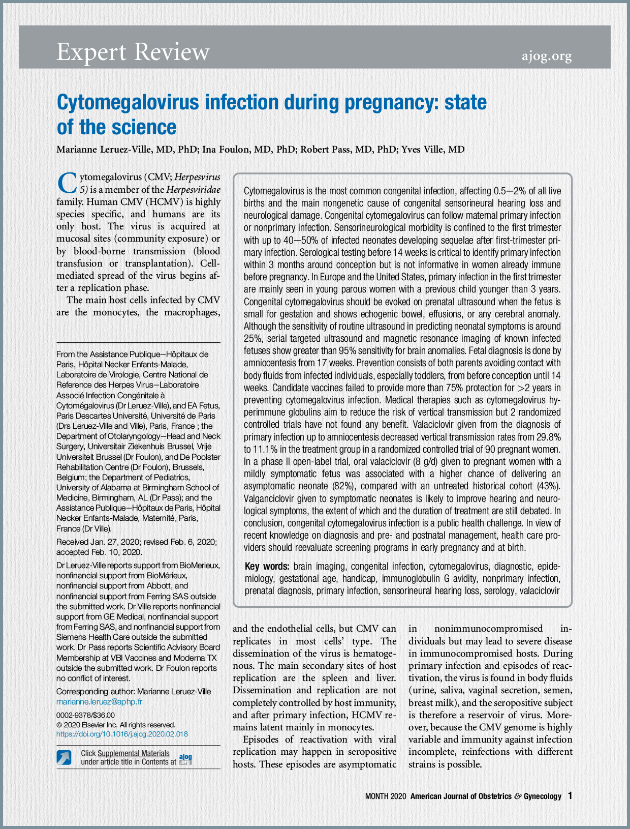
A critical aspect emphasized is that sequelae are primarily limited to cases where maternal infection occurred before 14 weeks of gestation. Despite advances in prenatal assessment using ultrasound and MRI, predicting neonatal status, especially in cases without overt severe cerebral features, remains challenging, underscoring the need for new prognostic biomarkers in amniotic fluid.

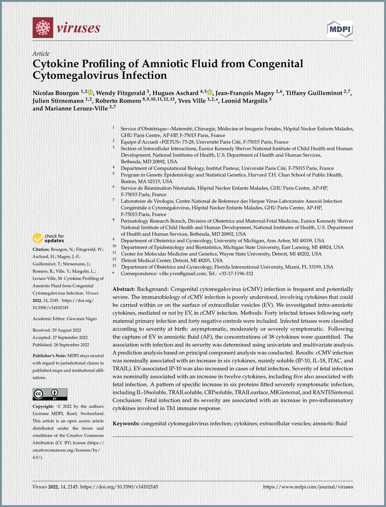
Background and Rationale: cCMV is a leading non-genetic cause of sensorineural hearing loss (SNHL) and a major contributor to neurological damage. Approximately 30% of infected newborns are symptomatic at birth, with 10–15% experiencing SNHL and vestibulitis, and 10–25% suffering more severe neurological damage, including intellectual disability or developmental delay. The immunobiology underlying cCMV infection is poorly understood. The study posits that cytokines, which are crucial mediators in innate immunity and immune control of cCMV, could serve as important biomarkers, particularly those transported within or on the surface of extracellular vesicles (EV). A previous study had already indicated elevated levels of interferon gamma-induced protein 10 (IP-10) in the amniotic fluid of infected fetuses.

Study Objectives: The primary objective of this study was to comprehensively investigate the concentrations and profiles of intra-amniotic cytokines, including their soluble, surface-associated, and internal (extracellular vesicle-associated) fractions, in cCMV infection. The goal was to correlate these cytokine profiles with the severity of the infection at birth, thereby identifying potential candidate biomarkers for prognosis.

Methodology: The study was conducted at the Fetal Medicine Unit at Necker Hospital in Paris, enrolling 80 pairs of women and fetuses/newborns between December 2011 and December 2017. This included 40 infected fetuses, whose mothers had primary CMV infection diagnosed within two months prior to conception or in the first trimester of pregnancy, and 40 non-infected, euploid controls, matched for fetal gender and gestational age at amniocentesis. Women with immune disorders or treatments affecting immunity were excluded.

Diagnosis and Classification:

  • Maternal Primary Infection (MPI): Timing was determined using an in-house algorithm based on CMV IgG and IgM antibody concentrations and IgG avidity.
  • Fetal Infection: Defined by a positive CMV DNA PCR on amniotic fluid (AF) samples. Amniocentesis was performed after 17 weeks of gestation and at least 8 weeks following MPI to ensure optimal diagnostic accuracy.
  • Neonatal Infection: Confirmed at birth by CMV DNA PCR on neonatal blood, urine, and saliva. All virological tests were performed by an expert virology laboratory.
  • Newborn Classification: Infected newborns were classified as symptomatic if they had at least one abnormal neonatal feature, or if they were deceased fetuses due to severe brain lesions or spontaneous intrauterine fetal death. Symptomatic cases were further divided into “non-severe” and “severe” subgroups. Asymptomatic newborns presented with no growth restriction, no abnormal clinical features, no biological abnormalities (like thrombocytopenia or liver enzyme elevation), and no abnormalities in ophthalmologic or audiologic assessments, or on transcranial ultrasound (TUS). Fetuses with only isolated minor cerebral features (e.g., subependymal cysts or lenticulostriate vasculopathy calcifications) were considered asymptomatic. Severe symptomatic fetuses were those with at least one severe cerebral feature such as cortical abnormalities, significant ventriculomegaly (>15 mm), or microcephaly. Postmortem examinations confirmed severe infection in lethal cases.

Cytokine Analysis: Amniotic fluid samples were stored and processed to separate different fractions: a “soluble fraction” from the supernatant, and “surface” and “internal” (luminal content) fractions from extracellular vesicles (EVs). The concentrations of 38 distinct cytokines were quantified using a Luminex immunoassay. Statistical analysis included filtering out proteins with insufficient non-zero values (77 of 114 remained) and then performing univariate and multivariate analyses to determine associations with infection and severity, with Bonferroni correction for multiple comparisons.

Key Findings:

  1. Cohort Characteristics: The study included 40 infected fetuses (9 asymptomatic, 31 symptomatic – 13 non-severe, 18 severe) and 40 controls, with similar clinical characteristics between groups.
  2. Cytokines Associated with Fetal Infection: Univariate analysis revealed a nominally significant increase in six proteins in the amniotic fluid of cCMV infected fetuses compared to controls. These included primarily soluble cytokines such as IP-10, IL-18, ITAC, and TRAIL. Notably, EV-associated IP-10 (both internal and surface-bound) was also significantly elevated. IP-10 was particularly robust, reaching the stringent Bonferroni-corrected p-value threshold for both infection and severity (p < 0.0003).
  3. Cytokines Associated with Symptomatic Status at Birth:
    • Twelve proteins showed a significant correlation with symptomatic status at birth.
    • These included four soluble proteins: IP-10, IL-18, TRAIL, and CRP.
    • Eight EV-associated proteins, predominantly within the EVs, were also linked: IP-10, IL-6, MCP1, MIG, and RANTES.
    • Five cytokines previously associated with fetal infection (soluble, surface, or internal IP-10, soluble IL-18, and soluble TRAIL) were also found to be related to symptomatic outcome.
    • A distinct pattern of increased levels of six specific mediators (soluble IL-18, soluble TRAIL, soluble CRP, surface TRAIL, internal MIG, and internal RANTES) was identified as characteristic of severe symptomatic infection.
    • Importantly, fetal thrombocytopenia was also significantly associated with severe fetal infection (p < 0.001).
    • In contrast, amniotic fluid CMV viral load and fetal liver tests were not found to be associated with fetal symptoms in this cohort.
  4. Prediction Analysis: The study demonstrated a strong potential for using cytokine profiles to predict both fetal infection and its severity. A model based on five principal components derived from all proteins showed an average AUC (area under the ROC curve) of 0.72 for predicting fetal infection (p = 0.037). For predicting severity, models based on soluble proteins and all proteins combined performed best, explaining up to 41% of the total variance.

Discussion and Conclusion: The findings of this study significantly enhance the understanding of the immunopathological processes in cCMV infection by highlighting changes in the amniotic fluid’s immunological signature. The increase in pro-inflammatory mediators (IP-10, IL-18, ITAC, and TRAIL) in cases of cCMV infection suggests a robust immune response within the intrauterine environment. The identification of distinct cytokine patterns linked to symptomatic status, and particularly severe infection, holds promise for developing new prognostic biomarkers. The confirmation that CRP is associated with severe fetal infection aligns with previous proteomic analyses.

The study’s strengths include its relatively large cohort size and the comprehensive investigation of cytokines across different fractions, including those associated with extracellular vesicles. The exploration of EV-associated cytokines is particularly novel, as EVs are increasingly recognized for their role in intercellular communication and as carriers of biomarkers in biological fluids, including amniotic fluid. Acknowledged limitations include the unbalanced ratio of cytokines to cases and potential sensitivity limitations of the ELISA method for detecting ultra-low concentrations of EV cytokines, suggesting that newer technologies like SiMoA might offer further insights.

In conclusion, the study firmly establishes that cCMV infection and its severity are associated with differential cytokine expression in amniotic fluid at mid-gestation. These proteins, predominantly soluble, emerge as promising candidate biomarkers for assessing the severity of fetal infection diagnosed by CMV-PCR, potentially improving prenatal counseling and intervention strategies.

The authors highlight cCMV as the most common congenital infection and the leading non-genetic cause of sensorineural hearing loss (SNHL) and neurological damage, emphasizing its significant public health burden. It accounts for up to 10% of all cerebral palsy cases and 8-21% of all congenital SNHL at birth, with this increasing to 25% by age four due to late-onset hearing loss.

Epidemiology and Transmission: CMV is ubiquitous, with seroprevalence in women of childbearing age ranging from 50% to 85% in Western Europe and the United States, increasing with age and lower socioeconomic status. Primary maternal infection (MPI) during pregnancy occurs in approximately 1-2% of pregnancies in these regions. Risk factors for MPI include being young and having at least one child, particularly toddlers who shed the virus for prolonged periods. Women seronegative at their first pregnancy and conceiving within two years face a significantly higher risk of MPI and related sequelae in subsequent infants.

The transplacental transmission rate after MPI is around 32%. While early reports suggested increased transmission with advancing gestation (26% in first, 28% in second, 65% in third trimester), long-term sequelae are primarily limited to maternal infections acquired in the first trimester. Infections occurring in the second or third trimester have not been reported to cause long-term sequelae in over 100 children in large studies. Maternal non-primary infection (MNPI) also contributes to cCMV burden, accounting for approximately 48-90% of cases in some settings, and can also lead to sequelae, though the risk of vertical transmission is generally lower compared to MPI (likely <3.5%).

Clinical Outcomes and Prognosis: About 20% of infected neonates experience neurodevelopmental impairment or permanent sequelae. Symptomatic neonates at birth, constituting about 10% of infected infants, have a higher risk of sequelae (40-58%), while even initially asymptomatic neonates face a 13.5% risk, mainly SNHL. The spectrum of disease, including hearing loss, is similar after MPI and MNPI. The critical prognostic factor is the gestational age at maternal infection, with long-term sequelae almost exclusively associated with first-trimester infections. Severe brain involvement detected early in pregnancy typically leads to a poor prognosis. Children with isolated SNHL are now recognized as symptomatic. Subtler problems in language development, concentration, and quality of life may also manifest at school age.

Diagnosis:

  • Maternal: Serological testing (IgG and IgM) is crucial, especially before 14 weeks of gestation, to identify MPI around conception. IgG avidity testing helps differentiate acute (low avidity, within 3 months) from past infections (high avidity). However, serology is not useful for women already immune before pregnancy.
  • Fetal: CMV DNA detection in amniotic fluid (AF) via PCR is the gold standard for prenatal diagnosis, as infected fetuses excrete the virus in urine. Amniocentesis is typically performed from 17 weeks gestation, at least 6-8 weeks after documented maternal seroconversion. Prenatal ultrasound and MRI are used to identify fetal abnormalities, which can range from subtle to severe, particularly in the brain. While ultrasound’s sensitivity for predicting neonatal symptoms is around 25%, serial targeted ultrasound and MRI in known infected fetuses have a >95% sensitivity for brain anomalies. Nevertheless, a normal ultrasound or non-severe findings at diagnosis can still miss cases that develop severe symptoms later in pregnancy (around 7% uncertainty). Fetal blood analysis, specifically platelet count and viral load, significantly enhances prognostic accuracy. A combination of ultrasound and fetal blood parameters can increase the negative predictive value for any symptoms at birth to 100%.
  • Neonatal: CMV PCR on saliva or urine collected within 3 weeks of birth is the recommended method for neonatal diagnosis. IgM testing in neonates has low sensitivity and is not recommended for diagnosis.

Pathophysiology and Neuropathogenesis: CMV causes neurodevelopmental sequelae, including mental retardation, cerebral palsy, and SNHL. The virus is neuropathic, preferentially infecting neural stem cells, which can inhibit their differentiation into neurons and astrocytes, potentially leading to structural and migratory abnormalities in the developing brain. Extensive necrosis in severely affected brains is associated with viral inclusions and inflammatory infiltrates. CMV infection of placental cells can also contribute to pathogenesis by altering placental formation, potentially leading to placental insufficiency, intrauterine growth restriction, and other fetal outcomes. Animal models, particularly guinea pig and rhesus macaque, are critical for understanding disease mechanisms and testing interventions, as their fetal infection pathogenesis closely mimics human cCMV.

Interventions and Management:

  • Primary Prevention:
    • Hygienic Measures: Avoiding contact with body fluids (saliva, urine, tears) from infected individuals, especially toddlers, from preconception until 14 weeks of gestation is recommended. Educational interventions, such as films and brochures, have improved hygiene adherence among pregnant women.
    • Screening: The article highlights that most European countries do not have routine serology screening in pregnancy, but there is an increasing recognition of its relevance, with some studies showing it to be cost-effective, especially when combined with valacyclovir treatment. Systematic maternal CMV serology in the first trimester is recommended as sequelae are limited to infections acquired during this period. Early serology allows for retesting seronegative women every 4 weeks until 14-16 weeks.
    • Vaccines: Several vaccine candidates are in early-phase clinical trials, including enveloped virus-like particle (eVLP) vaccines and mRNA vaccines expressing CMV gB and pentameric complex. These aim to prevent maternal and congenital CMV infection.
  • Secondary Prevention (to prevent vertical transmission after maternal infection):
    • Valacyclovir: Oral valacyclovir (8 g/day) administered as early as possible after diagnosis of MPI in the periconceptional period or first trimester, and continued until amniocentesis, significantly reduces vertical transmission rates. A meta-analysis confirmed a 70% reduction in vertical transmission and neonatal infection rates, with earlier treatment initiation being more effective. Side effects are minimal, primarily nausea, headache, and rarely, reversible acute kidney injury. Cessation of treatment after a negative amniocentesis is safe, as a negative PCR ensures absence of long-term sequelae.
    • Hyperimmune Globulin (HIG): Two randomized controlled trials did not find a benefit for intravenous HIG (100 IU/kg monthly) in preventing vertical transmission. However, a case-control study suggested possible benefit with higher doses (200 IU/kg biweekly) for very recent first-trimester infections, but this needs further confirmation. The European Congenital Infection Initiative (ECCI) recommends against HIG administration at doses of 100 IU/kg every 4 weeks.
  • Antenatal Treatment (for infected fetus):
    • Valacyclovir: A phase II open-label study demonstrated that oral valacyclovir (8 g/day) given to pregnant women with a moderately symptomatic infected fetus was associated with a significantly higher proportion of asymptomatic neonates (82%) compared to an untreated historical cohort (43%). Fetal blood viral loads decreased and platelet counts increased significantly with treatment. This dosage was well tolerated with high adherence. For women with confirmed fetal infection, discussion with an expert team regarding antenatal valacyclovir treatment (8g/day) may be considered.
  • Neonatal Treatment (for symptomatic neonates):
    • Valganciclovir/Ganciclovir: For newborns with significant CMV-related symptoms at birth, including CNS involvement or isolated SNHL, antiviral treatment with valganciclovir (or ganciclovir if enteral medication is not possible) is recommended for 6 months. Treatment should start as soon as possible, ideally before one month of age, though starting between 1-3 months may also be beneficial. Treatment is likely to improve hearing and neurological symptoms. Six weeks of treatment is recommended for isolated persistent thrombocytopenia. Routine treatment for isolated intrauterine growth restriction (IUGR) is not recommended.

Conclusion: The article concludes that cCMV infection presents a major public health challenge. Given the advancements in diagnosis and pre- and postnatal management, the authors advocate for reevaluating screening programs in early pregnancy and at birth to allow for earlier and more informed decision-making regarding prevention and treatment strategies.

Background and Rationale: Congenital CMV infection is a significant public health concern, affecting approximately 0.7% of live births globally. It is recognized as the leading non-genetic cause of sensorineural hearing loss (SNHL) and neurological damage, accounting for up to 10% of all cerebral palsy cases. While about 20% of infected children ultimately experience neurodevelopmental impairment with permanent sequelae, the severity of the disease varies greatly. Approximately 10% of infected neonates are symptomatic at birth, with their risk of long-term sequelae reaching up to 58%. Even those initially asymptomatic at birth face a 13% risk of sequelae, primarily progressive hearing loss.

Fetal CMV infection is typically diagnosed by detecting viral DNA in amniotic fluid (AF) via PCR, usually following documented maternal seroconversion or the appearance of suggestive ultrasound (US) features. Previous studies have established that severe brain abnormalities on prenatal US are associated with a poor prognosis. However, the prognosis for infected fetuses with no US features or only non-severe anomalies is challenging to determine until late in the second or third trimester. This uncertainty poses a significant challenge for parental counseling, particularly in regions where termination of pregnancy (TOP) is time-limited. The study aimed to provide earlier and more precise prognostic information by evaluating fetal parameters at the time of diagnosis.

Study Objective and Design: The primary objective was to evaluate the prognostic value of fetal US, amniotic fluid, and fetal blood analysis at the time of prenatal diagnosis of fetal CMV infection. The study reviewed cases of fetal CMV infection from 2008 to 2013 where amniotic fluid and/or fetal blood samples were analyzed for quantitative CMV PCR.

Study Population and Inclusion Criteria: The study included 82 fetuses diagnosed with CMV infection, primarily following maternal primary infection in the first trimester (62 out of 81 cases with known timing). Amniocentesis was performed at a median gestational age of 23 weeks. Fetal symptoms were classified based on prenatal ultrasound into three groups at the time of diagnosis:

  • Severe brain US abnormalities: (19 fetuses). These included ventriculomegaly ≥15mm, periventricular hyperechogenicity, hydrocephaly, microcephaly, mega-cisterna magna >10mm, vermian hypoplasia, porencephaly, lissencephaly, periventricular cysts, and abnormal corpus callosum. These cases typically led to TOP.
  • Non-severe US features: (22 fetuses). This group included at least one extracerebral abnormality (e.g., fetal growth restriction, abnormal amniotic fluid volume, ascites/pleural effusion, skin edema, hydrops, placentomegaly >40mm, hyperechogenic bowel, hepatomegaly >40mm, splenomegaly >30mm, liver calcifications) or one mild brain abnormality (e.g., moderate isolated ventriculomegaly <15mm, isolated cerebral calcification, isolated intraventricular adhesion, vasculopathy of lenticulostriate vessels).
  • Normal US examination: (41 fetuses).

Methods:

  • Maternal Infection Diagnosis: The timing of maternal primary infection was determined using CMV IgG/IgM and IgG avidity assays, with an algorithm to pinpoint the onset of infection.
  • Fetal Infection Diagnosis: Confirmed by positive CMV DNA PCR in amniotic fluid, collected at least 6 weeks after seroconversion and not earlier than 20 weeks’ gestation.
  • Prenatal Follow-up: Infected fetuses underwent serial US every 2-3 weeks. Fetal blood sampling via cordocentesis was offered to assess fetal platelets and viremia. Fetal cerebral MRI was performed at 32-34 weeks or earlier if US suspected brain lesions.
  • Classification of Neonatal Outcome: Neonates were classified as symptomatic or asymptomatic at birth based on a combination of clinical examination, laboratory assessment (platelet count, liver enzymes, bilirubin), audiometric assessment, and cerebral imaging (cranial US/MRI). Autopsy was performed in all TOP cases to confirm severe cerebral abnormalities.
  • Antenatal Treatment: Some women with non-severe US anomalies, fetal viremia >3000 copies/mL, or fetal platelets <100,000/mm³ received oral valacyclovir (8g/day) as part of an ongoing trial. However, in this specific study population, valacyclovir treatment was not associated with a better prognosis (P=1.0), suggesting it did not interfere with the interpretation of fetal parameters for outcome prediction.
  • Statistical Analysis: Univariate and bivariate logistic regression models were used to analyze the association between outcome (symptomatic/asymptomatic at birth or TOP) and US features, CMV DNA loads in AF and fetal blood, and fetal platelet counts.

Key Findings:

  1. Overall Prognostic Value:
    • Severe brain US abnormalities at prenatal diagnosis consistently predicted a dismal prognosis, with all 18 terminated fetuses in this group classified as symptomatic at autopsy.
    • 93% Negative Predictive Value (NPV) of US at diagnosis for severe brain abnormalities at birth or TOP. This means that if the US was normal at diagnosis, there was a high likelihood of a good outcome regarding severe brain issues.
    • However, a normal US examination or the presence of non-severe symptoms still missed 7% (3 out of 41 cases) that eventually progressed to severe symptoms in the third trimester. This indicates a remaining 7% uncertainty for parents.
  2. Added Value of Laboratory Parameters (for non-severe cases):
    • In the group without severe brain US abnormalities (63 fetuses), non-severe US features, higher DNA load in AF, lower fetal platelet count, and higher fetal blood DNA load were significantly associated with a symptomatic status at birth or TOP in univariate analysis.
    • Fetal blood parameters (viral load and platelet count) were better predictors of the outcome than amniotic fluid viral load.
    • A recursive partitioning model showed that if the platelet count was ≤114,000/mm³, there was a 62.5% risk of symptomatic status. If the platelet count was >114,000/mm³ but the CMV DNA load in fetal blood was ≥4.93 log10 IU/mL, the risk was 57%. If both were favorable, the risk dropped to 1%.
    • Combining US findings with fetal blood parameters (platelet count and viral load) increased the Negative Predictive Value (NPV) for any symptoms at birth to 100%. This suggests that if both US and fetal blood parameters are normal, there is virtually no risk of symptoms at birth.
    • The Positive Predictive Value (PPV) for non-severe US features alone was 60%. When combined with abnormal fetal blood results or high adjusted AF viral load, the PPV for any symptom at birth rose to 79% and 78%, respectively.
  3. Changes in Fetal Parameters:
    • CMV DNA levels in AF were found to increase with the time interval from seroconversion to amniocentesis, necessitating adjustment for this variable.
    • Symptomatic neonates consistently had significantly lower platelet counts at birth than asymptomatic neonates.

Strengths and Limitations:

  • Strengths: The study benefits from a relatively large cohort, extensive longitudinal prenatal assessment including serial imaging and invasive fetal testing, and the use of strict criteria for TOP based on confirmed brain lesions at autopsy. The detailed documentation of maternal primary infection timing and the adjustment of AF viral load for gestational age were also notable.
  • Limitations: The study had a subgroup where not all three laboratory parameters were consistently available. The duration of postnatal follow-up varied, and some children were lost to follow-up. The concurrent valacyclovir treatment in some cases was a potential confounder, though analysis suggested it did not impact the prognostic findings in this specific cohort. The authors also note the need to validate CMV DNA load cut-off values in different clinical and laboratory settings.

Conclusion and Implications: The study concludes that a comprehensive prognostic assessment of CMV-infected fetuses is feasible as early as the time of prenatal diagnosis in the second trimester. This is achieved by combining targeted ultrasound examination with viral load measurements in amniotic fluid and fetal blood, alongside fetal platelet count. While ultrasound alone provides valuable information, integrating fetal blood parameters significantly enhances the predictive accuracy, reducing the uncertainty regarding neonatal outcomes. The authors emphasize that the decision to undergo additional invasive procedures like cordocentesis, given the increased predictive value, remains an individual choice for well-informed women. This research provides crucial data for improved counseling and potentially for guiding early antenatal treatment decisions for cCMV.

Background and Rationale: Congenital cytomegalovirus (cCMV) infection is identified as a major cause of morbidity and mortality, affecting approximately 0.7% of live births globally. It stands as the leading cause of congenital neurological disease of infectious origin. While some cCMV cases are asymptomatic, about 10% of infected neonates are symptomatic at birth, with their risk of long-term sequelae, including sensorineural hearing loss (SNHL) and cognitive or motor deficits, reaching up to 58%. Even those initially asymptomatic at birth face a notable 13% risk of sequelae, predominantly progressive hearing loss.

The authors highlight that cCMV disease is progressive, with early symptoms potentially appearing as extracerebral findings on prenatal ultrasound, while brain involvement often manifests several weeks later. Although severe brain lesions on prenatal ultrasound predict a dismal prognosis, there is a critical window of opportunity for intervention in symptomatic fetuses without such severe brain involvement.

Existing management options have limitations:

  • A cCMV vaccine is not yet available.
  • The use of CMV-specific hyperimmune globulin to prevent vertical transmission has yielded conflicting results.
  • While neonatal antiviral treatments with ganciclovir or valganciclovir show promise in improving auditory and neurological outcomes in symptomatic newborns, these drugs are highly genotoxic in vitro and are not approved for use during pregnancy.
  • Previous pilot studies indicated that high-dose valacyclovir, though less effective than ganciclovir against CMV in vitro, could achieve therapeutic concentrations in fetal blood and significantly reduce viral load in infected fetuses. Valacyclovir also has a favorable safety profile, with thousands of recorded exposures during pregnancy showing no increased risk of birth defects.
  • A key challenge noted was the impracticality of conducting a randomized controlled trial (RCT) comparing valacyclovir to placebo in symptomatic infected fetuses, as many pregnant women declined randomization, unwilling to accept a placebo arm given the severe potential outcomes. This underscored the need for alternative study designs to evaluate potential treatments.

Study Objective and Design: Given these challenges, the study was designed as a multicenter, open-label, phase II, single-arm trial, employing Simon’s optimal 2-stage design. The primary objective was to evaluate the proportion of asymptomatic neonates born to mothers treated with valacyclovir, specifically targeting fetuses with moderately symptomatic cCMV infection who were at high risk for neurosensory and neurological impairment.

Inclusion and Exclusion Criteria:

  • Eligible participants were pregnant women with a confirmed fetal CMV infection via positive CMV PCR in amniotic fluid obtained after 21 weeks of gestation.
  • Fetal symptoms for inclusion comprised at least one of the following:
    • Extracerebral abnormalities: such as fetal growth restriction, abnormal amniotic fluid volume, ascites/pleural effusion, skin edema, hydrops, placentomegaly (>40mm), hyperechogenic bowel, hepatomegaly (>40mm), splenomegaly (>30mm), or liver calcifications.
    • Isolated cerebral abnormalities: including moderate isolated ventriculomegaly (<15mm), isolated cerebral calcification, isolated intraventricular adhesion, or vasculopathy of lenticulostriate vessels.
    • Abnormal laboratory findings in fetal blood: specifically fetal viremia >3000 copies/mL or fetal platelet count <100,000/mm³.
  • Excluded were fetuses with severe brain abnormalities (e.g., ventriculomegaly ≥15mm, microcephaly, lissencephaly, cortical abnormalities, or periventricular cysts), as their prognosis was considered dismal and unlikely to be altered by treatment. Cases with no ultrasound features or laboratory abnormalities were also excluded, as their outcome was typically good without intervention.

Intervention and Endpoints: All participants received oral valacyclovir at a dose of 8 grams daily (administered as 2 grams, four times a day). Treatment continued until delivery or for a maximum of 24 weeks, whichever occurred first.

  • The primary endpoint was the proportion of asymptomatic neonates at birth, defined by normal growth, clinical examination, laboratory findings, cerebral imaging, funduscopy, and audiology.
  • Secondary endpoints included monitoring for maternal adverse events, assessing adherence to treatment, and evaluating changes in fetal blood viral load and platelet counts.
  • A historical comparator group of 47 similarly symptomatic fetuses from three published studies was used to contextualize the treatment’s efficacy, with 42.55% of neonates in this untreated group being asymptomatic at birth.

Results:

  • Enrollment: The study enrolled 41 pregnant women with 43 fetuses (39 singletons, 2 pairs of twins) from January 2012 to December 2014. The median gestational age at enrollment was 26 weeks, and almost all maternal infections (40 of 41) were primary infections, with a median timing of 10 weeks gestation. Most fetuses (54%) presented with at least two qualifying ultrasound symptoms, 39% with one, and 7% (3 fetuses) only with high fetal viremia (>3000 copies/mL).
  • Primary Endpoint Achievement: The study successfully met its predefined efficacy criteria based on Simon’s 2-stage design. In the first stage, 8 of the initial 11 treated women delivered an asymptomatic neonate, meeting the threshold to proceed. The final analysis, including all 43 cases, revealed that 34 fetuses (79.1%, approximately 82% based on unbiased estimation) were asymptomatic at birth, surpassing the required 31 for the treatment to be considered effective.
  • Comparison to Historical Cohort: The results showed a significant increase in the proportion of asymptomatic neonates when compared to the historical untreated cohort. While only 43% of neonates in the historical group were asymptomatic, this figure rose to 82% in the valacyclovir-treated group.
  • Safety and Adherence:
    • Valacyclovir was exceptionally well tolerated by mothers. Only two women reported headaches, and mild, clinically irrelevant increases in maternal liver enzymes were observed. Creatinine levels remained unchanged.
    • Despite the high pill burden (16 pills/day), adherence to treatment was consistently high, exceeding 90%.
  • Fetal Parameters:
    • Significant reductions were observed in fetal blood viral loads, with a median decrease from 4.0 to 3.05 log10 IU/mL (P=.01) from pretreatment sampling to birth.
    • Concurrently, fetal platelet counts significantly increased from a median of 173,000/mm³ to 245,000/mm³ (P<.001).
    • These changes were attributed to the antiviral effects of valacyclovir. While the duration of maternal treatment did not correlate with changes in viral loads, longer treatment duration was associated with higher platelet counts at birth. Symptomatic neonates had significantly lower platelet counts at birth.

Conclusion and Implications: The authors conclude that high-dose oral valacyclovir administered during pregnancy is effective in improving the outcome of moderately symptomatic fetuses infected with CMV. The study’s findings demonstrate a clear benefit in reducing symptoms at birth, supported by objective changes in fetal viral load and platelet counts. Despite not being a randomized controlled trial, the observed improvements, coupled with the drug’s excellent safety profile and high adherence rates, provide strong evidence for its clinical utility. This study is considered foundational for future research into new and potentially more potent anti-CMV drugs, using valacyclovir as a proven baseline treatment in pregnancy. The authors also reiterate the importance of re-evaluating current screening programs for CMV in early pregnancy, as highlighted in related expert recommendations.

The Enduring Burden of Congenital Cytomegalovirus Infection: The letter emphasizes that cCMV is the leading cause of congenital infection globally, affecting approximately 0.5–2% of all live births worldwide. It is also recognized as the primary non-genetic cause of sensorineural hearing loss (SNHL), accounting for about one-third of congenital hearing loss cases and 8–21% of all congenital SNHL at birth, a figure that increases to 25% by the age of four due to late-onset hearing loss. Beyond auditory impairments, cCMV is a major cause of neurological damage, contributing to up to 10% of all cerebral palsy cases.

The severity of cCMV infection is highly variable, ranging from asymptomatic presentations to severe neurological disabilities or even perinatal death. Sequelae, including neurodevelopmental impairment, are almost exclusively linked to maternal primary infections (MPI) acquired before 14 weeks of gestation. Around 20–30% of children exposed in utero following maternal seroconversion in early pregnancy will develop neurological sequelae. Specifically, about 30% of newborns are symptomatic at birth, with 10–15% experiencing SNHL and 10–25% suffering more severe neurological damage, including intellectual disability or developmental delay. Even in neonates who appear asymptomatic at birth, there remains a significant risk of long-term sequelae, estimated at around 13.5%, primarily due to progressive hearing loss. The cumulative impact is substantial, with an estimated 30,000 cases resulting in related disabilities annually in the United States and a similar number in the European Union, alongside overall mortality and sequelae rates of 0.5% and 17–20%, respectively. A critical point highlighted is that cCMV is the only congenital infection whose worldwide prevalence has not decreased over the past 60 years. Furthermore, primary CMV infection poses a particular risk to seronegative women who have a child aged 2–3 years, with infection rates reaching up to 10% in this demographic.

Current Management Practices and Identified Gaps: The authors note that while the potential severity of primary maternal infection in early pregnancy is well-established, practical challenges render primary prevention measures, such as hygiene counseling, less effective, particularly if initiated late in pregnancy (e.g., at 11–12 weeks) when crucial windows of vulnerability have already passed. Currently, prenatal diagnosis of cCMV in France involves amniocentesis for CMV genome PCR, performed either after documented maternal seroconversion or when suggestive fetal abnormalities are observed on routine ultrasound examinations. These suggestive findings include intrauterine growth restriction, microcephaly, echogenic bowel, hepatosplenomegaly, placentomegaly, oligohydramnios or polyhydramnios, and various cerebral anomalies such as ventriculomegaly, abnormal midline structures, or posterior fossa and cerebellar abnormalities. Amniocentesis is typically recommended at or after 21 weeks of gestation, and at least 6 weeks following the presumed date of maternal primary infection, though recent studies suggest reliability from 17 weeks if performed at least 8 weeks post-MPI. A negative CMV PCR result in amniotic fluid following a timely amniocentesis generally assures the absence of long-term sequelae.

However, the authors point out significant limitations in the current approach:

  • Lack of Universal Screening: While Italy and Greece have implemented universal serological screening for CMV in early pregnancy based on expert recommendations, France and most other European countries have not, though the issue is currently under scrutiny.
  • Limited Prognostic Information: The number of pregnancy terminations related to fetal CMV infection has remained stable and notably low before 20 weeks of gestation, a period when only fetal CMV infection status is available without comprehensive prognostic information. This uncertainty hinders informed parental decision-making regarding potential pregnancy termination, even when severe fetal findings are identified later.
  • Incomplete Protection and Awareness: Serological testing is not informative for women who are already immune before pregnancy. Moreover, the fact that only about 10% of infected fetuses are symptomatic at birth complicates early prognosis and decisions regarding prenatal treatment. Crucially, normal prenatal imaging findings (ultrasound or MRI) do not rule out the development of SNHL and minor neurodevelopmental abnormalities in children born with cCMV.

Arguments for Universal Screening and Early Intervention: The letter strongly advocates for universal screening based on several key arguments:

  • Empowering Pregnant Women and Ensuring Equity: Universal screening would address the current disparity where many women are unaware of their CMV risk, thus empowering them to make informed decisions. It would uphold the principle of horizontal equity, ensuring that all pregnant women receive equal access to information about severe fetal CMV, allowing them to consider pregnancy termination if permitted by national laws.
  • Efficacy of Valacyclovir in Secondary Prevention: The emergence of effective antiviral treatment, specifically valacyclovir, for secondary prevention of vertical transmission strengthens the case for early screening.
    • Recent randomized controlled trials and quasi-randomized studies have demonstrated that oral valacyclovir at a dose of 8 g/day significantly reduces the vertical transmission rate of CMV by 70–71% in women with primary CMV infection acquired periconceptionally or during the first trimester of pregnancy. This reduction is observed for both periconceptional and first-trimester infections.
    • The meta-analysis cited indicates that valacyclovir also reduces the rate of neonatal infection (adjusted odds ratio, 0.30).
    • Significantly, valacyclovir treatment has been shown to reduce the rate of termination of pregnancy due to CMV-associated severe fetal findings by 77% (adjusted odds ratio, 0.23).
    • The efficacy of treatment is highly dependent on early initiation, with earlier commencement leading to better prevention outcomes.
    • Prior research, including a phase II study, supported these findings, demonstrating that high-dose valacyclovir administered to women with moderately symptomatic infected fetuses increased the proportion of asymptomatic neonates from 43% in an untreated historical cohort to 82% in the treated group. This study also indicated that fetal blood viral loads decreased and platelet counts increased significantly between treatment initiation and birth, suggesting a direct antiviral effect.
  • Favorable Safety Profile and Adherence: High-dose valacyclovir (8 g/day) has demonstrated an exceptionally good safety profile for mothers, with minimal side effects such as nausea or headache (reported in about 20% of women) and rare instances of mild to moderate acute kidney injury (2.1%), which typically resolved upon treatment cessation. The risk of acute kidney injury can be further mitigated by fragmenting the daily administration (e.g., 2g four times daily) and ensuring adequate hydration. Despite the high pill burden (16 pills a day), adherence to treatment was consistently high (>90%).
  • Cost-Effectiveness: Research has demonstrated that universal screening for first-trimester CMV infection is cost-effective and even cost-saving when combined with timely valacyclovir treatment.

Conclusion and Call to Action: In summary, the authors assert that the scientific understanding of cCMV has significantly advanced, particularly regarding the efficacy of antiviral treatment in preventing vertical transmission following primary maternal infection. They advocate that the benefits of early and accurate maternal diagnosis through serological tests, combined with the proven efficacy and safety of valacyclovir, are compelling enough to warrant a shift towards universal CMV screening in early pregnancy. This would not only enhance risk management and prevention strategies but also allow for refined personalized postnatal care based on risk assessments, ensuring targeted interventions for affected families. The letter serves as a crucial call to action for public health authorities and healthcare professionals to re-evaluate existing screening policies and implement a more proactive approach to mitigate the severe consequences of congenital CMV infection.

  1. Background and Burden of cCMV Infection: Congenital CMV infection is identified as the leading cause of birth defects and childhood disorders in the United States, affecting an estimated 40,000 children annually, with around 400 fatal cases. While only a small percentage (10-15%) of infected children exhibit clinical signs at birth, a substantial proportion – 60-90% of symptomatic infants and 10-15% of asymptomatic infants – develop one or more long-term neurological sequelae, such such as mental retardation, psychomotor retardation, and sensorineural hearing loss (SNHL). This makes cCMV a more common cause of birth defects and childhood disabilities than conditions like Down syndrome or spina bifida.

CMV is a ubiquitous herpesvirus that establishes lifelong latency with periodic reactivations. The greatest risk for cCMV is associated with a primary maternal infection (infection in a seronegative individual) during pregnancy, with approximately one-third of these cases leading to transplacental transmission. About half of these intrauterine infections can result in a symptomatic clinical syndrome. The timing of primary infection is crucial, with neurological outcomes being more severe when infection occurs during the first trimester, although transmission can occur throughout gestation. While maternal seropositivity prior to conception offers some protection, it is not absolute, and reinfection with different CMV strains can still lead to fetal infection and sequelae. Maternal antibodies play a critical role in preventing transplacental transmission, and their qualitative aspects (e.g., neutralizing, high-avidity antibodies) are key indicators of fetal protection. Quantitative analysis of viral load in amniotic fluid has been identified as the best predictor for neurological damage in cCMV infection, with higher loads correlating with symptomatic infections.

  1. Pathology of cCMV Infection: The CNS is the most severely affected organ system in cCMV, with injuries generally considered irreversible. Common symptoms at birth include intrauterine growth retardation, purpura, jaundice, hepatosplenomegaly, microencephaly, hearing impairment, and thrombocytopenia. While some signs like hepatosplenomegaly are transient, neurological deficits often persist for life or become evident later in early childhood, such as SNHL.
  • Brain Structural Abnormalities:
    • Neonatal imaging (CT, MRI) almost always reveals structural brain abnormalities in symptomatic infants, including intracranial calcifications (50% of cases), ventriculomegaly, white matter changes, polymicrogyria, and cysts.
    • Subtle patterns of white matter lesions, with or without polymicrogyria and anterior temporal lobe cysts, are highly suggestive of CMV infection.
    • While normal imaging does not entirely rule out future hearing loss, the extent to which white matter lesions correlate with SNHL or other neurological syndromes needs further investigation.
  • Auditory Abnormalities (SNHL):
    • CMV is the most common infectious disease associated with SNHL in children.
    • The frequency of SNHL is higher in symptomatic cCMV cases (30-65%) than in asymptomatic cases (7-15%), where it is often the only sequela.
    • SNHL often progresses with age, meaning universal newborn hearing screening may miss a majority of cases, as hearing loss can develop later or worsen over time.
    • Persistent viral replication and high viral burden, indicated by increased levels and longer duration of urinary CMV excretion, correlate with the development of hearing loss.
    • CMV-induced hearing loss is believed to result from virus-induced labyrinthitis, involving damage to vestibular endolymphatic structures, with the virus potentially entering the endolymph via the stria vascularis.

III. Pathogenic Mechanisms of CNS Injury: The article delves into various proposed mechanisms by which CMV injures the developing fetal CNS:

  • CMV as a Teratogen: While a direct teratogenic role is not definitively proven, studies suggest CMV may cause birth defects through direct chromosomal injury (e.g., breaks on chromosome 1, potentially linked to SNHL and visual impairment) or by modulating developmental gene expression.
  • Apoptosis and Cell Cycle Changes: CMV has evolved mechanisms to delay apoptosis in infected cells, using viral proteins (IE1, IE2, UL36, UL37) to allow for its replication. However, neuronal damage via apoptosis is observed in cCMV brains, often in uninfected “bystander” cells, suggesting indirect mechanisms like neuroinflammatory responses.
  • Neural Stem Cells (NSCs) and Developmental Susceptibility:
    • CMV preferentially infects cells in the ventricular or subventricular regions of the brain, where neural stem/precursor cells reside.
    • Infection of human neural precursor cells inhibits their proliferation and alters their differentiation profiles into neurons and astrocytes. This disruption may account for many structural and migratory abnormalities.
    • The fetal brain’s higher proportion of actively dividing immature neural stem cells may explain why neurological symptoms are more severe in congenital infection compared to adults.
  • Neuroinflammatory Processes:
    • Immune responses in the CNS, involving resident glial cells (astrocytes, microglia) and infiltrating immune cells, play a critical role.
    • CMV infection induces the production of various cytokines and chemokines (e.g., CCL2, CXCL10, TNF-α, IL-6), which can have both protective (inhibiting viral replication) and deleterious effects (neurotoxicity).
    • An attenuated interferon (IFN) response in neonates compared to adults is observed in mouse models.
    • Fetal immune responses, particularly CD8 T cell effectors, are robust but may have decreased cytokine production, potentially influencing neurological outcomes.
    • The balance between viral control and immunopathology is delicate; dysregulated inflammatory responses can lead to severe damage.
  • Placental Insufficiency: The placenta acts as an amplifying reservoir and conduit for viral transmission. CMV infection of placental cells can alter placental formation, leading to placental insufficiency, which may contribute to fetal pathologies by causing vascular damage, hypoxia, and altered permeability.
  1. Animal Models for Studying cCMV: Animal models are crucial for understanding CMV pathogenesis, despite host specificity.
  • Guinea Pig Model (GPCMV): Highly valuable as it naturally crosses the placental barrier and can cause neurological disease, labyrinthitis, and SNHL, mirroring human cCMV. Studies have shown that systemic immunity can protect against SNHL, supporting vaccine development.
  • Mouse Model (MCMV): While MCMV typically does not cross the placental barrier naturally (except in severely immunodeficient mice), it provides insights due to its genetic similarity to HCMV and the availability of numerous genetic tools and reagents. It helps study brain susceptibility based on gestational age, effects on neural stem cells, and physiological changes in neurons.
  • Rhesus Macaque Model (RhCMV): Considered highly relevant due to its remarkably similar pathogenesis to human cCMV, including CNS and cochlear injury, and close genome organization and protein-coding content.
  1. Prospects for Intervention: The article also touches upon existing and developing intervention strategies:
  • Antiviral Drugs:
    • Ganciclovir and its prodrug valganciclovir are the primary antiviral agents for cCMV disease.
    • A randomized controlled trial showed that ganciclovir therapy significantly improved hearing or maintained normal hearing in symptomatic congenital CMV disease involving the CNS, and notably, none of the ganciclovir recipients had worsening of hearing compared to 41% in controls at 6 months. This benefit was sustained at 12 months.
    • Other antivirals like foscarnet and cidofovir are second-line options but carry significant toxicities (e.g., nephrotoxicity).
    • CMV Immune Globulin (HIG): While used for prophylaxis in transplant patients, its efficacy in preventing fetal CMV infection in pregnancy has shown conflicting results in studies, though some uncontrolled trials suggested benefits in reducing transmission or improving ultrasonic abnormalities.
  • Vaccines:
    • Vaccine development is a top national priority for public health.
    • Early vaccine candidates like the Towne strain failed to protect seronegative mothers from CMV infection.
    • Novel candidates, including enveloped virus-like particle (eVLP) vaccines, replication-defective virion vaccines, and messenger RNA (mRNA) vaccines, are in early clinical trials, aiming for broader immune responses.
    • Animal models, particularly the guinea pig, have demonstrated the potential of gB subunit DNA vaccines to reduce congenital infection rates and that adjuvanted glycoprotein vaccines can protect against congenital infection and disease.
    • The concept of herd immunity through universal immunization is highlighted as a potential strategy to significantly reduce the incidence of cCMV, drawing parallels with rubella elimination.

In conclusion, the article underscores the complexity of cCMV neuropathogenesis, involving direct viral effects, immune responses, and impacts on neural stem cell development. It calls for continued research into disease mechanisms and an increased sense of urgency in developing and implementing effective interventions, including better awareness, funding for pathogenesis studies, and clinical vaccine trials.

1/ Objective and Methods: The study aimed to determine the short- and long-term outcomes of pregnancies with documented primary maternal CMV infection in the first trimester and evidence of fetal infection. The cohort included patients referred to the Fetal Medicine Unit at Sheba Medical Center, Tel-Aviv, Israel, between January 2011 and January 2018.

  • Diagnosis of Maternal and Fetal Infection:
    • Primary maternal CMV infection was diagnosed by IgG seroconversion or the presence of IgM antibodies combined with low IgG avidity. Israel’s common practice of serial CMV screening in the first trimester facilitated the availability of data on infection onset and natural history.
    • Fetal infection (vertical transmission) was confirmed by CMV PCR in amniotic fluid, collected at least 7 weeks after seroconversion and not earlier than 21 completed weeks’ gestation.
    • Neonatal infection was re-tested by CMV PCR in urine or saliva samples within the first few days after birth, with the CMV genome detected in all cases post-birth.
  • Prenatal Assessment:
    • All patients underwent serial prenatal ultrasound (US) scans every 3-4 weeks until delivery, assessing fetal growth, amniotic fluid volume, placental appearance, and markers of fetal disease (e.g., ventriculomegaly, echogenic foci, cysts, posterior fossa/cerebellar anomalies, liver/bowel echogenicity). US findings were classified as severe brain, mild-to-moderate brain, or extracerebral abnormalities.
    • Fetal Magnetic Resonance Imaging (MRI) was performed at 32–33 weeks’ gestation, or earlier if brain lesions were suspected on ultrasound. MRI findings were graded from 1 (normal) to 5 (migration disorders, cerebellar hypoplasia, microcephaly), with Grade 4/5 indicating anatomical findings.
  • Postnatal Assessment and Follow-up:
    • All neonates underwent ocular fundus examination, a neonatal brain ultrasound scan (TCUS), and hearing evaluation (transient evoked optoacoustic emission (TEOAE) and brainstem evoked response audiometry (BERA)) within the first week of life.
    • Follow-up was periodic for a median of 2 years (range: 6 months to 10 years), obtained from hospital charts and parent interviews.
    • CMV-associated sequelae were defined as bilateral/unilateral/partial sensorineural hearing loss (SNHL), neurodevelopmental abnormality (mainly hypotonia or motor delay), composite clinical outcome (any SNHL or neurodevelopmental abnormality), and composite outcome (additionally including termination of pregnancy (TOP)).
    • No patients in this study received fetal therapy (CMV hyperimmune globulin (HIG) or antiviral therapy) during pregnancy. Neonates with abnormal findings (clinical, lab, hearing, imaging) received ganciclovir or valganciclovir for 12 months.

2/ Results:

  • Prenatal Imaging Findings:
    • Out of 123 patients with first-trimester CMV infection and proven fetal infection, 30.9% (38/123) had abnormal ultrasound findings. This included 9.8% with severe brain findings, 12.2% with mild-to-moderate brain findings, and 8.9% with extracerebral findings (e.g., intrauterine growth restriction, hepatomegaly, splenomegaly, echogenic bowel).
    • Abnormal MRI findings were observed in 30.1% (37/123) of cases overall. Crucially, 14.1% (12/85) of patients with normal ultrasound findings still had an abnormal MRI, with 5.9% (5/85) of these showing true anatomical findings (e.g., periventricular cystic lesions, arachnoid cysts, temporal lobe cysts).
    • Abnormal MRI findings were significantly associated with abnormal sonographic results (P < 0.001). New ultrasound findings, mainly lenticulostriate vasculopathy (LSV) but also cystic lesions and calcifications, were detected at 34-36 weeks’ gestation in five cases where previous serial US and MRI were normal until 32 weeks.
  • Pregnancy Outcomes:
    • Fifteen patients chose to terminate the pregnancy, primarily due to abnormal US (13 cases, 8 with severe brain findings) or MRI findings (2 cases with normal US but Grade-4 MRI findings).
    • Of the 108 live-born infants, 35.2% (38/108) had abnormal initial neonatal TCUS, with 24/38 having new findings not diagnosed prenatally.
    • The overall rate of CMV-associated postnatal and childhood sequelae was 27.8%.
    • SNHL was observed in 16.7% (18/108) of live-born children.
      • Three children categorized as deaf had normal prenatal imaging.
      • Seven additional children developed SNHL after an initial normal BERA examination, with three of these having normal prenatal imaging.
      • Progressive SNHL was frequent.
    • Neurodevelopmental abnormalities occurred in 11.1% (12/108) of children, mostly slight motor or verbal delay.
    • Approximately half of the cases with CMV-associated sequelae did not have any abnormal prenatal imaging findings. Specifically, of the 73 cases with both normal US and MRI, 10 (13.7%) developed SNHL and 5 (6.8%) developed neurodevelopmental abnormality.
  • Correlation between Imaging and Outcomes:
    • Abnormal prenatal US findings were not significantly associated with SNHL, neurodevelopmental delay, or composite clinical outcome (P > 0.05). However, there was a non-significant trend for a higher rate of SNHL in the abnormal US group.
    • Abnormal prenatal US findings were significantly associated with the composite outcome including TOP (P < 0.001).
    • Abnormal MRI findings correlated significantly only with neurodevelopmental abnormality and composite outcome (P = 0.014 and P < 0.001, respectively).
    • In the subgroup with normal US, no correlation was found between abnormal MRI and neurodevelopmental abnormality (P = 0.16).
    • Multivariable logistic regression analysis did not identify any significant predictor of SNHL, but abnormal MRI findings were significantly associated with neurodevelopmental abnormality (P = 0.05).

3/ Discussion and Clinical Implications:

  • The study highlights that while severe cerebral injuries on prenatal imaging often lead to discussions about TOP, the prognostic value of non-severe abnormalities or MRI-only anomalies remains challenging.
  • Despite prior studies suggesting a correlation, this large cohort did not find a significant association between US findings and SNHL or neurodevelopmental abnormality, nor between MRI findings and SNHL. The high rate of TOP in cases with abnormal imaging might have contributed to this lack of statistical significance, as these severe cases were excluded from live-born outcome analysis.
  • The study’s most critical finding for clinical practice is that normal prenatal imaging (both ultrasound and MRI) does not rule out the development of SNHL and minor neurodevelopmental abnormalities. This means that a significant proportion of adverse outcomes (half of SNHL and neurodevelopmental impairment cases in this cohort) occurred in children with initially normal prenatal scans.
  • Compared to previous studies, this cohort showed a higher rate of hearing loss and neurodevelopmental abnormality in children with normal prenatal imaging, potentially due to its larger size and focus on first-trimester infections, which carry a higher risk of sequelae.
  • The authors emphasize that the current practice of offering serial US and third-trimester fetal cerebral MRI remains important, but they also suggest that additional larger multicenter studies are necessary to confirm these findings regarding outcomes in cases with normal imaging.
  • It’s noted that most neonates with abnormal imaging findings were treated with antiviral therapy, which may have lowered the rate of later childhood sequelae in those specific cases. This study, however, did not directly evaluate the impact of antenatal antiviral treatment.

4/ Strengths and Limitations:

  • Strengths include the relatively large cohort, strict inclusion criteria for the timing of maternal infection, routine US follow-up, and availability of MRI examinations for all cases.
  • Limitations include the high rate of TOP, which prevents a full statistical exploration of the association between prenatal findings and clinical sequelae in the live-born population.

The study specifically focused on infections acquired during the periconceptional period or the first trimester of pregnancy, as these are the crucial periods associated with a higher risk of serious long-term sequelae for the fetus.

  1. Objective The primary objective of this meta-analysis was to evaluate whether oral valacyclovir, administered at a dose of 8 g/day, could substantially reduce the rate of vertical CMV transmission in pregnant women who had a primary CMV infection periconceptionally or in their first trimester. A secondary objective was to assess the maternal safety profile of this high-dose valacyclovir regimen.
  2. Methods The study was designed as an IPD meta-analysis, following the Preferred Reporting Items for Systematic Reviews and Meta-Analyses (PRISMA) statement, and was registered with the International Prospective Register of Systematic Reviews (CRD42022370458).
  • Study Selection and Inclusion Criteria:
    • Design: Randomized controlled trials (RCTs) and quasi-randomized studies were included.
    • Intervention: Studies had to involve the administration of 8 g/day of oral valacyclovir to pregnant women.
    • Participants: Pregnant women with primary CMV infection acquired periconceptionally or during the first trimester of pregnancy. This specific focus is critical given that the risk of major sequelae is largely limited to maternal infection in the first trimester of pregnancy.
    • Diagnosis of Fetal Infection: Included studies must have performed and reported results from amniocentesis for prenatal diagnosis of congenital CMV (cCMV).
    • No restrictions were placed on publication date or language.
  • Data Sources and Search Strategy: Electronic databases (MEDLINE, Scopus, Cochrane Central Register of Controlled Trials, US registry of clinical trials) and gray literature were searched until March 2023. The search used various combinations of terms related to valacyclovir, secondary prevention, and CMV infection. Two independent reviewers assessed study eligibility and resolved discrepancies through consensus.
  • Data Collection: Anonymized individual patient data were obtained directly from the corresponding authors of eligible studies. This comprehensive data included:
    • Gestational age at maternal primary infection, at treatment initiation, and at amniocentesis.
    • Details of the valacyclovir regimen and duration of treatment.
    • Results of amniocentesis and presence of CMV infection at birth.
    • Information on maternal side effects and termination of pregnancy (TOP) due to CMV-associated severe fetal findings on ultrasound or MRI.
  • Risk of Bias Assessment: The methodological quality of the included studies was rigorously assessed using specific tools: RoB 2 for RCTs and ROBINS-I for non-randomized studies. All three studies included in the meta-analysis were classified as having a low risk of bias.
  • Outcome Measures:
    • Primary Outcome: The rate of vertical transmission of CMV, determined by a positive CMV PCR result in amniotic fluid, collected at or after 17 weeks’ gestation and at least 6 weeks after maternal infection.
    • Secondary Outcomes:
      • Positive CMV infection at birth.
      • Termination of pregnancy due to CMV-associated central nervous system (CNS) findings or multiorgan involvement on prenatal imaging (ultrasound or MRI).
      • Maternal side effects related to valacyclovir administration.
  • Statistical Analysis: A one-stage IPD meta-analysis was employed, using a generalized linear mixed model clustered by individual trials. Subgroup analyses were performed to separately evaluate the effects of valacyclovir for periconceptional versus first-trimester infections.

III. Results The meta-analysis included three studies with a total of 527 pregnant women. Among these, 218 women received valacyclovir (8 g/day), while 309 received placebo or no intervention. The mean maternal age was 32.2 years, and the mean gestational age at seroconversion was 3.68 weeks (0.2 weeks for periconceptional cases and 7.9 weeks for first-trimester cases).

  • Primary Outcome – Vertical Transmission (Amniocentesis):
    • In the placebo/no intervention group, 25.5% (76/298) of amniocenteses were positive for CMV.
    • In the valacyclovir group, only 11.1% (24/217) of amniocenteses were positive for CMV.
    • The adjusted odds ratio (aOR) for valacyclovir treatment was 0.34 (95% CI, 0.18–0.61), indicating a substantial reduction in vertical transmission.
    • Gestational age at the initiation of treatment was a significant predictive factor (OR, 1.28; 95% CI, 1.05–1.57), demonstrating that earlier treatment initiation was associated with lower rates of vertical transmission.
  • Secondary Outcomes:
    • CMV Infection at Birth:
      • In the placebo/no intervention group, 41.1% (90/219) of neonates tested positive for CMV at birth.
      • In the valacyclovir group, 19.2% (34/177) of neonates tested positive for CMV at birth.
      • The aOR for valacyclovir treatment was 0.30 (95% CI, 0.19–0.47).
    • Maternal Side Effects:
      • Among 139 women who received valacyclovir, 20.8% reported nausea and 20.8% reported headache.
      • Acute kidney injury occurred in 2.1% (3 women), which resolved upon treatment cessation.
      • The overall prevalence of severe side effects was notably low at 2.1%.
    • Termination of Pregnancy (TOP) due to Severe Fetal Findings:
      • In the placebo/no intervention group, 4.5% (14/309) of women terminated their pregnancies due to severe CMV-associated fetal findings.
      • In the valacyclovir group, only 0.9% (2/218) terminated their pregnancies for these reasons.
      • The aOR for valacyclovir treatment was 0.23 (95% CI, 0.22–0.24), indicating a significant reduction in TOP due to severe fetal findings.
  • Subgroup Analyses (Periconceptional vs. First-Trimester Infection):
    • Periconceptional Infection: Valacyclovir significantly reduced positive amniocentesis rates (14.6% vs. 6.5%; aOR, 0.34) and neonatal infection rates (22.9% vs. 9.5%; aOR, 0.30). The impact on TOP for severe findings was not statistically significant in this subgroup (1.9% vs. 1.6%; aOR, 0.62).
    • First-Trimester Infection: Valacyclovir significantly reduced positive amniocentesis rates (36.7% vs. 17.0%; aOR, 0.35) and neonatal infection rates (57.9% vs. 30.5%; aOR, 0.30). Notably, none of the 94 women in the valacyclovir group terminated their pregnancy due to severe fetal findings, compared to 7.1% in the control group (aOR, 0.55).
  1. Discussion and Conclusion This IPD meta-analysis provides compelling evidence that oral valacyclovir at a dose of 8 g/day is highly effective in the secondary prevention of congenital CMV infection following primary maternal infection acquired periconceptionally or in the first trimester of pregnancy. The study demonstrates a significant reduction in both vertical transmission rates (as assessed by amniocentesis and neonatal positivity) and the incidence of terminations of pregnancy due to severe fetal manifestations of CMV.

The findings underscore the critical importance of early initiation of valacyclovir treatment, as earlier intervention was consistently associated with better preventative outcomes. This reinforces the recommendation for early maternal CMV serology in pregnancy. The observed side effects were mostly mild (nausea, headache) or, in the case of acute kidney injury (2.1%), resolvable, confirming the overall safety profile of high-dose valacyclovir for mothers. The authors also highlight that discontinuing valacyclovir after a negative amniocentesis is a safe practice, as late fetal infections (those occurring after the first trimester, post-amniocentesis) are not associated with long-term sequelae.

The strength of this study lies in its design as an IPD meta-analysis, utilizing data exclusively from randomized or quasi-randomized trials classified as low risk of bias. This approach allowed for robust statistical power to detect effects even for less common events like severe side effects, and to account for confounding factors such as the timing of treatment initiation. This study’s findings are a significant contribution to evidence-based counseling and management strategies for pregnancies complicated by primary CMV infection, particularly given the substantial burden of congenital CMV infection and its potential for serious long-term effects on children.

Podcast

Course Outline:
Management of Congenital Cytomegalovirus (cCMV) Infection

  • A. Burden and Epidemiology (5 min)
    • cCMV is the most common congenital infection, affecting 0.5–2% of live newborns worldwide, with a global prevalence of 0.64%.
    • It is the leading non-genetic cause of sensorineural hearing loss (SNHL) and a major cause of neurological damage and congenital malformations in high-income countries.
    • Approximately 17–20% of infected children develop serious long-term effects (permanent sequelae).
    • The risk of major sequelae is limited to maternal infection acquired during the periconceptional period or in the first trimester of pregnancy (before 14 weeks of gestation).
    • Incidence of primary maternal infection (MPI) is 1-2% with a vertical transmission rate of between 10% and 32% at the end of first trimester.
  • B. Pathophysiology Overview (5 min)
    • CMV can infect various fetal cell types, including neural stem cells, disrupting normal cellular differentiation and migration, which contributes to central nervous system (CNS) injury.
    • The immune response to cCMV involves cytokines, with evidence of increased pro-inflammatory cytokines in amniotic fluid during infection.
  • A. Importance of Early Maternal Diagnosis (10 min)
    • Maternal CMV serology should be performed in the first trimester of pregnancy. This early screening is critical to identify primary infection around conception and enhances risk management and prevention strategies.
    • Diagnosis of primary infection is established by IgG seroconversion or the concomitant presence of CMV-specific IgM antibodies and low IgG avidity.
    • Earlier treatment initiation is consistently associated with lower rates of vertical transmission.
  • B. Primary Prevention Strategies (5 min)
    • Hygienic modifications (e.g., avoiding contact with body fluids, especially from toddlers who are a significant source of infection) are recommended for all pregnant women.
    • There is currently no uniform EU policy on the prevention of CMV infection during pregnancy.
    • Vaccine candidates are in development, aiming to prevent maternal and congenital CMV infection.
  • A. Amniocentesis: The Gold Standard (10 min)
    • CMV PCR on amniotic fluid (AF) is the gold standard for diagnosing fetal CMV infection.
    • Timing for optimal diagnosis: Amniocentesis should be performed at or after 17 weeks’ gestation and at least 6 weeks after the estimated time of maternal primary infection.
    • A negative CMV PCR in amniotic fluid reliably ensures the absence of long-term sequelae in the child.
  • B. Fetal Imaging (Ultrasound and MRI) (7 min)
    • Serial targeted fetal ultrasound assessment is recommended for pregnancies with confirmed fetal infection.
    • Fetal magnetic resonance imaging (MRI) in the third trimester (30-32 weeks) is recommended as it provides complementary prognostic information regarding CMV-associated findings.
    • Ultrasound findings can include extracerebral features (e.g., echogenic bowel, hepatosplenomegaly, placentomegaly, IUGR, ascites) and cerebral features (e.g., ventriculomegaly, calcifications, subependymal cysts, abnormal gyration).
    • Brain lesions develop only following maternal infection in the first trimester of pregnancy.
    • Crucially, normal prenatal imaging findings (ultrasound and MRI) do not rule out the development of childhood sequelae, essentially SNHL and mild neurodevelopmental abnormalities.
  • C. Prognostic Biomarkers (3 min)
    • Analysis of fetal blood parameters (platelet count and CMV DNA load) can refine the prognosis, showing better predictive value for symptomatic status at birth than AF viral load.
    • Studies suggest that cCMV infection and its severity are associated with increased levels of pro-inflammatory cytokines in amniotic fluid (e.g., IP-10, IL-18, TRAIL, CRP, MIG, RANTES), which could serve as candidate biomarkers of severity.
    • Severe fetal infection is notably associated with fetal thrombocytopenia.
  • A. Valacyclovir for Secondary Prevention (Preventing Vertical Transmission) (15 min)
    • Key Recommendation: Oral valacyclovir at a dose of 8 g/day should be administered as early as possible after the diagnosis of primary maternal infection (periconceptional or in the first trimester) and continued until amniocentesis.
    • Efficacy: An individual patient data (IPD) meta-analysis demonstrated a significant reduction in the vertical transmission rate of CMV (adjusted Odds Ratio [aOR], 0.34) and neonatal infection rates (aOR, 0.30). This represents a 70% reduction in vertical transmission.
    • Safety: Valacyclovir is generally well tolerated by mothers. Mild side effects such as nausea and headache were reported in approximately 21% of women. Acute kidney injury occurred in a low percentage (2.1%) but resolved after cessation of treatment. The 2g four times per day regimen is recommended to minimize the risk of renal side effects.
    • Impact on Termination of Pregnancy (TOP): Valacyclovir significantly reduced the rate of TOP due to CMV-associated severe fetal findings (aOR, 0.23).
  • B. Hyperimmune Globulin (HIG) for Prevention (3 min)
    • Recommendation: The administration of HIG at a dose of 100 IU/kg every 4 weeks is not recommended for prevention, as randomized controlled trials (RCTs) found no benefit.
  • C. Valacyclovir for Fetal Treatment (After Confirmed Fetal Infection) (2 min)
    • Consideration: Valacyclovir 8 g/day may be considered for treatment of the infected fetus after discussion with an expert team.
    • Evidence: A phase II open-label study indicated that valacyclovir (8g/day) administered to mothers carrying a moderately symptomatic fetus was associated with a higher proportion of asymptomatic neonates (82% vs. 43% in an untreated historical cohort). Fetal viral loads decreased and platelet counts increased significantly with treatment.
  • A. Neonatal Diagnosis (3 min)
    • CMV PCR on urine or saliva samples should be performed within 3 weeks of birth to distinguish congenital from postnatal infection. Positive saliva results should be confirmed with a urine sample.
  • B. Neonatal Treatment (7 min)
    • Valganciclovir is the treatment of choice. Intravenous ganciclovir may be used for infants unable to take enteral medication or in very severe cases, switching to oral as soon as possible.
    • Recommendation: 6 months of antiviral treatment is recommended for newborns with significant CMV-related symptoms at birth (including CNS-related symptoms or isolated SNHL).
    • Treatment should be started as soon as possible and before 1 month of age. Treatment initiated between 1 and 3 months of age may also be beneficial.
    • Full blood count and liver function tests should be checked regularly during antiviral treatment.
  • C. Postnatal Follow-up (5 min)
    • Long-term follow-up, up to at least 6 years of age, is recommended for children with cCMV where transmission was confirmed in the first trimester or timing of transmission is unknown.
    • This follow-up should include a complete anthropometric and physical examination, ophthalmologic assessment, audiologic assessment at birth, and regular follow-ups to monitor for potential sequelae.
    • MRI is recommended in infants with clinical manifestations at birth, SNHL, chorioretinitis, or abnormalities detected on cranial ultrasound.
    • Ophthalmological follow-up is only recommended for infants with retinitis at birth.
    • The aim is to identify neurodevelopmental, behavioral, learning, and late hearing problems in high-risk children.
  • Key Takeaways: Early maternal serology, timely and appropriate use of valacyclovir for secondary prevention, and comprehensive prenatal and postnatal management are crucial for improving outcomes in cCMV infection.
  • The continued burden of cCMV infection highlights the need for a uniform EU policy on prevention.
  • Ongoing research into new prognostic biomarkers in amniotic fluid, novel vaccine candidates, and improved antiviral treatment strategies is essential for further advancements.

Simulated Clinical Cases

Clinical Presentation: Mrs. Dupont, a 30-year-old G2P1, presents at 8 weeks + 3 days of gestation for her first prenatal visit. Routine serology performed at 6 weeks + 0 days of gestation showed negative CMV IgG and negative CMV IgM. Repeat serology at 12 weeks + 0 days of gestation now shows positive CMV IgM positive IgG and low CMV IgG avidity, confirming a recent primary maternal CMV infection. She reports mild flu-like symptoms a few weeks ago. Fetal ultrasound at this visit shows normal findings.

Commented Answer:

  1. Immediate Management Recommendation:
    • Oral Valacyclovir Administration: We recommend the immediate administration of oral valacyclovir at a dose of 8 g/day (preferably as 2 g four times per day to minimize the risk of renal side effects). This treatment should be started as early as possible after the diagnosis of primary maternal infection (periconceptional period or first trimester) and continued until the result of the CMV PCR in amniotic fluid.
    • Rationale: An individual patient data (IPD) meta-analysis has demonstrated that oral valacyclovir at 8 g/day significantly reduces the vertical transmission rate of CMV by 70% (adjusted Odds Ratio [aOR], 0.34) and neonatal infection rates (aOR, 0.30) following primary maternal infection acquired periconceptionally or in the first trimester. Earlier treatment initiation is consistently associated with lower vertical transmission rates. This dosage is generally well tolerated, with mild side effects (nausea, headache) reported in about 21% of women, and acute kidney injury (2.1%) resolving after cessation of treatment, particularly minimized by the 2g four times a day regimen.
  2. Vertical Transmission Rate and Influence of Timing:
    • The incidence of primary maternal infection (MPI) is 1-2% with a vertical transmission rate of 32% in the first trimester.
    • The risk of serious long-term effects (permanent sequelae) in the child (17–20% of infected children) is primarily limited to maternal infection acquired during the periconceptional period or in the first trimester of pregnancy (before 14 weeks of gestation). Brain lesions, which are a major concern, generally develop only following maternal infection in the first trimester. Infections later in pregnancy (second or third trimester) are typically not associated with long-term sequelae.
  3. Fetal Diagnosis and Reliability:
    • Timing: Amniocentesis for fetal CMV PCR should be performed at 17 weeks’ gestation and at least 6 weeks after the estimated time of maternal primary infection. Given her 8-week gestation and recent seroconversion, amniocentesis would be appropriate when she reaches this gestational age and post-infection interval.
    • Method: CMV PCR on amniotic fluid (AF) is the gold standard for diagnosing fetal CMV infection.
    • Reliability: Dupont should be reassured that a negative CMV PCR in amniotic fluid reliably ensures the absence of long-term sequelae in the child. Even if late fetal infection occurs after a timely negative amniocentesis (approximately 8% of cases), it is not associated with long-term sequelae.

Clinical Presentation: Mrs. Chen, a 35-year-old G3P2, had confirmed primary CMV infection at 10 weeks + 0 days of gestation. She started valacyclovir 8g/day promptly. Amniocentesis at 17 weeks + 0 days confirmed fetal CMV infection. Serial ultrasound at 24 weeks + 0 days now reveals moderate ventriculomegaly (12 mm), echogenic bowel, and splenomegaly.

Commented Answer:

  1. Further Prenatal Investigations:
    • Fetal MRI: Fetal magnetic resonance imaging (MRI) in the third trimester (30-32 weeks) is recommended as it provides complementary prognostic information regarding CMV-associated findings. It can help classify neuroimaging findings into severe and mild categories. MRI can be performed earlier if brain lesions are suspected on ultrasound.
    • Fetal Blood Sampling: Analysis of fetal blood parameters (platelet count and CMV DNA load) can refine the prognosis, showing better predictive value for symptomatic status at birth than amniotic fluid viral load. Severe fetal infection is notably associated with fetal thrombocytopenia. A high viral load in fetal blood has also been associated with a higher risk of symptomatic neonates.
    • Amniotic Fluid Biomarkers: While not routinely used for prognosis at the time of the provided source’s consensus, analysis of pro-inflammatory cytokines in amniotic fluid (e.g., IP-10, IL-18, TRAIL, CRP, MIG, RANTES) has been shown to be associated with cCMV infection and its severity, potentially serving as candidate biomarkers. These findings could be discussed as emerging prognostic markers.
  2. Prognostic Significance of Ultrasound Findings:
    • Ventriculomegaly (12 mm): This falls into the category of “mild-to-moderate ventriculomegaly (<15 mm)”. Isolated mild-to-moderate ventriculomegaly, along with subependymal cysts or calcification of lenticulostriate vessels, if strictly isolated, may carry a prognosis similar to an asymptomatic neonate, though partial SNHL may still develop.
    • Extracerebral Features (Echogenic Bowel, Splenomegaly): These are considered extracerebral findings. Isolated extracerebral features carry a 30% risk of sequelae without treatment. With timely given valaciclovir, the risk is around half, relating to SNHL.
    • Combined Findings: The presence of both mild cerebral (ventriculomegaly) and extracerebral features (echogenic bowel, splenomegaly) suggests a symptomatic fetus. While severe brain abnormalities on prenatal imaging (e.g., cortical abnormalities, ventriculomegaly >15 mm, microcephaly) are associated with a poor prognosis, fetuses with non-severe US abnormalities (which includes extracerebral or mild brain abnormalities) are specifically targeted for potential antenatal treatment in one study.
  3. In Utero Valacyclovir Treatment:
    • Recommendation: Valacyclovir 8 g/day may be considered for treatment of the infected fetus after discussion with an expert team.
    • Evidence: A phase II open-label study (CYMEVAL II) specifically evaluated valacyclovir (8g/day) for mothers carrying a moderately symptomatic fetus, defined by the presence of measurable extracerebral or mild cerebral ultrasound symptoms, or specific laboratory findings (fetal platelet count <100,000/mm3 or CMV DNA viral load >3000 copies/mL). This study showed that such treatment was associated with a higher proportion of asymptomatic neonates (82% vs. 43% in an untreated historical cohort). Fetal viral loads decreased and platelet counts increased significantly with treatment. However, it is generally not recommended for fetuses with severe brain anomalies or those completely asymptomatic at presentation. Given Mrs. Chen’s fetus has moderately symptomatic features, this would be a strong consideration, but needs expert discussion.
  4. Counseling on Overall Prognosis and Long-Term Outcomes:
    • Uncertainty: Normal prenatal imaging (ultrasound and MRI) does not rule out the development of childhood sequelae, particularly sensorineural hearing loss (SNHL) and mild neurodevelopmental abnormalities. Even in the CYMEVAL II study where 82% of treated neonates were asymptomatic, some symptomatic outcomes still occurred, including unilateral/bilateral hearing loss and growth restriction.
    • Follow-up: Regardless of prenatal imaging findings or treatment, long-term follow-up up to at least 6 years of age is recommended for children with cCMV, especially when transmission was confirmed in the first trimester or timing is unknown. This follow-up should include audiologic assessment at birth and regularly thereafter, ophthalmologic assessment (if signs of retinitis), and neurodevelopmental assessments to monitor for potential issues such as SNHL, intellectual disability, epilepsy, visual impairment, cerebral palsy, autism, attention-deficit/hyperactivity, and behavioral problems.

Clinical Presentation: Ms. Rodriguez, a 25-year-old G2P1, had confirmed primary CMV infection at 12 weeks + 0 days of gestation. She declined valacyclovir. Amniocentesis at 20 weeks + 0 days confirmed fetal CMV infection. Serial ultrasounds have consistently been normal, including a detailed fetal MRI at 32 weeks + 0 days. She is now at 38 weeks + 0 days of gestation and is concerned about the baby’s health despite the normal scans.

Commented Answer:

  1. Counseling on Current Prognosis:
    • Reassurance (Partial): While normal prenatal imaging (ultrasound and MRI) provides a negative predictive value of close to 100% for moderate to severe sequelae, Ms. Rodriguez should be counseled that this does not completely rule out the development of childhood sequelae.
    • Residual Risk: The sources explicitly state that even with normal prenatal imaging, there is a 17% residual risk of unilateral sensorineural hearing loss (SNHL). Minor neurodevelopmental abnormalities also remain a possibility.
    • Context: Since the maternal infection occurred in the first trimester, this is the period associated with the highest risk of major sequelae. Therefore, while no severe signs have been detected prenatally, continued vigilance is necessary.
  2. Specific Long-Term Sequelae at Risk:
    • The primary risks despite normal prenatal imaging are:
      • Sensorineural Hearing Loss (SNHL): This is the leading non-genetic cause of SNHL and can be progressive and fluctuating, even in children with asymptomatic congenital CMV infection. The residual risk of unilateral SNHL is 17%. SNHL is the main long-term sequela in children with no apparent disease at birth, occurring in 13.5% of cases.
      • Mild Neurodevelopmental Abnormalities: While infants with no apparent disease at birth have long-term sequelae in 13.5% of cases, mainly SNHL, the sources also indicate a need to monitor for subtle developmental outcomes, including neurodevelopmental, behavioral, learning, and late hearing problems. Some studies suggest increased incidence of autism, attention-deficit/hyperactivity, and behavioral problems, particularly for those infected in the first trimester.
  1. Recommended Postnatal Management and Follow-up:
    • Neonatal Diagnosis: CMV PCR on urine or saliva samples should be performed within 3 weeks of birth (ideally ASAP) to confirm congenital infection and distinguish it from postnatal infection. A positive saliva result should be confirmed with a urine sample.
    • Initial Neonatal Assessment (Regardless of Symptoms):
      • Complete anthropometric and physical examination at birth.
      • Full blood count, liver enzymes, and bilirubin (total and conjugated) at birth.
      • Ophthalmologic assessment at birth (fundoscopic examination).
      • Audiologic assessment (otoacoustic emissions (OAE) and/or automated auditory brainstem response (AABR) in case of abnormal OAE) at birth.
      • Neonatal transcranial ultrasound (TCUS) examination. MRI is recommended if abnormalities are detected on TCUS.
    • Treatment: Since the fetus appears asymptomatic based on prenatal imaging, antiviral treatment (valganciclovir) is not recommended at birth if the infant remains completely asymptomatic. Treatment is primarily for those with significant CMV-related symptoms or isolated SNHL.
    • Long-Term Follow-up: Long-term follow-up, up to at least 6 years of age, is recommended for children with cCMV where transmission was confirmed in the first trimester or timing of transmission is unknown. This follow-up should include:
      • Regular audiologic assessment (BERA test repeated every 3–4 months until 1 year, then twice yearly until 3 years, or more often if needed).
      • Formal neurodevelopmental assessment at 24–36 months for children at risk for long-term sequelae (i.e., infection during first trimester or unknown timing, apparent manifestations at birth, SNHL, chorioretinitis or presence of neuroimaging abnormalities). Monitoring until school entry to identify neurodevelopmental, behavioral, learning, and late hearing problems.

Clinical Presentation: Baby Leo, born at term (39 weeks) to a mother with no known history of CMV infection during pregnancy, failed the universal newborn hearing screening at 2 days of life. Repeat screening at 1 week also failed. CMV PCR performed on a saliva swab at 1 week of life is positive. Physical examination reveals petechiae and hepatosplenomegaly. Initial laboratory tests show thrombocytopenia (platelet count: 70,000/mm³) and elevated liver enzymes.

Commented Answer:

  1. Urgent Next Step for Diagnosis Confirmation:
    • Confirm Saliva PCR with Urine: A positive CMV PCR result on a saliva sample should be confirmed with a CMV PCR on a urine sample. This is crucial to differentiate congenital from postnatal infection, especially since false positive results in saliva can occur due to contamination (e.g., from the maternal genital tract during birth or recent breastfeeding). The sample should be collected within 3 weeks of birth, ideally as soon as possible.
  2. Classification of Baby Leo’s Congenital CMV Infection:
    • Baby Leo’s congenital CMV infection would be classified as symptomatic.
    • Rationale: Symptomatic cCMV infection is defined by the presence of abnormal neonatal features. Baby Leo exhibits multiple significant CMV-related symptoms at birth, including:
      • Failed newborn hearing screening (SNHL). SNHL is a compelling indicator for CMV testing.
      • Petechiae.
      • Hepatosplenomegaly.
      • Thrombocytopenia (platelet count: 70,000/mm³). Severe fetal infection is notably associated with fetal thrombocytopenia.
      • Elevated liver enzymes.
    • Infants with cCMV and clinically-apparent disease at birth are more likely to have central nervous system (CNS) involvement and a higher risk of long-term neurological disabilities.
  3. Recommended Treatment Plan for Baby Leo:
    • Drug of Choice: Valganciclovir is the treatment of choice. Intravenous ganciclovir may be used if the infant is unable to take enteral medication or in very severe cases, switching to oral valganciclovir as soon as possible.
    • Duration: 6 months of antiviral treatment is recommended for newborns with significant CMV-related symptoms at birth, which includes CNS-related symptoms (such as SNHL) or multi-organ involvement.
    • Initiation: Treatment should be started as soon as possible and before 1 month of age. Treatment initiated between 1 and 3 months of age may also be beneficial.
    • Monitoring: Full blood count and liver function tests should be checked regularly during antiviral treatment.
  4. Long-Term Follow-up for Baby Leo:
    • Comprehensive Follow-up: Long-term follow-up, up to at least 6 years of age, is recommended for children with cCMV, especially those with symptomatic infection or where the timing of maternal transmission is unknown. This is to identify neurodevelopmental, behavioral, learning, and late hearing problems.
    • Specific Assessments:
      • Audiologic: Regular follow-up is critical, as SNHL can be progressive or fluctuate. The BERA test should be repeated every 3–4 months until 1 year of age, and then twice yearly until the age of 3 years, or more often if necessary.
      • Ophthalmologic: Regular follow-up is only recommended if retinitis was present at birth.
      • Neuroimaging: An MRI is recommended in infants with clinical manifestations at birth (like Baby Leo), SNHL, chorioretinitis, or abnormalities detected on cranial ultrasound.
      • Neurodevelopmental: Formal neurodevelopmental assessment at 24–36 months is recommended for children at risk for long-term sequelae (which includes those with apparent manifestations at birth or SNHL). Continued monitoring until school entry is important.

Clinical Presentation: Mrs. Garcia, a 32-year-old G2P1, presented with a confirmed primary CMV infection at 9 weeks + 0 days of gestation. Amniocentesis at 17 weeks + 0 days confirmed fetal infection. Serial ultrasound show progressive severe ventriculomegaly (now 18mm), periventricular calcifications, and micro-encephaly. Cordocentesis at 20 weeks + 0 days reveals fetal thrombocytopenia (platelet count: 85,000/mm³) and a high CMV DNA load in fetal blood (6.0 log10 IU/mL).

Commented Answer:

  1. Prognostic Significance of Combined Prenatal Imaging Findings:
    • Dismal Prognosis: The observed severe cerebral abnormalities, including severe ventriculomegaly (>15mm), periventricular calcifications, and microcephaly, are associated with a dismal or poor prognosis. Severe cerebral findings consistently predict severe neurodevelopmental outcomes. These are considered “severe brain anomalies”.
    • Fetal MRI: A fetal MRI (ideally at 30-32 weeks or earlier if brain lesions are suspected) is recommended to provide complementary prognostic information and classify neuroimaging findings. It would likely confirm and further delineate the extent of these severe lesions, reinforcing the poor prognosis.
  2. Prognostic Value of Fetal Blood Parameters and Amniotic Fluid Cytokine Levels:
    • Fetal Blood Parameters:
      • Thrombocytopenia: Fetal thrombocytopenia (platelet count: 85,000/mm³) is a significant prognostic indicator. Severe fetal infection is notably associated with fetal thrombocytopenia. A platelet count <114,000/mm³ was associated with symptomatic status at birth. A platelet count <50,000/mm³ had an 80% predictive value for poor outcome.
      • CMV DNA Load in Fetal Blood: A high CMV DNA load in fetal blood (6.0 log10 IU/mL) also has significant prognostic value. CMV DNA loads in fetal blood >4.93 log IU/mL were associated with symptomatic status at birth. Higher viral loads (≥5,000 copies) were predictive of symptomatic infections.
      • Combined, these fetal blood findings strongly indicate a high likelihood of severe symptomatic disease at birth and a poor prognosis.
  1. In Utero Valacyclovir Treatment Recommendation:
    • Recommendation: In utero valacyclovir treatment is generally NOT recommended for fetuses with severe brain anomalies.
    • Justification: The phase II study on valacyclovir for in utero treatment specifically excluded fetuses with severe brain anomalies because treatment was deemed unlikely to modify such outcomes. The high odds ratio for a poor outcome (40.64) associated with severe cerebral lesions suggests that these are unlikely to be reversible with antiviral treatment. The goal of valacyclovir treatment in utero is to improve the outcome of moderately symptomatic fetuses, not those with already severe, established brain damage.
  2. Counseling on Prognosis and Options:
    • Severe Prognosis: Garcia should be counseled that the combination of severe prenatal imaging findings (ventriculomegaly, calcifications, microcephaly) and abnormal fetal blood parameters (thrombocytopenia, high viral load), along with the elevated inflammatory cytokines in the amniotic fluid, indicates a very high likelihood of severe long-term neurological sequelae and poor outcome.
    • Options: The options presented should include:
      • Continuation of Pregnancy with Expectant Management: Emphasize the high probability of severe neurodevelopmental impairment and other sequelae, including SNHL, intellectual disability, epilepsy, visual impairment, and cerebral palsy.
      • Termination of Pregnancy (TOP): In countries where legal, TOP should be discussed for fetuses with cerebral abnormalities on ultrasound or MRI. The valacyclovir meta-analysis indicated that valacyclovir significantly reduced the rate of TOP due to CMV-associated severe fetal findings, implying that TOP is a relevant option in such severe cases. This decision would be based on the principle of horizontal equity, ensuring information about severe fetal CMV is available to enable informed decision-making.
    • No “Good Outcome” expectation: It is important to clearly convey that a “good outcome” (asymptomatic neonate) is highly unlikely in this specific case, differentiating it from moderately symptomatic cases where valacyclovir might offer benefit.

Clinical Presentation: Mrs. Silva, a 33-year-old G1P0, is known to be CMV IgG positive since before her current pregnancy. At her 22-week anomaly scan, the ultrasound technician notes mild hepatomegaly and a small, isolated subependymal cyst in the fetal brain. Mrs. Silva is concerned about the possibility of congenital CMV infection due to the new findings.

Commented Answer:

  1. Relevance of Non-Primary Maternal CMV Infection to Fetal Sequelae:
    • Lower Risk: Congenital CMV (cCMV) can occur after non-primary maternal infection (NMPI). However, the risk of serious long-term effects (permanent sequelae) is primarily limited to maternal infection acquired during the periconceptional period or in the first trimester of pregnancy.
    • Limited Sequelae from NMPI: While the epidemiology of NMPI is poorly documented, a meta-analysis reported CMV shedding in 21.5% of seropositive pregnant women, with a vertical transmission rate probably low, less than 3.5%. Importantly, one source states that maternal primary infection and non-primary infection have similar impact on the long-term outcomes of infants. However, other sources emphasize that severe sequelae are limited to maternal infection in the first trimester. This indicates a potential contradiction or nuance in the sources: while type of maternal infection (primary vs. non-primary) might not inherently change the impact if fetal infection occurs, the likelihood of fetal infection with sequelae is much lower for NMPI due to lower vertical transmission rates and generally less severe fetal disease if it does occur (as severe disease is linked to first-trimester exposure). For a DIU level, recognizing this nuance and potential contradiction is important. Given the context of the ECCI guidelines, the emphasis is often on primary infection in the first trimester as the major risk for severe outcomes.
    • Pre-existing Immunity: Pre-existing maternal immunity provides substantial protection against materno-fetal transmission, though it is not absolute due to possibilities of reinfection with different strains.
  2. Utility of Maternal CMV PCR in Blood and Urine:
    • Not Useful for Diagnosis/Prognosis in Seropositive Women: The sources explicitly state: “We do not recommend testing for CMV PCR in blood or in urine since it is not helpful for diagnosing maternal primary infection or for predicting fetal outcome“. Furthermore, in known seropositive pregnant women, serology is not useful and can be misleading. CMV DNAemia (positive PCR in blood) can be reported in 24-66% of seropositive women with pre-existing immunity, but there is no valid lab test to identify women at risk of giving birth to an infected neonate.
    • Conclusion: Therefore, the negative maternal CMV PCR results in blood and urine in Mrs. Silva are not useful for determining whether a congenital infection has occurred or for predicting fetal outcome.
  3. Recommended Approach to Diagnose or Exclude Fetal CMV Infection:
    • Amniocentesis: CMV PCR on amniotic fluid (AF) is the gold standard for diagnosing fetal CMV infection. Given the suspicious ultrasound findings (even if isolated), amniocentesis is the recommended diagnostic step.
    • Timing: Amniocentesis should be performed at or after 17 weeks’ gestation and at least 8 weeks after the estimated time of maternal infection (if a specific timing for NMPI could be approximated, which is often difficult, or after the appearance of the suspicious findings). Since Mrs. Silva is 22 weeks, the gestational age criterion is met. If the primary infection timing is truly unknown, performing the amniocentesis at 22 weeks would be reasonable given the current guidelines.
  4. Interpretation of Ultrasound Findings:
    • Isolated Subependymal Cyst: This is classified as a “mild brain abnormality”. When such a feature remains strictly isolated, the prognosis is generally that of an asymptomatic neonate, though there remains a possibility of partial SNHL.
    • Hepatomegaly: This is an extracerebral finding. Isolated extracerebral features carry a 30% risk of sequelae.
    • Combined Interpretation: The presence of a mild brain abnormality (subependymal cyst) AND an extracerebral finding (hepatomegaly) suggests the fetus is symptomatic from a diagnostic classification standpoint. While these are not “severe” brain anomalies typically associated with dismal prognoses, they warrant careful evaluation and may indicate a moderately symptomatic infection that could benefit from potential in utero valacyclovir treatment (if fetal infection is confirmed). However, it’s important to remember that normal prenatal imaging does not rule out SNHL or mild neurodevelopmental abnormalities.

Clinical Presentation: Mrs. Lee, a 31-year-old G2P1, was diagnosed with primary CMV infection at 11 weeks + 0 days of gestation and started on valacyclovir 8g/day (4g twice daily). She is now at 18 weeks + 0 days of gestation and reports significant nausea and persistent headaches since starting the medication, impacting her daily life. She is considering stopping the treatment. Amniocentesis for fetal CMV PCR is scheduled for next week.

Commented Answer:

  1. Recommended Valacyclovir Dose Regimen and Importance for Adherence:
    • Recommended Regimen: Lee should be advised to switch her valacyclovir regimen from 4g twice daily to 2g four times per day.
    • Rationale: The 2g four times per day regimen is specifically recommended to minimize the risk of renal side effects. One study reported acute renal failure in 4% of women treated with the 4gx2/day regimen compared to 0% with 2gx4/day, suggesting the latter is preferable.
    • Adherence: High pill burden (16 pills a day) was noted in one study, but adherence was >90%. Changing the regimen may help improve tolerability and thus adherence, which is crucial for the efficacy of the treatment, as earliest possible initiation and continued administration are important for reducing vertical transmission.
  2. Importance of Continuing Valacyclovir until Amniocentesis:
    • Efficacy: The sources explicitly recommend that oral valacyclovir 8 g/day be administered “as early as possible after the diagnosis and until the result of the CMV PCR in amniocentesis”. This strategy has been shown to significantly reduce the vertical transmission rate of CMV.
    • Critical Period: Lee’s primary infection was at 11 weeks, placing her firmly within the first trimester window where the risk of major sequelae is highest. The treatment aims to prevent vertical transmission during this critical period. Discontinuing treatment prematurely would negate the preventive effect during the time leading up to the definitive fetal diagnostic test.
  3. Common and Severe Side Effects and Management:
    • Common Side Effects: Mild side effects (nausea or headache) were reported by approximately 21% of women in a meta-analysis. Lee’s symptoms align with these common side effects.
      • Management: Management typically involves symptomatic relief and, if severe enough to impact quality of life, adjusting the dose regimen (as discussed above). If a specific side effect is severe, temporary suspension may be considered, as seen in a case where headache led to a 10-day suspension.
    • Severe Side Effects: Acute kidney injury occurred in a low percentage (2.1%) of women but resolved after cessation of treatment. This risk is related to crystallization of a by-product in the renal tubules and can be minimized by the 2g four times a day regimen and ensuring adequate hydration.
      • Monitoring: Maternal plasma levels of aspartate aminotransferase, alanine aminotransferase, and creatinine should be assessed once a month during treatment to monitor for renal or hepatic issues.
  1. Subsequent Management if Amniocentesis Results are Negative:
    • Discontinuation of Valacyclovir: If the CMV PCR in amniotic fluid is negative, valacyclovir preventive therapy should be discontinued.
    • Reassurance: Lee should be reassured that a negative CMV PCR in amniotic fluid reliably ensures the absence of long-term sequelae in the child. Even if a delayed vertical transmission followed by a late fetal infection (after the amniocentesis) were to occur, it would likely have no clinically relevant consequences or long-term sequelae. She should then return to usual antenatal care.

Clinical Presentation: You are reviewing the file of a 4-year-old boy, Daniel, who was diagnosed with congenital CMV infection at birth because his mother had a primary CMV infection at 12 weeks of gestation. Daniel was asymptomatic at birth with normal physical exam, hearing screen, and cranial ultrasound. He did not receive antiviral treatment. At his 4-year well-child visit, his parents express concern about his development, noting difficulties with language acquisition and increasing attention problems. Audiology re-screening confirms bilateral sensorineural hearing loss.

Commented Answer:

  1. Surprise Factor for SNHL and Neurodevelopmental Concerns:
    • SNHL: The development of SNHL is not surprising and is a known sequela of congenital CMV infection, even in children who are asymptomatic at birth. Sensorineural hearing loss (SNHL) can be progressive and fluctuating. Infants with no apparent disease at birth (as Daniel was) have long-term sequelae in 13.5% of cases, mainly SNHL. The risk of major sequelae is primarily limited to maternal infection in the first trimester, as was Daniel’s case.
    • Neurodevelopmental Concerns: While asymptomatic infants generally perform equally well on neurodevelopmental assessments when compared with healthy controls in some studies, the sources also highlight the need for better long-term prospective studies to clarify more subtle developmental outcomes until school age. Recent studies suggest that autism, attention-deficit/hyperactivity, and behavioral problems may have a higher incidence among children with cCMV, particularly those infected in the first trimester. Therefore, while isolated SNHL is a more classic “asymptomatic” sequela, the emergence of language and attention problems, although not universal, is within the spectrum of potential long-term issues.
  2. Recommended Management for Daniel’s Newly Diagnosed SNHL:
    • Antiviral Treatment: For infants with cCMV and isolated hearing loss, antiviral treatment is recommended. The recommended duration of antiviral treatment is 6 months.
    • Drug of Choice: Valganciclovir is the treatment of choice. Treatment should ideally be started as soon as possible, but treatment initiated between 1 and 3 months of age may also be beneficial. Daniel is 4 years old, so the timing window for optimal benefit, particularly for hearing, needs to be discussed, acknowledging that earlier initiation is generally preferred. However, the sources also discuss “long-term assessment of SNHL” with prolonged treatment.
    • Monitoring: Full blood count and liver function tests should be checked regularly during antiviral treatment.
  3. Comprehensive Long-Term Follow-up Plan and Further Assessments:
    • Should-Have-Been-In-Place Follow-up: For children with cCMV and confirmed transmission in the first trimester (like Daniel), long-term follow-up from birth, through treatment (if indicated), at 6 and 12 months of age, then annually to school age (by Pediatric Infectious Diseases or General Pediatrics) is recommended. This follow-up should include:
      • Complete anthropometric and physical examination.
      • Regular audiologic assessments (e.g., BERA test repeated every 3–4 months until 1 year of age, then twice yearly until 3 years, or more often if necessary).
      • Ophthalmologic assessment (at birth, and if retinitis detected, then follow-up).
      • Formal neurodevelopmental assessment at 24–36 months for children at risk for long-term sequelae (which includes Daniel due to first-trimester infection).
    • Further Assessments Now Indicated:
      • Detailed Audiologic Evaluation: To precisely characterize the extent and configuration of the bilateral SNHL and guide intervention (e.g., hearing aids, cochlear implants if needed).
      • Comprehensive Neurodevelopmental Assessment: A formal assessment by a pediatric neurologist or developmental specialist is now essential to fully evaluate the language acquisition difficulties, attention problems, and other potential neurodevelopmental impairments. This should include evaluation for autism spectrum disorder, ADHD, and behavioral problems.
      • Brain MRI: An MRI is recommended in infants with SNHL or clinical manifestations at birth (even if late onset). While Daniel’s initial cranial ultrasound was normal, MRI provides complementary and more detailed information for prognostic assessment. It could be undertaken where timing of transmission is not known, or where primary infection occurred in the first trimester.
      • Ophthalmologic Follow-up: While not indicated at birth if the initial exam was normal, a re-evaluation might be considered if visual concerns arise, though sources specifically say it’s only recommended for retinitis at birth.
  1. Limitations of Predicting Outcomes in Asymptomatic Cases at Birth:
    • Masked Sequelae: The most significant limitation is that normal prenatal imaging findings (ultrasound and MRI) and an asymptomatic presentation at birth do not rule out the development of childhood sequelae, particularly SNHL (which can be progressive or late-onset) and mild neurodevelopmental abnormalities.
    • Subtle Deficits: As highlighted, children asymptomatic at birth may still present with subtler problems related to processing of visual, auditory, or even sensory information, as well as language development, concentration, and quality of life issues later in childhood.
    • Need for Long-Term Follow-up: This inherent uncertainty underscores why long-term follow-up until school age is crucial for high-risk children, as initial screenings at birth, even if normal, are insufficient to capture the full spectrum of potential long-term outcomes.