Bibliographic and Educational Resources in Anatomic Pathology and Cytopathology

This platform is designed to serve as a comprehensive educational and bibliographic resource for healthcare professionals involved in Anatomic Pathology and Cytopathology. Covering a wide range of up-to-date topics within the field, it offers structured access to recent scientific literature and a variety of pedagogical tools tailored to clinicians, educators, and trainees.

Each topic is grounded in a curated selection of recent publications, accompanied by in-depth summaries that go far beyond traditional abstracts—offering clear, clinically relevant insights without the time burden of reading full articles. These summaries act as gateways to the original literature, helping users identify which articles warrant deeper exploration.

In addition to these detailed reviews, users will find a rich library of supplementary materials: topic overviews, FAQs, glossaries, synthesis sheets, thematic podcasts, fully structured course outlines adaptable for teaching, and ready-to-use PowerPoint slide decks. All resources are open access and formatted for easy integration into academic or clinical training programs.

By providing practical, well-structured content, the platform enables members of the cytogenomics community to efficiently update their knowledge on selected topics. It also offers educational materials that are easily adaptable for instructional use.

Chronic Histiocytic Intervillositis (CHI): Histopathology, Diagnosis, and Obstetrical Outcomes

Chronic histiocytic intervillositis (CHI), also referred to as chronic intervillositis or massive chronic intervillositis, is a rare inflammatory placental disorder characterized by the accumulation of maternal mononuclear inflammatory cells—predominantly histiocytes—within the intervillous space. Although uncommon, CHI carries major clinical significance because of its strong association with severe obstetrical complications, including recurrent miscarriage, intrauterine growth restriction (IUGR), intrauterine fetal demise, and prematurity related to placental insufficiency. The condition is also notable for its high recurrence rate in subsequent pregnancies, making accurate diagnosis and understanding of its pathology essential for both pathologists and clinicians.

Over the past decade, CHI has gained increasing attention as evidence has accumulated supporting an immunologically mediated mechanism, potentially analogous to antibody-mediated rejection in solid-organ transplantation. The knowledge base underlying this course highlights the central role of placental histopathology, immunohistochemical markers such as CD68 and C4d, and the correlation between lesion severity and pregnancy outcome.

The defining histological feature of CHI is the presence of an inflammatory infiltrate occupying the intervillous space, composed predominantly of maternal macrophages. These cells are identified by strong expression of CD68 on immunohistochemistry, confirming their histiocytic origin. By convention, macrophages must constitute the majority—often at least 80%—of the infiltrate to fulfill diagnostic criteria.

The infiltrate may be diffuse or focal and is frequently associated with perivillous or intervillous fibrin deposition. In severe cases, fibrin accumulation can be extensive and confluent, contributing directly to placental dysfunction by impairing maternal–fetal exchange. The syncytiotrophoblast overlying affected villi often shows signs of injury or necrosis, reflecting ongoing inflammatory damage.

Importantly, CHI is diagnosed in the absence of identifiable infectious agents. The exclusion of infection is a critical diagnostic step, as infectious intervillositis or placentitis may present with overlapping features but differs substantially in pathogenesis and clinical implications.

Several grading systems have been proposed to quantify the severity of CHI, although no universally accepted standard exists. Most approaches rely on estimating the proportion of the intervillous space involved by the inflammatory infiltrate:

  • Focal or mild CHI: involvement of less than 10% of the intervillous space
  • Moderate CHI: involvement of approximately 10–50%
  • Severe or massive CHI: involvement exceeding 50% of the intervillous space

Higher grades of CHI are consistently associated with worse obstetrical outcomes. Severe and diffuse lesions are particularly linked to early fetal losses and severe growth restriction, whereas focal or mild forms may occasionally be observed in placentas from uncomplicated pregnancies. This variability underscores the importance of careful quantification and standardized reporting in pathology practice.

Immunohistochemistry plays a central role in confirming the diagnosis of CHI and in exploring its underlying pathophysiology. CD68 staining is essential to identify and quantify macrophages within the intervillous space. In addition, staining for complement split products—particularly C4d—has emerged as a key diagnostic and mechanistic marker.

C4d is a stable byproduct of classical complement pathway activation. In CHI, C4d deposition is typically localized to the microvillous surface of the syncytiotrophoblast. Studies in the KB demonstrate that C4d staining is significantly more frequent and more extensive in placentas with chronic intervillositis than in those with villitis alone. This pattern mirrors that observed in antibody-mediated rejection of transplanted organs, where C4d serves as a footprint of antibody-driven complement activation.

The extent of C4d positivity may vary from focal to diffuse. Strong and widespread staining—particularly involving more than half of the syncytiotrophoblastic surface—has been associated with severe disease and poor pregnancy outcomes. Although not all cases of CHI show C4d positivity, its presence provides strong support for an immune-mediated mechanism.

The histological diagnosis of CHI requires careful distinction from other placental inflammatory lesions. The most important differential diagnosis is villitis of unknown etiology (VUE), which primarily affects the villous stroma rather than the intervillous space and is characterized by lymphohistiocytic infiltrates within villi. While intervillous macrophages may be present in VUE, they are not the dominant feature.

Other conditions to exclude include infectious placentitis, malaria-associated intervillositis, and acute inflammatory processes dominated by neutrophils. Correlation with clinical data, microbiological investigations, and the composition of the inflammatory infiltrate is essential to avoid misclassification.

CHI is strongly associated with adverse obstetrical outcomes. Across multiple series summarized in the KB, the most frequent complications include recurrent early miscarriages, severe IUGR, intrauterine fetal death, and medically indicated prematurity. The severity of placental lesions correlates closely with clinical outcome: massive and diffuse CHI is rarely compatible with normal fetal growth or survival.

One of the most striking features of CHI is its tendency to recur. Recurrence rates reported in the literature range from approximately 30% to nearly 100%, particularly in women who have experienced severe disease. Importantly, recurrence may occur after an initial normal pregnancy, suggesting sensitization to fetal or paternal antigens over time.

Recent studies in the KB place CHI within a broader conceptual framework of maternal–fetal alloimmune conflict. The demonstration of complement-fixing anti–paternal HLA antibodies, C4d deposition, macrophage-rich infiltrates, and shared molecular signatures with antibody-mediated rejection of kidney allografts strongly supports this paradigm. From a histopathological perspective, CHI can thus be viewed as a form of antibody-mediated placental injury, with direct implications for diagnosis, prognosis, and future therapeutic strategies.

CHI is a rare inflammatory placental disorder defined by an abnormal accumulation of maternal macrophages within the intervillous space, often associated with severe obstetrical complications.

Diagnosis is based on placental histology showing a predominantly macrophagic (CD68-positive) infiltrate occupying the intervillous space, in the absence of infection.

The infiltrate is mainly composed of maternal CD68-positive histiocytes (macrophages), with only small numbers of lymphocytes.

CD68 immunostaining confirms the histiocytic nature of the infiltrate and helps distinguish CHI from other inflammatory placental lesions.

C4d is a complement split product indicating activation of the classical complement pathway. Its deposition on the syncytiotrophoblast supports an antibody-mediated mechanism.

C4d staining is usually seen along the microvillous surface of the syncytiotrophoblast, reflecting complement activation at the maternal–fetal interface.

C4d is not entirely specific, but it is significantly more frequent and extensive in CHI than in villitis of unknown etiology, strengthening diagnostic confidence.

Severity is estimated by the proportion of the intervillous space involved (focal, moderate, or massive) and by the presence and extent of fibrin deposition.

Fibrin accumulation contributes to placental insufficiency by reducing effective maternal–fetal exchange surfaces.

The main differentials include villitis of unknown etiology, infectious placentitis, malaria-associated intervillositis, and acute inflammatory placental lesions.

CHI primarily affects the intervillous space with macrophages, whereas VUE involves lymphohistiocytic inflammation within the villous stroma.

CHI is associated with recurrent miscarriages, intrauterine growth restriction, intrauterine fetal death, and prematurity due to placental insufficiency.

No. Mild or focal CHI lesions may occasionally be found in placentas from uncomplicated pregnancies, although severe forms are strongly linked to poor outcomes.

Recurrence rates are high, often around one-third or higher, particularly after severe or massive lesions.

In many cases, lesions appear earlier and are more severe in subsequent pregnancies, suggesting immune sensitization.

Findings include maternal anti–paternal HLA antibodies, complement activation with C4d deposition, macrophage-rich infiltrates, and parallels with transplant rejection.

Infectious causes can mimic intervillositis histologically but require different management and have different implications for recurrence.

Currently, CHI is usually diagnosed retrospectively on placental examination, as there are no validated antenatal biomarkers.

Greater extent and severity of intervillous infiltration correlate with earlier gestational losses and more severe growth restriction.

Because of its prognostic implications and high recurrence risk, accurate identification and detailed reporting are essential for counseling and future pregnancy management.

Bendon RW, Coventry S, Thompson M, Rudzinski ER, Williams EM, Oron AP.
Significance of C4d immunostaining in placental chronic intervillositis. Pediatric and Developmental Pathology. 2015;18(5):362–368.

Background and Rationale

Chronic intervillositis, particularly in its massive form, is a rare placental lesion associated with severe obstetrical outcomes, including fetal growth restriction, intrauterine fetal demise, and recurrent pregnancy loss. Despite its recognized clinical severity, the underlying pathophysiology has long remained unclear. Prior to this study, hypotheses included infectious, inflammatory, autoimmune, or alloimmune mechanisms, but objective pathological markers supporting any specific mechanism were lacking.

In transplantation pathology, deposition of the complement split product C4d is a well-established marker of antibody-mediated rejection, reflecting classical complement activation triggered by donor-specific antibodies. Importantly, the placenta represents a unique immunological interface in which fetal tissues expressing paternal antigens are directly exposed to maternal blood. The authors hypothesized that chronic intervillositis may represent an antibody-mediated process analogous to transplant rejection and that C4d immunostaining could provide histological evidence supporting this mechanism.

This study was designed as an exploratory pathological investigation to assess whether C4d deposition is associated with chronic intervillositis and whether its presence differs from that observed in villitis with intervillous monocytes.

Objectives

The primary objective of the study was to evaluate the presence and extent of C4d immunostaining in placentas with intervillous mononuclear cell infiltration and to determine whether C4d deposition is more strongly associated with chronic intervillositis than with villitis.

Secondary objectives included:

  • Comparing C4d staining patterns between complicated and uncomplicated pregnancies
  • Exploring correlations between histological categories and pregnancy outcomes
  • Describing recurrence patterns in affected mothers

Study Design and Methods

This was a retrospective, multicenter pathological study conducted at two pediatric pathology centers in the United States: Kosair Children’s Hospital (Louisville, Kentucky) and Seattle Children’s Hospital (Seattle, Washington). Institutional review board approval was obtained at both sites.

Case Selection

Archived placentas were identified through pathology databases using the terms “intervillositis” and “villitis.” A total of 64 placentas were included and categorized into four groups:

  1. Intervillositis with complicated pregnancy outcomes
  2. Intervillositis with uncomplicated pregnancy outcomes
  3. Villitis with complicated pregnancy outcomes
  4. Villitis with uncomplicated pregnancy outcomes

Intervillositis was further subclassified into:

  • Massive chronic intervillositis (MCI)
  • Few intervillous monocytes (FIM)

Villitis cases often had accompanying intervillous monocytes but were primarily villous-based inflammatory processes.

Immunohistochemistry

Representative placental tissue blocks were selected for immunohistochemical staining using a rabbit polyclonal anti-C4d antibody. Each institution used its routine antigen retrieval and staining protocols.

C4d staining was assessed exclusively at the microvillous surface of the syncytiotrophoblast, consistent with patterns seen in antibody-mediated rejection.

Scoring System

Five pathologists independently evaluated each slide, blinded to clinical data and original diagnoses. C4d staining was scored according to the proportion of villi showing positive staining:

  • Score 0: 0–5%
  • Score 1: 5–25%
  • Score 2: 25–75%
  • Score 3: >75%

Consensus scores were then derived by grouping individual assessments into four categories, ensuring interobserver agreement within one scoring category.

Statistical Analysis

Associations between C4d staining, histological diagnosis, and pregnancy outcome were analyzed using proportional odds logistic regression, adjusted for institutional differences.

Results

Association Between C4d and Intervillositis

The most significant finding of the study was a strong association between C4d deposition and chronic intervillositis. C4d staining was markedly more frequent and more extensive in placentas with intervillositis than in those with villitis.

Statistically, the odds ratio for significant C4d staining in intervillositis compared to villitis was 6.3, with a confidence interval of 2.1–18.7 and a highly significant p-value (p = 0.001). This demonstrated that C4d is not merely a nonspecific placental finding but is preferentially associated with intervillous inflammatory pathology.

Staining Patterns

C4d localized predominantly to the microvillous border of the syncytiotrophoblast, mirroring the distribution seen in antibody-mediated rejection of transplanted organs. In several cases, C4d staining was focal and spatially correlated with areas of dense intervillous macrophage accumulation.

Both diffuse and focal staining patterns were observed. Importantly, focal massive intervillositis cases showed C4d deposition limited to affected regions, reinforcing the biological specificity of the finding.

Pregnancy Outcomes

When pregnancies were grouped by outcome (complicated vs. uncomplicated), no statistically significant difference in C4d staining intensity was observed. This indicated that while C4d is strongly associated with the presence of intervillositis, it does not independently predict clinical outcome severity.

Recurrence Data

The study documented several mothers with recurrent massive chronic intervillositis across multiple pregnancies. In one illustrative case, decreasing C4d staining intensity was observed in a pregnancy treated with corticosteroids, suggesting a possible therapeutic modulation of the immune process, although this observation was purely descriptive.

Interpretation and Pathophysiological Implications

This study provided the first systematic evidence linking chronic intervillositis with complement activation at the maternal–fetal interface. The demonstration of C4d deposition on the syncytiotrophoblast supports the hypothesis that chronic intervillositis represents an antibody-mediated placental injury, analogous to humoral rejection in transplantation.

The authors propose that maternal antibodies directed against paternal antigens expressed on the syncytiotrophoblast activate the classical complement pathway, resulting in C4d deposition and recruitment of monocytes/macrophages into the intervillous space. Complement-derived cytokines may further enhance macrophage adhesion and persistence, amplifying placental injury.

Strengths and Limitations

Strengths

  • Blinded, multi-observer scoring
  • Inclusion of both intervillositis and villitis comparison groups
  • Application of transplant pathology concepts to placental disease

Limitations

  • Retrospective design
  • Lack of direct serological data on maternal antibodies
  • Absence of standardized histological criteria across institutions

Educational and Clinical Implications

For teaching purposes, this article is foundational because it:

  • Introduces C4d as a meaningful placental biomarker
  • Establishes the conceptual bridge between placental pathology and transplant immunology
  • Provides a reproducible immunohistochemical approach for pathology reporting

This work laid the groundwork for later studies that would definitively frame CHI as a form of maternal–fetal alloimmune rejection.

Mekinian A, Costedoat-Chalumeau N, Carbillon L, et al.
Chronic histiocytic intervillositis: diagnosis and management. La Revue de Médecine Interne. 2018;39(2):117–121

Background and Clinical Context

Chronic histiocytic intervillositis (CHI) is an uncommon placental inflammatory disorder that has long posed diagnostic and therapeutic challenges for clinicians. Prior to the publication of this review, the condition was primarily described in pathology literature, with limited integration into internal medicine, obstetrics, and multidisciplinary clinical care. Mekinian and colleagues sought to address this gap by providing a comprehensive synthesis of existing knowledge on CHI, with particular emphasis on clinical presentation, histological diagnosis, recurrence risk, associated diseases, and management strategies.

This article occupies a pivotal position within the knowledge base because it bridges placental pathology and clinical medicine. Unlike purely pathological studies, it frames CHI as a systemic obstetrical disorder with immunological implications and positions internists—especially those managing autoimmune diseases—as key contributors to patient care.

Objectives

The objectives of this narrative review were:

  1. To summarize current knowledge regarding the definition and histological diagnosis of chronic histiocytic intervillositis
  2. To describe clinical manifestations and obstetrical outcomes associated with CHI
  3. To review data on recurrence rates and prognostic factors
  4. To explore associated autoimmune and inflammatory diseases
  5. To discuss current, albeit non-standardized, therapeutic management strategies

Definition and Histopathological Criteria

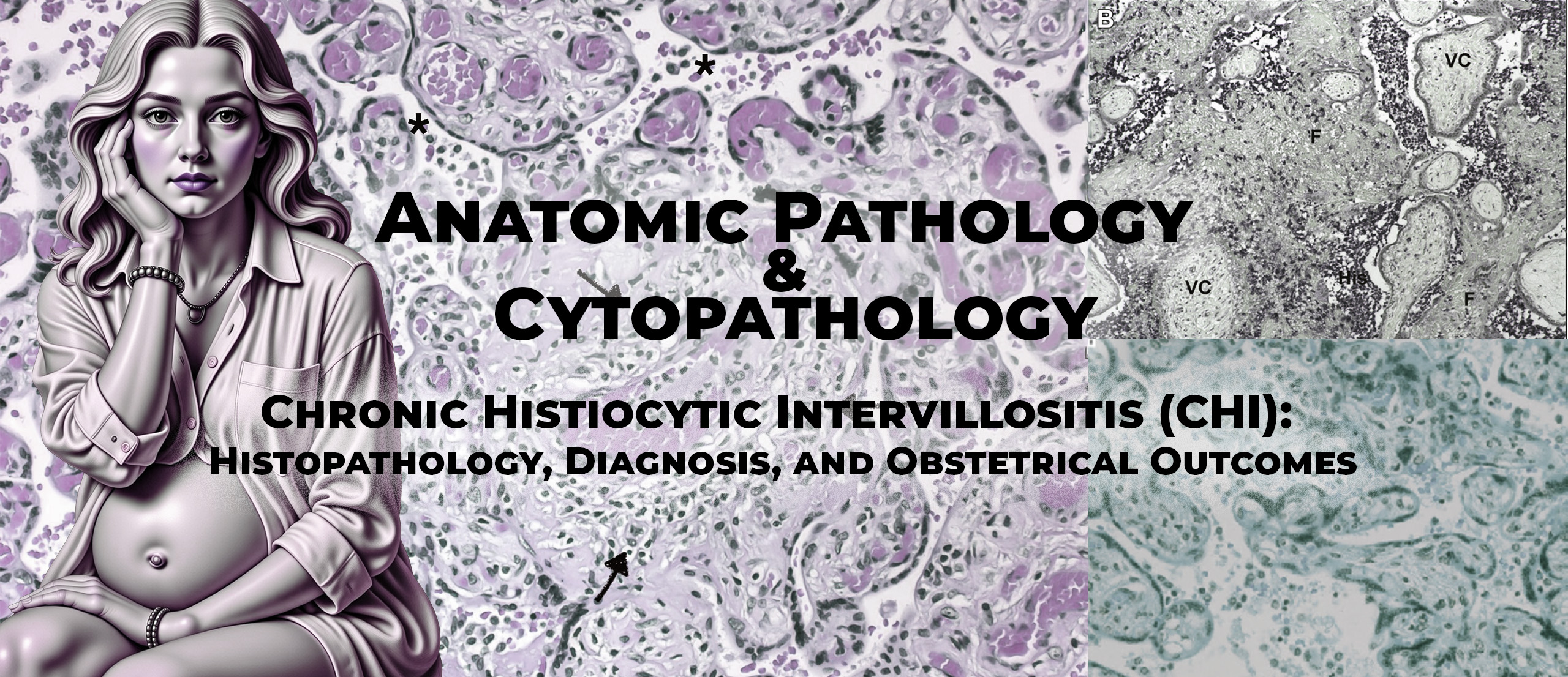
The authors define CHI as a placental lesion characterized by maternal histiocytic infiltration of the intervillous space, often accompanied by fibrinoid deposits. These macrophages are of maternal origin and are typically identified by CD68 immunostaining.

A major issue highlighted is the lack of standardized histological criteria. Several unresolved questions remain, including:

  • Whether immunohistochemistry is mandatory for diagnosis
  • How to quantify lesion extent reproducibly
  • Whether C4d staining should be included in routine evaluation

The review summarizes different histological scoring systems proposed in the literature. These systems generally assess:

  • Extent of intervillous involvement (focal, moderate, massive)
  • Distribution (diffuse versus multifocal)
  • Intensity of macrophage infiltration
  • Presence and extent of fibrin deposition

Importantly, the authors emphasize that massive and diffuse CHI lesions are strongly associated with adverse outcomes, whereas limited lesions may occasionally be found in placentas from uncomplicated pregnancies.

Epidemiology and Frequency

CHI is described as a rare condition, with reported prevalence varying widely depending on the population studied. In unselected placental series, CHI is identified in approximately 0.3–0.5% of placentas. However, among placentas examined following fetal loss or severe growth restriction, prevalence may rise significantly, reaching up to 20% in selected cohorts.

The authors underline that the true incidence of CHI is likely underestimated due to:

  • Limited awareness among clinicians
  • Inconsistent placental examination practices
  • Variability in histological diagnostic thresholds

Clinical Presentation and Obstetrical Outcomes

From a clinical standpoint, CHI is associated with a severe obstetrical phenotype. The most frequently reported complications include:

  • Recurrent early miscarriages, often in the first trimester
  • Intrauterine growth restriction (IUGR), frequently severe and early-onset
  • Intrauterine fetal death (IUFD)
  • Prematurity, typically secondary to placental insufficiency

The review highlights that IUGR is the most characteristic complication, reported in approximately 50% of affected pregnancies. Growth restriction is often severe (<3rd percentile) and may be associated with abnormal Doppler findings, although uterine Dopplers can remain normal in some cases, emphasizing the placental rather than vascular origin of pathology.

The authors also note that different adverse outcomes may occur in successive pregnancies in the same woman, reflecting variability in lesion timing and severity.

Recurrence Risk and Prognosis

One of the most clinically relevant aspects of CHI is its high recurrence rate. Across published series summarized in the review, recurrence rates range from 18% to over 70%, with an average around 30–40%.

Several important observations are emphasized:

  • Recurrence risk is higher after severe or massive lesions
  • Recurrent CHI often presents earlier in gestation than the index pregnancy
  • Lesions may become progressively more severe, suggesting immunological memory

Despite these trends, the authors stress that no reliable predictive biomarker exists to identify women at highest risk of recurrence.

Interestingly, placental alkaline phosphatase elevation has been reported in some cases and may correlate with fibrin deposition and severe lesions, but this marker lacks specificity and validation.

Associated Diseases and Etiological Considerations

In most cases, no clear etiology is identified. However, Mekinian et al. emphasize the importance of a systematic etiological workup, particularly in cases of recurrent pregnancy loss.

Autoimmune Diseases

A significant proportion of women with CHI have associated autoimmune conditions. Reported associations include:

  • Antiphospholipid syndrome
  • Systemic lupus erythematosus
  • Sjögren’s syndrome
  • Autoimmune thyroid disease
  • Celiac disease

In a prospective French cohort cited by the authors, approximately 29% of women with CHI had a documented autoimmune disease. This observation supports an immune-mediated pathogenesis and justifies involvement of internists and immunologists in patient management.

Infectious Causes

Although CHI is defined as a non-infectious lesion, certain infections—most notably placental malaria—can produce similar intervillous macrophage infiltrates. The authors stress the importance of excluding infections, including TORCH pathogens, particularly in endemic or high-risk settings.

Genetic Causes

In cases of recurrent early miscarriage, chromosomal abnormalities must also be excluded, as abnormal fetal karyotypes may coexist with CHI-like lesions.

Pathophysiological Hypotheses

The review strongly supports an alloimmune rejection hypothesis, in which maternal immune responses are directed against paternal antigens expressed by the placenta. Supporting arguments include:

  • Predominance of maternal macrophages
  • Presence of complement activation (C4d) in some cases
  • Detection of anti-paternal HLA antibodies in affected women
  • High recurrence rates resembling immune memory

The authors also discuss potential roles of innate immunity, including Toll-like receptor activation and inflammatory cytokine production, which may promote macrophage recruitment and adhesion to the syncytiotrophoblast.

Management and Therapeutic Strategies

A major contribution of this article is its discussion of management, despite the absence of randomized controlled trials. The authors clearly state that no treatment is currently validated, and all strategies are empirical.

Therapeutic approaches reported in the literature include:

  • Low-dose aspirin
  • Low-molecular-weight heparin
  • Corticosteroids
  • Hydroxychloroquine
  • Combination regimens

These treatments are rationalized by the presumed immune-mediated pathophysiology and are particularly considered in women with:

  • Recurrent CHI
  • Severe obstetrical complications
  • Associated autoimmune disease

The review emphasizes the need for multidisciplinary management, involving obstetricians, internists, pathologists, and immunologists.

Strengths and Limitations

Strengths

  • Comprehensive synthesis of pathological and clinical data
  • Integration of internal medicine perspective
  • Clear identification of unmet clinical needs

Limitations

  • Narrative, non-systematic review
  • Reliance on heterogeneous retrospective studies
  • Lack of therapeutic efficacy data

Educational Value

For teaching purposes, this article is essential because it:

  • Frames CHI as a multisystem clinical problem, not just a pathological curiosity
  • Highlights diagnostic uncertainty and real-world decision-making
  • Provides a rationale for immunomodulatory therapies despite limited evidence

It sets the stage for later mechanistic studies that firmly establish CHI as a form of maternal–fetal alloimmune rejection.

Benachi A, Rabant M, Martinovic J, et al.
Chronic histiocytic intervillositis: manifestation of placental alloantibody-mediated rejection. American Journal of Obstetrics and Gynecology. 2021.

Background and Conceptual Framework

By 2021, accumulating clinicopathological evidence suggested that chronic histiocytic intervillositis (CHI) might represent a form of maternal–fetal immune rejection, yet definitive mechanistic proof was still lacking. Benachi and colleagues approached CHI from a novel and rigorous perspective by explicitly applying the diagnostic criteria used in solid-organ transplantation, particularly the Banff classification for antibody-mediated rejection (AMR), to placental pathology.

This article represents a conceptual turning point in the understanding of CHI. Rather than describing associations alone, the authors sought to demonstrate that CHI fulfills all three cardinal criteria required to diagnose antibody-mediated rejection:

  1. Histological evidence of tissue injury
  2. Evidence of antibody–tissue interaction (complement activation)
  3. Serological evidence of donor-specific antibodies

The study therefore reframes CHI not merely as an inflammatory placental lesion, but as a true alloantibody-mediated placental rejection syndrome.

Objectives

The primary objective was to determine whether CHI satisfies the Banff criteria for antibody-mediated rejection by demonstrating:

  • The presence of fetus-specific anti-HLA antibodies in maternal serum
  • Complement activation within placental tissue (C4d and C5b-9 deposition)
  • Histological features of placental injury consistent with immune-mediated damage

A secondary objective was to explore why CHI often recurs after a normal first pregnancy, by reconstructing maternal HLA sensitization history.

Study Design and Clinical Material

This was a highly detailed translational case-based study involving two couples who experienced recurrent severe fetal losses after an initial uncomplicated pregnancy.

Clinical Context

Both women had:

  • A first pregnancy with a healthy live-born infant
  • Subsequent pregnancies complicated by severe, early-onset fetal growth restriction, leading to second-trimester pregnancy termination or fetal demise
  • Histological evidence of chronic histiocytic intervillositis in affected placentas

The recurrence pattern—normal first pregnancy followed by repeated losses—was a central clinical clue suggesting immune sensitization rather than congenital or infectious pathology.

Histopathological Findings

Placental examination revealed classic features of severe CHI:

  • Diffuse infiltration of the intervillous space by CD68-positive histiocytes
  • Prominent intervillous and perivillous fibrin deposition
  • Evidence of syncytiotrophoblast injury, consistent with immune-mediated damage

Immunohistochemistry demonstrated:

  • Strong C4d deposition along the syncytiotrophoblastic surface
  • Presence of the terminal complement complex C5b-9, indicating full activation of the classical complement cascade

These findings satisfied the Banff criterion of current or recent antibody–tissue interaction.

Immunological Investigations

A major strength of this study lies in its extensive immunological workup, rarely performed in placental disease.

Detection of Fetus-Specific Antibodies (FSAs)

Maternal sera were analyzed for anti-HLA antibodies using single-antigen bead assays. The authors demonstrated:

  • High titers of complement-fixing anti-HLA antibodies
  • Specificity for paternal HLA alleles inherited by the fetus
  • Ability of these antibodies to bind C1q, confirming complement-activating capacity

These antibodies met the transplant definition of donor-specific antibodies, here termed fetus-specific antibodies.

HLA Typing and Epitope Analysis

Both parents and offspring underwent high-resolution HLA typing. Using HLA Matchmaker software, the authors reconstructed maternal sensitization at the epitope (eplet) level.

A key finding was that:

  • Sensitization likely occurred during the first normal pregnancy
  • Antibodies were directed against shared or public HLA epitopes
  • This led to broad cross-reactivity against multiple paternal HLA alleles in subsequent pregnancies

This elegant analysis explained why later pregnancies—even with different paternal haplotypes—were affected.

Placental Expression of HLA Molecules

A critical conceptual barrier in placental immunology has been the assumption that syncytiotrophoblasts do not express polymorphic HLA antigens. This study challenged that dogma.

The authors demonstrated:

  • Expression of polymorphic HLA class I molecules on inflamed trophoblastic villi
  • Induction of HLA class II expression, which is absent in normal placentas

This inflammatory upregulation provided a direct target for maternal anti-HLA antibodies, making antibody-mediated placental injury biologically plausible.

Integration with Banff Criteria

The authors explicitly mapped their findings onto the Banff classification:

  1. Histological injury
    • Diffuse macrophage-rich infiltrate
    • Syncytiotrophoblast damage
    • Extensive fibrin deposition
  2. Evidence of antibody interaction
    • Strong C4d and C5b-9 deposition
    • Complement activation at the villous surface
  3. Serological evidence
    • High-level complement-fixing fetus-specific anti-HLA antibodies

All three criteria were fulfilled, allowing the authors to formally diagnose antibody-mediated placental rejection.

Clinical Correlation and Phenotype

Clinically, pregnancies were characterized by:

  • Very early and severe fetal growth restriction
  • Disproportionately small placentas
  • Normal uterine Doppler findings, suggesting a placental rather than vascular pathology
  • Increasing severity and earlier onset in successive pregnancies

Postmortem fetal examination showed:

  • Delayed bone maturation
  • Global growth impairment
  • No genetic or structural abnormalities

These findings excluded alternative diagnoses such as skeletal dysplasia or chromosomal disease.

Interpretation and Pathophysiological Model

The authors propose a compelling model in which:

  1. A first pregnancy induces maternal alloimmunization against fetal HLA epitopes
  2. Long-lived plasma cells produce complement-fixing antibodies
  3. In subsequent pregnancies, antibody binding to inflamed syncytiotrophoblast triggers:
    • Complement activation
    • Macrophage recruitment
    • Placental destruction
  4. Resulting placental insufficiency leads to fetal growth restriction and demise

This model explains:

  • High recurrence rates
  • Worsening severity
  • Early gestational onset
  • Poor response to standard obstetrical interventions

Strengths and Limitations

Strengths

  • Application of transplant immunology criteria
  • Comprehensive immunological and pathological analysis
  • Elegant epitope-level HLA reconstruction
  • Strong clinicopathological correlation

Limitations

  • Small sample size (two index cases)
  • High technical complexity limits routine clinical application
  • Lack of interventional therapeutic data

Educational and Clinical Implications

For teaching purposes, this article is pivotal because it:

  • Definitively reframes CHI as alloantibody-mediated rejection
  • Introduces transplant immunology concepts into placental pathology
  • Provides a mechanistic explanation for recurrence
  • Opens the door to personalized immunological monitoring and therapy

This work laid the intellectual foundation for subsequent molecular studies confirming shared pathways between CHI and kidney allograft rejection.

Albersammer L, Leon J, Martinovic J, et al.
Histologic and molecular features shared between antibody-mediated rejection of kidney allografts and chronic histiocytic intervillositis support common pathogenesis. Journal of Pathology. 2025;266:177–191.

Background and Rationale

By the mid-2020s, the hypothesis that chronic histiocytic intervillositis (CHI) represents a form of maternal–fetal alloimmune rejection had gained strong support from histological and serological studies. However, a crucial question remained unresolved: does CHI share not only conceptual similarities but also concrete cellular and molecular signatures with antibody-mediated rejection (AMR) of solid-organ allografts? Albersammer and colleagues addressed this question by directly comparing CHI to kidney AMR, the best-characterized model of antibody-mediated tissue rejection.

This study represents the most advanced and integrative contribution within the knowledge base. It moves beyond descriptive pathology and serology to incorporate multiplex immunofluorescence phenotyping and transcriptomic profiling, thereby providing molecular-level evidence that CHI and kidney AMR share a common pathogenic program.

Objectives

The primary objectives of the study were:

  1. To precisely characterize the immune cell composition of the intervillous inflammatory infiltrate in severe CHI using multiplex immunofluorescence
  2. To compare the molecular signatures of CHI placentas with those observed in antibody-mediated rejection of kidney allografts
  3. To determine whether CHI fulfills molecular criteria of AMR, as defined by the Banff classification

Secondary objectives included refining the pathological phenotype of severe CHI and strengthening the maternal–fetal rejection paradigm.

Study Design and Population

This was a retrospective translational pathology study conducted within French tertiary referral centers specializing in placental pathology, obstetrics, immunology, and transplantation medicine.

Inclusion Criteria

The authors deliberately selected a highly specific and homogeneous CHI cohort, including only patients with:

  • Severe CHI (grade II or III, involving ≥10% of the intervillous space)
  • Strong C4d positivity (>50% of the syncytiotrophoblastic surface)
  • Presence of fetus-specific anti-HLA antibodies with high mean fluorescence intensity

This strict selection ensured that all cases represented unequivocal alloimmune-mediated CHI.

Control Group

Five gestational age–matched control placentas without inflammatory lesions were included for comparison, allowing differentiation between disease-specific and physiological placental immune signatures.

Histopathological and Immunophenotypic Analysis

Conventional Histology

All placental samples underwent centralized review by expert fetal pathologists. Consistent histological features included:

  • Dense intervillous macrophage infiltration
  • Frequent and often extensive fibrin deposition
  • Evidence of syncytiotrophoblast injury, including necrosis

These features confirmed the diagnosis of severe CHI and established a structural substrate for placental insufficiency.

Multiplex Immunofluorescence: Cellular Composition

A major methodological innovation of this study was the use of multiplex immunofluorescence, allowing simultaneous identification of multiple immune cell populations within the same tissue section.

Macrophage Phenotype

The intervillous infiltrate was dominated by CD68-positive macrophages, but crucially, these cells were predominantly CD206-negative, corresponding to an M1-like (pro-inflammatory) macrophage phenotype. This finding contrasts with the tolerogenic, M2-skewed macrophage populations typically found in normal placentas.

The predominance of M1 macrophages aligns with:

  • Pro-inflammatory cytokine production
  • Tissue-destructive immune responses
  • Patterns observed in kidney AMR

Lymphocyte Populations

The study identified:

  • A relative increase in CD8-positive T lymphocytes compared with controls
  • Very low numbers of CD4-positive T cells
  • Minimal presence of natural killer (NK) cells

This immune profile mirrors that seen in antibody-mediated rejection, where macrophages and CD8 T cells play key effector roles, whereas helper T cells are less prominent.

Molecular Signature Analysis

NanoString Transcriptomic Profiling

To investigate molecular pathways, the authors used NanoString® technology with the B-HOT panel, a gene set specifically recommended by the Banff classification to assess AMR in kidney allografts.

This approach allowed direct comparison between CHI placentas and kidney transplant biopsies with established AMR.

Key Molecular Findings

CHI placentas demonstrated:

  • Overexpression of HLA class II genes, indicating antigen presentation and immune activation
  • A strong interferon-gamma (IFN-γ) signature, characteristic of alloimmune rejection
  • Upregulation of cytokine and chemokine gene sets, promoting macrophage recruitment and activation

These molecular signatures were strikingly similar to those observed in kidney AMR, providing direct evidence of shared pathogenic pathways.

HLA Expression by Syncytiotrophoblast

Consistent with earlier work, the authors confirmed that in severe CHI:

  • The syncytiotrophoblast aberrantly expresses HLA class I and class II molecules
  • This expression is inflammation-induced and absent in normal placentas

This finding is crucial because it establishes a molecular target for maternal anti-HLA antibodies, completing the pathogenic loop of antibody-mediated placental injury.

Integration with the Banff Classification

One of the most important contributions of this article is its explicit integration of CHI into the Banff framework for antibody-mediated rejection.

The authors demonstrate that severe CHI fulfills:

  1. Histological criteria: macrophage-rich infiltrate and tissue injury
  2. Immunopathological criteria: strong C4d deposition
  3. Serological criteria: high-titer fetus-specific anti-HLA antibodies
  4. Molecular criteria: AMR-associated transcriptomic signature

This is the first study to show that CHI meets all four levels—morphological, immunohistochemical, serological, and molecular—used to diagnose AMR in transplantation.

Clinical Correlations

Clinically, the cohort was characterized by:

  • High rates of recurrent pregnancy loss, severe IUGR, and intrauterine fetal death
  • Earlier gestational onset in recurrent cases
  • Poor placental growth and severe placental insufficiency

These clinical features closely parallel graft failure in kidney AMR, reinforcing the translational relevance of the findings.

Strengths and Limitations

Strengths

  • Highly selective and well-defined cohort
  • Use of advanced multiplex and molecular techniques
  • Direct comparison with kidney AMR using standardized tools
  • Multidisciplinary collaboration across pathology, immunology, and obstetrics

Limitations

  • Small sample size due to rarity of severe CHI
  • Focus on severe C4d-positive cases may not capture the full disease spectrum
  • Limited immediate applicability to routine diagnostic laboratories

Educational and Conceptual Impact

This article represents the culmination of the CHI rejection paradigm. For teaching purposes, it is invaluable because it:

  • Provides molecular proof of maternal–fetal alloimmune rejection
  • Demonstrates how placental pathology can be interpreted through transplant immunology
  • Explains why CHI is recurrent, progressive, and clinically severe
  • Justifies immunomodulatory strategies as biologically rational

In an educational curriculum, this paper should be presented as the definitive mechanistic study that transforms CHI from a descriptive pathological entity into a clearly defined immune-mediated disease.

Antibody-mediated rejection (AMR)
A form of immune injury caused by antibodies directed against target antigens, leading to complement activation and tissue damage; a paradigm increasingly applied to CHI.

C4d
A stable split product of complement component C4, deposited after activation of the classical complement pathway and used as a tissue marker of antibody-mediated injury.

CD68
An immunohistochemical marker expressed by macrophages and histiocytes, used to identify the predominant inflammatory cells in CHI.

Chronic histiocytic intervillositis (CHI)
A rare placental inflammatory disorder characterized by maternal macrophage infiltration of the intervillous space, often associated with severe obstetrical outcomes and high recurrence risk.

Complement activation
A cascade of immune reactions leading to inflammation and cell injury; in CHI, activation of the classical pathway is suggested by C4d deposition.

Diffuse intervillositis
A pattern of CHI in which inflammatory cells occupy a large proportion of the intervillous space, usually associated with severe placental dysfunction.

Fibrinoid (fibrin) deposition
Accumulation of fibrin within the intervillous or perivillous space, frequently accompanying CHI and contributing to placental insufficiency.

Focal intervillositis
Limited involvement of the intervillous space by inflammatory infiltrates, typically affecting less than 10% of the placental surface.

Histopathology
The microscopic examination of tissues to identify disease-related structural and cellular changes; the cornerstone of CHI diagnosis.

Histiocyte
A tissue macrophage derived from circulating monocytes; the predominant inflammatory cell type in CHI.

Intervillous space
The placental compartment filled with maternal blood, surrounding chorionic villi and enabling maternal–fetal exchange.

Intrauterine fetal death (IUFD)
Fetal demise occurring in utero, frequently associated with severe and diffuse forms of CHI.

Intrauterine growth restriction (IUGR)
Failure of the fetus to achieve its genetic growth potential, commonly resulting from placental insufficiency caused by CHI.

Macrophage-rich infiltrate
An inflammatory infiltrate dominated by macrophages rather than lymphocytes, a defining feature of CHI.

Maternal–fetal interface
The anatomical and immunological boundary between maternal tissues and the placenta, where immune tolerance and rejection phenomena may occur.

Placental insufficiency
Inadequate placental function leading to impaired fetal growth or survival; a central consequence of severe CHI.

Recurrence
The reappearance of CHI in subsequent pregnancies, often with similar or increasing severity.

Syncytiotrophoblast
The outer multinucleated layer of the chorionic villi in direct contact with maternal blood; the site of C4d deposition in CHI.

Villitis of unknown etiology (VUE)
A chronic inflammatory placental lesion affecting the villous stroma, included in the differential diagnosis of CHI.

Chronic Histiocytic Intervillositis (CHI): Histopathology, Diagnosis, and Obstetrical Outcomes

Time: 10 minutes

1.1. Why CHI matters (5 min)

  • Rarity of CHI but disproportionate clinical impact
  • Association with recurrent miscarriage, IUGR, fetal death
  • High recurrence rates and counseling implications
  • Importance of placental pathology in unexplained adverse outcomes

1.2. Historical evolution of the concept (5 min)

  • First pathological descriptions (Labarrere)
  • Initial descriptive pathology phase
  • Emergence of immune-mediated hypotheses
  • Transition toward the rejection paradigm

Time: 10 minutes

2.1. Normal intervillous space physiology (5 min)

  • Maternal blood flow and exchange
  • Role of syncytiotrophoblast
  • Immune tolerance at the maternal–fetal interface

2.2. Normal immune cells in the placenta (5 min)

  • Physiological macrophages and lymphocytes
  • Tolerogenic vs inflammatory profiles
  • Why inflammation in the intervillous space is pathological

Time: 20 minutes

3.1. Core histological criteria (10 min)

  • Intervillous inflammatory infiltrate
  • Predominance of maternal histiocytes
  • CD68 immunostaining as diagnostic confirmation
  • Absence of infectious features

3.2. Lesion patterns and grading (10 min)

  • Focal vs diffuse involvement
  • Mild, moderate, and massive CHI
  • Role of fibrinoid and intervillous fibrin deposition
  • Relationship between extent of infiltration and placental insufficiency

Time: 15 minutes

4.1. CD68 and macrophage characterization (5 min)

  • Confirmation of histiocytic nature
  • Maternal origin of infiltrating cells
  • Distinction from lymphocyte-predominant lesions

4.2. C4d: marker of complement activation (10 min)

  • Biology of C4d and classical complement pathway
  • Localization to syncytiotrophoblastic microvillous surface
  • Evidence from Bendon et al.
  • Comparison with transplant pathology
  • Interpretation of focal vs diffuse C4d positivity

Time: 15 minutes

5.1. Villitis of unknown etiology (VUE) (7 min)

  • Villous vs intervillous localization
  • Cellular composition differences
  • Overlapping and coexisting lesions

5.2. Other differentials (8 min)

  • Infectious placentitis (TORCH, malaria)
  • Acute inflammatory lesions
  • Placental lesions associated with chromosomal abnormalities
  • Importance of clinicopathological correlation

Time: 10 minutes

6.1. Spectrum of adverse outcomes (5 min)

  • Early recurrent miscarriages
  • Severe and early-onset IUGR
  • Intrauterine fetal demise
  • Prematurity due to placental insufficiency

6.2. Prognostic value of histology (5 min)

  • Severity and extent as outcome predictors
  • Why mild CHI may be incidental
  • Recurrence patterns across pregnancies

Time: 8 minutes

7.1. Evidence supporting rejection (4 min)

  • C4d deposition
  • Fetus-specific anti-HLA antibodies
  • Macrophage-rich infiltrates
  • Parallels with kidney antibody-mediated rejection

7.2. Integration of molecular data (4 min)

  • Shared transcriptomic signatures
  • IFN-γ and HLA class II expression
  • Final conceptual model of placental rejection

Time: 7 minutes

8.1. What the pathologist should report (4 min)

  • Extent and severity of CHI
  • Presence of fibrin deposition
  • C4d status and distribution
  • Differential diagnosis explicitly addressed

8.2. Clinical implications (3 min)

  • Importance for counseling
  • Need for multidisciplinary discussion
  • Implications for future pregnancy management

Time: 5 minutes

  • CHI is a rare but severe placental disease
  • Diagnosis relies on careful histopathology and immunohistochemistry
  • C4d is a crucial marker linking CHI to antibody-mediated injury
  • Severity correlates strongly with obstetrical outcome
  • CHI represents a paradigm of maternal–fetal alloimmune rejection
Slide 1 – Title and scope of the lecture
  • Definition of chronic histiocytic intervillositis (CHI)
    → Introduce CHI as a rare but severe placental inflammatory disease.
  • Why CHI is clinically important
    → Emphasize recurrence risk and severe obstetrical outcomes.
  • Target audience and level
    → Pathologists, obstetricians, maternal–fetal medicine specialists.
  • Structure of the lecture
    → Histology → diagnosis → outcomes → immunological paradigm.

Illustration: None required

Slide 2 – Historical background of CHI
  • First pathological descriptions (1980s)
    → Initial recognition as a distinct intervillous inflammatory lesion.
  • Early uncertainty regarding etiology
    → Infectious vs inflammatory vs immune hypotheses.
  • Progressive recognition of recurrence patterns
    → Key clue pointing toward immune memory.
  • Shift toward immune-mediated interpretation
    → Set the stage for later C4d and alloimmune studies.

Illustration: None required

Slide 3 – Normal placental intervillous space
  • Structure of the intervillous space
    → Maternal blood bathes chorionic villi.
  • Role of syncytiotrophoblast
    → Interface for exchange and immune tolerance.
  • Normal immune environment
    → Limited, tightly regulated immune cell presence.
  • Importance of immune tolerance
    → Necessary for fetal survival despite semi-allogenicity.

Illustration: Optional schematic (general placental anatomy, not article-specific)

Slide 4 – Definition of CHI
  • Core diagnostic concept
    → Chronic inflammatory infiltrate of the intervillous space.
  • Maternal origin of infiltrating cells
    → Cells circulate in maternal blood.
  • Predominance of histiocytes
    → Macrophages constitute the majority of infiltrate.
  • Non-infectious nature
    → Infection must be excluded.

Illustration:

  • Histology image from Mekinian et al., 2018, Fig. 1 (intervillous histiocytic infiltration)
Slide 5 – Cellular composition of the infiltrate
  • CD68-positive macrophages
    → Diagnostic hallmark of CHI.
  • Minor lymphocyte component
    → Mostly CD8⁺ T cells.
  • Absence of neutrophilic dominance
    → Helps exclude acute infection.
  • Maternal immune origin
    → Localization within maternal blood space.

Illustration:

  • CD68 immunostaining from Mekinian et al., 2018, Fig. 2
Slide 6 – Histological patterns of CHI
  • Focal vs diffuse distribution
    → Lesions may be patchy or widespread.
  • Mild, moderate, massive forms
    → Based on percentage of intervillous involvement.
  • Association with fibrin deposition
    → Contributes to placental insufficiency.
  • Importance of thorough sampling
    → Focal lesions can be missed.

Illustration:

  • Histological comparison images from Bendon et al., 2015, Fig. 1–2
Slide 7 – Grading and severity assessment
  • Quantification of lesion extent
    → <10%, 10–50%, >50% of intervillous space.
  • Lack of international consensus
    → Multiple scoring systems exist.
  • Correlation with outcomes
    → Severe lesions → worse prognosis.
  • Reporting implications
    → Severity should always be stated.

Illustration:

  • Histological grading examples discussed in Mekinian et al., 2018
Slide 8 – Fibrin deposition and placental insufficiency
  • Intervillous and perivillous fibrin
    → Common accompaniment of CHI.
  • Mechanical obstruction of exchange
    → Reduces maternal–fetal transfer.
  • Association with growth restriction
    → Explains IUGR phenotype.
  • Marker of lesion chronicity
    → Reflects ongoing inflammation.

Illustration:

  • Placental histology with fibrin from Bendon et al., 2015, Fig. 2
Slide 9 – Immunohistochemistry in CHI
  • Role of CD68 staining
    → Confirms histiocytic infiltrate.
  • Utility of C4d staining
    → Identifies complement activation.
  • Added diagnostic confidence
    → Especially in equivocal cases.
  • Bridge between pathology and immunology
    → Introduces rejection paradigm.

Illustration:

  • CD68 + C4d images from Bendon et al., 2015
Slide 10 – C4d: biological meaning
  • C4d as a complement split product
    → Marker of classical pathway activation.
  • Stability in tissue
    → “Footprint” of antibody-mediated injury.
  • Established role in transplant pathology
    → Used in kidney rejection diagnostics.
  • Translational relevance to placenta
    → Same principles applied to CHI.

Illustration: None required

Slide 11 – C4d staining patterns in CHI
  • Localization to syncytiotrophoblast
    → Microvillous surface staining.
  • Focal vs diffuse positivity
    → Mirrors lesion distribution.
  • Strong association with intervillositis
    → More frequent than in villitis.
  • Diagnostic but not prognostic alone
    → Presence ≠ outcome severity.

Illustration:

  • C4d immunostaining images from Bendon et al., 2015, Fig. 2–3
Slide 12 – Differential diagnosis: VUE
  • Villitis of unknown etiology (VUE)
    → Inflammation of villous stroma.
  • Different topography
    → Villous vs intervillous.
  • Different cellular composition
    → More lymphocytes.
  • Possible coexistence with CHI
    → Requires careful interpretation.

Illustration:

  • Comparative histology discussed in Bendon et al., 2015
Slide 13 – Other differential diagnoses
  • Infectious placentitis
    → TORCH, bacterial infections.
  • Malaria-associated intervillositis
    → Macrophage-rich but infectious.
  • Genetic causes of fetal loss
    → CHI may be incidental.
  • Importance of clinicopathological correlation
    → Diagnosis is contextual.

Illustration: None required

Slide 14 – Obstetrical outcomes associated with CHI
  • Recurrent early miscarriage
    → Often first manifestation.
  • Severe intrauterine growth restriction
    → Hallmark clinical feature.
  • Intrauterine fetal death
    → Especially in severe forms.
  • Prematurity due to placental failure
    → Often iatrogenic.

Illustration:

  • Outcome summary tables from Mekinian et al., 2018, Table 1
Slide 15 – Recurrence risk and prognosis
  • High recurrence rates
    → ~30–70% depending on severity.
  • Earlier onset in subsequent pregnancies
    → Suggests immune memory.
  • Variable clinical expression
    → Miscarriage, IUGR, or IUFD.
  • Counseling implications
    → Critical role of placental diagnosis.

Illustration:

  • Recurrence data table from Mekinian et al., 2018, Table 1
Slide 16 – CHI as maternal–fetal alloimmune rejection
  • Semi-allogeneic nature of pregnancy
    → Paternal antigens expressed by fetus.
  • Presence of anti-HLA antibodies
    → Directed against paternal HLA.
  • Complement activation
    → Explains C4d deposition.
  • Conceptual shift
    → From inflammation to rejection.

Illustration:

  • Conceptual framework from Benachi et al., 2021 (AJOG at a Glance)
Slide 17 – Serological and immunological evidence
  • Fetus-specific anti-HLA antibodies
    → High titers, complement-fixing.
  • HLA typing of parents and fetus
    → Confirms antigen specificity.
  • Epitope spreading
    → Explains recurrence after normal pregnancy.
  • Parallel with transplant rejection
    → Strong mechanistic analogy.

Illustration:

  • HLA sensitization figures from Benachi et al., 2021, immunology section
Slide 18 – Cellular and molecular parallels with kidney AMR
  • Macrophage-dominant infiltrate
    → M1-like phenotype.
  • IFN-γ–driven molecular signature
    → Shared with kidney AMR.
  • Upregulation of HLA class II
    → Abnormal placental expression.
  • Molecular confirmation of rejection
    → Beyond morphology alone.

Illustration:

  • Multiplex immunofluorescence images from Albersammer et al., 2025
  • Gene expression heatmaps from same article
Slide 19 – Practical implications for pathology reports
  • Always report extent and severity
    → Prognostic relevance.
  • Mention fibrin deposition
    → Marker of placental insufficiency.
  • Report C4d status
    → Supports alloimmune mechanism.
  • Explicitly address differentials
    → Guides clinical management.

Illustration: None required

Slide 20 – Key messages and conclusions
  • CHI is rare but severe
    → High clinical impact.
  • Diagnosis is histopathological
    → Requires careful placental examination.
  • C4d links CHI to rejection
    → Conceptual breakthrough.
  • Multidisciplinary management is essential
    → Pathologists play a central role.

Illustration: None required

Case description
A 32-year-old woman (G4P0) is referred for evaluation of three consecutive first-trimester miscarriages at 8–10 weeks’ gestation. Fetal karyotyping from two losses was normal. Infectious screening is negative. Placental histology from the most recent miscarriage shows focal accumulation of CD68⁺ histiocytes in the intervillous space with associated fibrin deposition. No villitis is present.

Answer and discussion
This presentation is highly suggestive of early, focal chronic histiocytic intervillositis. Although CHI is classically associated with second- or third-trimester complications, the KB clearly demonstrates that CHI can occur in the first trimester and manifest as recurrent early miscarriage. The absence of infection and normal fetal karyotype strengthen the pathological significance of the lesion.

The focal nature of the infiltrate does not exclude clinical relevance, especially in the context of recurrence. CD68 positivity confirms the histiocytic nature of the infiltrate, fulfilling diagnostic criteria. This case illustrates that CHI should be considered in unexplained recurrent pregnancy loss and that even limited lesions may be pathogenic in early gestation.

Key learning points:

  • CHI can present as recurrent early miscarriage
  • Mild or focal lesions may still be clinically significant
  • Placental histology is essential in unexplained losses

Case description
A 29-year-old primigravida presents at 22 weeks with severe fetal growth restriction (<3rd percentile). Uterine and umbilical artery Dopplers are normal. Amniotic fluid volume is reduced. Delivery occurs at 26 weeks for fetal compromise. Placental examination shows diffuse intervillous infiltration by CD68⁺ macrophages involving >50% of the intervillous space, extensive fibrin deposition, and strong C4d staining of the syncytiotrophoblast.

Answer and discussion
This case represents severe, massive CHI, the classic phenotype associated with early-onset IUGR. The normal Doppler studies point away from uteroplacental vascular disease and toward a primary placental parenchymal pathology, as emphasized in the KB.

Diffuse macrophage infiltration and extensive fibrin deposition explain the placental insufficiency. Strong C4d positivity supports complement activation and an antibody-mediated mechanism. This constellation predicts poor prognosis and high recurrence risk.

Key learning points:

  • Severe CHI causes IUGR independent of vascular Doppler abnormalities
  • Extent of intervillous involvement correlates with severity
  • C4d positivity supports alloimmune injury

Case description
A placenta from a stillbirth at 30 weeks shows chronic inflammatory infiltrates. Histology reveals lymphohistiocytic inflammation within villous stroma and scattered intervillous macrophages. CD68 highlights macrophages, while CD3 highlights lymphocytes within villi. C4d staining is minimal.

Answer and discussion
This lesion is more consistent with villitis of unknown etiology (VUE) rather than CHI. The primary localization within villi, lymphocyte-rich infiltrate, and minimal C4d staining argue against CHI. Intervillous macrophages may coexist with VUE but are not dominant.

This case highlights the importance of topography and cellular composition in differentiating chronic placental inflammatory lesions.

Key learning points:

  • CHI is intervillous; VUE is villous
  • Cellular composition matters
  • C4d is supportive but not mandatory

Case description
A woman delivers a healthy term infant in her first pregnancy. Her second and third pregnancies are complicated by severe IUGR at 18–20 weeks, leading to pregnancy termination. Placental histology from both losses shows diffuse CHI with strong C4d positivity.

Answer and discussion
This pattern is characteristic of alloimmune sensitization following a first normal pregnancy, as described in Benachi et al. The first pregnancy likely induced maternal anti–paternal HLA antibodies. Subsequent pregnancies triggered antibody-mediated placental rejection, leading to earlier and more severe disease.

Key learning points:

  • Normal first pregnancy does not exclude immune-mediated disease
  • Immune memory explains recurrence and worsening severity
  • CHI behaves analogously to transplant rejection

Case description

A 36-year-old woman (G3P1) is referred after an intrauterine fetal death at 24 weeks’ gestation. Her medical history is significant for primary Sjögren’s syndrome, diagnosed 6 years earlier, with anti-Ro/SSA positivity and sicca symptoms. She is not known to have antiphospholipid syndrome. Her first pregnancy resulted in a healthy term infant.

During the index pregnancy, fetal biometry was normal until 18 weeks. From 20 weeks onward, ultrasound demonstrated progressive growth restriction, with estimated fetal weight falling below the 5th percentile. Umbilical and uterine artery Dopplers remained within normal limits. At 24 weeks, fetal demise was diagnosed.

Placental examination shows a small placenta (<3rd percentile). Histology reveals moderate intervillous infiltration by CD68-positive macrophages, involving approximately 30–40% of the intervillous space, associated with patchy fibrin deposition. No villitis is present. Microbiological studies and special stains for infection are negative. C4d staining shows focal positivity along the syncytiotrophoblast.

Answer and discussion

This case illustrates moderate chronic histiocytic intervillositis occurring in a patient with an underlying autoimmune disease. Although Sjögren’s syndrome is not causative per se, autoimmune diseases are overrepresented among women with CHI, reflecting global immune dysregulation.

The normal Doppler studies argue against a primary vascular disorder and support a placental parenchymal disease. The predominance of intervillous CD68-positive macrophages, absence of infection, and focal C4d deposition support a diagnosis of CHI with an immune-mediated mechanism.

Importantly, CHI should not be misclassified as autoimmune placentitis. The pathology reflects alloimmune injury directed against the fetal–placental unit, rather than maternal autoantigens.

Key learning points

  • Autoimmune diseases are frequent comorbidities in CHI
  • They support immune dysregulation but do not redefine CHI as autoimmune
  • Moderate CHI can be sufficient to cause IUFD
  • C4d positivity strengthens the alloimmune hypothesis

Case description

A 28-year-old woman delivers a healthy 3,400 g neonate at 39 weeks after an uncomplicated pregnancy. Placental examination is requested for non-specific reasons. Gross examination is unremarkable.

Histological sampling reveals a small, focal aggregate of intervillous macrophages, occupying less than 5% of the intervillous space. CD68 immunostaining confirms macrophage identity. There is no fibrin deposition, no villitis, and no evidence of trophoblastic injury. C4d immunostaining is negative.

Answer and discussion

This case represents incidental, focal CHI without clinical significance. The KB clearly indicates that limited macrophage accumulations can be observed in normal placentas, particularly when lesions are minimal, lack fibrin deposition, and are C4d-negative.

Overinterpretation of such findings should be avoided. In the absence of adverse outcomes or recurrence history, this lesion does not carry prognostic weight and should be described cautiously.

Key learning points

  • Not all CHI lesions are clinically relevant
  • Extent, associated fibrin, and clinical context determine significance
  • Avoid overdiagnosis in asymptomatic pregnancies

Case description

A 34-year-old woman (G2P0) experiences a second-trimester pregnancy loss at 19 weeks. She reports no fever or infectious symptoms. TORCH serologies are negative. Placental histology shows dense intervillous inflammatory infiltrates, initially raising concern for infection.

Closer examination reveals that the infiltrate is composed almost exclusively of CD68-positive macrophages, with very few neutrophils. There is associated fibrin deposition and syncytiotrophoblastic damage. Gram stain, PAS, Giemsa, and PCR testing for common pathogens are negative. C4d immunostaining shows patchy positivity.

Answer and discussion

This case emphasizes the critical distinction between CHI and infectious placentitis. Although both can involve the intervillous space, infectious processes are typically neutrophil-rich, often with identifiable organisms or positive microbiology.

The macrophage-dominant infiltrate, negative infectious workup, and C4d positivity strongly support non-infectious chronic histiocytic intervillositis.

Key learning points

  • Cell composition is essential for diagnosis
  • CHI is macrophage-predominant, not neutrophilic
  • Infection must be rigorously excluded before diagnosing CHI

Case description

A 31-year-old woman with a history of one unexplained fetal loss at 22 weeks delivers a growth-restricted infant at 34 weeks. Placental histology shows intermediate-density intervillous histiocytic infiltration, involving approximately 15–20% of the intervillous space. The lesion is patchy, and initial assessment is inconclusive.

CD68 confirms macrophage predominance. There is limited fibrin deposition. C4d immunostaining demonstrates clear focal positivity on the syncytiotrophoblast.

Answer and discussion

This case illustrates the diagnostic value of C4d in equivocal CHI. While the extent of infiltration alone might be borderline, the presence of syncytiotrophoblastic C4d deposition provides strong evidence of antibody-mediated complement activation.

In such cases, C4d acts as a pathophysiological discriminator, tipping the diagnosis toward CHI with alloimmune features.

Key learning points

  • C4d is especially useful in borderline cases
  • Focal positivity can still be biologically meaningful
  • Diagnosis should integrate morphology and immunopathology

Case description

A woman experiences a second-trimester fetal loss at 21 weeks. The pathology report describes “chronic placental inflammation with fibrin” but does not specify localization, cell type, or diagnosis. No immunohistochemistry is performed.

Two years later, she presents with a recurrent pregnancy complicated by severe IUGR at 17 weeks. Repeat placental examination now reveals diffuse CHI with extensive fibrin deposition and strong C4d positivity.

Answer and discussion

This case highlights the direct clinical consequences of inadequate placental reporting. Failure to recognize and name CHI in the initial pregnancy prevented appropriate counseling regarding recurrence risk and closer surveillance in the subsequent pregnancy.

Clear identification and quantification of CHI are essential because the diagnosis itself carries prognostic information, independent of treatment availability.

Key learning points

  • CHI must be explicitly named in pathology reports
  • Extent and severity should always be stated
  • Diagnostic clarity affects future pregnancy management

Case description

A multidisciplinary meeting is convened for a patient with two consecutive fetal losses associated with CHI. Obstetricians question whether the lesion represents non-specific inflammation, while immunologists suggest an alloimmune mechanism. Pathology demonstrates diffuse CD68-positive intervillositis, fibrin deposition, and strong C4d staining. Maternal serum contains complement-fixing anti–paternal HLA antibodies.

Answer and discussion

This case encapsulates the conceptual evolution of CHI. Based on the KB, CHI fulfills all criteria for antibody-mediated rejection, including histological injury, complement activation, and serological evidence of fetus-specific antibodies.

Viewing CHI as a rejection phenomenon provides a coherent explanation for recurrence, severity escalation, and early gestational onset. It also aligns placental pathology with established transplant immunology principles.

Key learning points

  • CHI is best understood as maternal–fetal alloimmune rejection
  • This paradigm integrates pathology, immunology, and clinical recurrence
  • Conceptual framing has implications for future therapies and research
  1. Definition (Must-know)

Chronic histiocytic intervillositis (CHI) is a rare, non-infectious placental inflammatory disease characterized by maternal macrophage (CD68⁺) infiltration of the intervillous space, frequently associated with fibrin deposition, placental insufficiency, and severe adverse obstetrical outcomes.

  1. Key Histopathological Features

Core diagnostic criteria

  • Inflammatory infiltrate located in the intervillous space
  • Predominance of histiocytes/macrophages (maternal origin)
  • Confirmation with CD68 immunohistochemistry
  • Exclusion of infection is mandatory

Associated features

  • Intervillous and/or perivillous fibrin deposition
  • Syncytiotrophoblast injury
  • Variable lesion distribution (focal to diffuse)
  1. Grading and Severity

Although no universal grading system exists, severity is commonly assessed by extent of intervillous involvement:

  • Mild / focal: <10%
  • Moderate: 10–50%
  • Severe / massive: >50%

👉 Severity strongly correlates with poor obstetrical outcome

  1. Immunohistochemistry (High-yield)

CD68

  • Confirms macrophage-rich infiltrate
  • Essential for diagnosis

C4d

  • Complement split product
  • Marker of classical complement pathway activation
  • Localized to syncytiotrophoblastic microvillous surface
  • Strongly supports antibody-mediated injury
  • Particularly useful in equivocal cases

⚠️ C4d is supportive but not mandatory for diagnosis

  1. Differential Diagnosis (Very frequently tested)

Villitis of Unknown Etiology (VUE)

Feature

CHI

VUE

Main location

Intervillous space

Villous stroma

Dominant cells

Macrophages

Lymphocytes

CD68

+++

+

CD3

Minimal

Prominent

C4d

Often positive

Usually absent

Other differentials

  • Infectious placentitis (TORCH, malaria)
  • Acute inflammatory lesions (neutrophils)
  • Chromosomal abnormalities (CHI may be incidental)
  1. Obstetrical Outcomes

Strongly associated with:

  • Recurrent early miscarriages
  • Severe intrauterine growth restriction (IUGR)
  • Intrauterine fetal death (IUFD)
  • Prematurity (often iatrogenic)

👉 Normal uterine and umbilical Dopplers are common, emphasizing placental—not vascular—pathology

  1. Recurrence (Key exam point)
  • Recurrence rate: ~30–70%
  • Often earlier onset and more severe in subsequent pregnancies
  • May follow a normal first pregnancy

👉 Suggests maternal immune memory

  1. Pathophysiology: The Rejection Paradigm (Essential concept)

CHI is now considered a form of maternal–fetal alloimmune rejection, supported by:

  • Fetus-specific anti–paternal HLA antibodies
  • Complement activation (C4d, C5b-9)
  • Macrophage-dominant infiltrate
  • Shared molecular signatures with kidney antibody-mediated rejection
  • Aberrant expression of HLA class I and II by inflamed syncytiotrophoblast

🧠 Think of CHI as the placental equivalent of antibody-mediated graft rejection

  1. Associated Conditions
  • Autoimmune diseases (APS, SLE, Sjögren’s, thyroid disease)
  • These reflect immune dysregulation, not causation
  • CHI itself is alloimmune, not purely autoimmune
  1. Diagnosis in Practice
  • Usually retrospective, on placental examination
  • No validated antenatal biomarker
  • Requires:
    • Adequate placental sampling
    • CD68 immunostaining
    • C4d when available
    • Clinicopathological correlation
  1. Management (What to know for exams)
  • No validated treatment
  • Empirical strategies sometimes used:
    • Low-dose aspirin
    • Low-molecular-weight heparin
    • Corticosteroids
    • Hydroxychloroquine
  • Evidence is limited → multidisciplinary care essential
  1. What Must Be in a Pathology Report (Very important)
  • Explicit diagnosis: “Chronic histiocytic intervillositis”
  • Extent and severity
  • Presence and extent of fibrin deposition
  • C4d status and distribution
  • Differential diagnoses addressed

👉 Direct impact on recurrence counseling and future pregnancy management

  1. Common Pitfalls

❌ Confusing CHI with VUE
❌ Underestimating focal lesions in recurrent miscarriage
❌ Failing to exclude infection
❌ Omitting severity and extent in reports
❌ Considering CHI as nonspecific inflammation

  1. Absolute Take-Home Messages
  • CHI is rare but severe
  • Diagnosis is histopathological
  • Macrophages + intervillous space = think CHI
  • C4d links CHI to antibody-mediated rejection
  • Severity predicts outcome
  • Accurate diagnosis changes patient counseling
Chronic histiocytic intervillositis is primarily defined by:

A. Neutrophilic infiltration of chorionic villi
B. Maternal macrophage accumulation in the intervillous space
C. Lymphocytic infiltration of decidua
D. Fetal macrophage infiltration of villous stroma
E. Granulomatous inflammation of membranes

Correct answer: B
Explanation: CHI is defined by maternal CD68⁺ macrophages in the intervillous space.

The predominant immune cell type in CHI is:

A. CD4⁺ T lymphocytes
B. CD8⁺ T lymphocytes
C. CD68⁺ histiocytes
D. Neutrophils
E. NK cells

Correct answer: C
Explanation: Macrophages constitute the vast majority of the infiltrate.

Which immunohistochemical marker is essential to confirm the diagnosis of CHI?

A. CD3
B. CD20
C. CD68
D. CD56
E. MPO

Correct answer: C
Explanation: CD68 confirms histiocytic (macrophage) origin.

C4d deposition in CHI reflects:

A. Alternative complement pathway activation
B. Lectin pathway activation
C. Classical complement pathway activation
D. Coagulation cascade activation
E. Apoptotic debris accumulation

Correct answer: C
Explanation: C4d is a stable product of classical complement activation.

The typical localization of C4d staining in CHI is:

A. Villous stroma
B. Decidual arteries
C. Chorionic plate
D. Syncytiotrophoblastic microvillous surface
E. Amniotic epithelium

Correct answer: D

Which placental compartment is primarily affected in CHI?

A. Villous stroma
B. Intervillous space
C. Decidua basalis
D. Chorionic plate
E. Amnion

Correct answer: B

Massive CHI is generally defined by involvement of:

A. <5% of intervillous space
B. <10% of intervillous space
C. 10–25% of intervillous space
D. 25–50% of intervillous space
E. >50% of intervillous space

Correct answer: E

Which feature most strongly correlates with poor obstetrical outcome in CHI?

A. Maternal age
B. Placental weight alone
C. Extent of intervillous macrophage infiltration
D. Fetal sex
E. Mode of delivery

Correct answer: C

The most characteristic obstetrical complication of CHI is:

A. Preterm premature rupture of membranes
B. Placenta accreta
C. Severe intrauterine growth restriction
D. Gestational diabetes
E. Shoulder dystocia

Correct answer: C

Which outcome is frequently associated with severe CHI?

A. Macrosomia
B. Intrauterine fetal death
C. Polyhydramnios
D. Twin-to-twin transfusion
E. Congenital malformations

Correct answer: B

CHI is best distinguished from villitis of unknown etiology (VUE) by:

A. Presence of lymphocytes
B. Localization of inflammation
C. Gestational age at delivery
D. Placental weight
E. Fetal sex

Correct answer: B
Explanation: CHI is intervillous; VUE is villous.

In VUE, inflammation primarily involves:

A. Intervillous space
B. Decidua
C. Chorionic plate
D. Villous stroma
E. Amnion

Correct answer: D

Which condition must be excluded before diagnosing CHI?

A. Preeclampsia
B. Placental infarction
C. Infectious placentitis
D. Gestational diabetes
E. Cord prolapse

Correct answer: C

Which infection can mimic CHI histologically?

A. Listeriosis
B. Syphilis
C. Malaria
D. Toxoplasmosis
E. Rubella

Correct answer: C

Recurrence of CHI in subsequent pregnancies is:

A. Rare
B. Impossible
C. Approximately 1–5%
D. Approximately 30–70%
E. Universal (100%)

Correct answer: D

Recurrence of CHI is best explained by:

A. Persistent placental infection
B. Maternal vascular disease
C. Genetic fetal abnormalities
D. Maternal immune sensitization
E. Environmental toxins

Correct answer: D

CHI is increasingly considered a form of:

A. Acute inflammation
B. Autoimmune vasculitis
C. Antibody-mediated placental rejection
D. Infectious placentitis
E. Degenerative placental disease

Correct answer: C

Which antibodies are implicated in severe recurrent CHI?

A. Anti-phospholipid antibodies
B. Anti-dsDNA antibodies
C. Anti-HLA paternal antibodies
D. Anti-thyroid antibodies
E. Anti-platelet antibodies

Correct answer: C

Fetus-specific antibodies (FSAs) target:

A. Maternal HLA antigens
B. Decidual stromal cells
C. Paternal HLA antigens expressed by the fetus
D. Viral antigens
E. Placental hormones

Correct answer: C

Which complement complex indicates terminal pathway activation in CHI?

A. C1q
B. C3b
C. C4d
D. C5b-9
E. Factor B

Correct answer: D

In severe CHI, syncytiotrophoblast may aberrantly express:

A. CD68
B. HLA class I and II molecules
C. CD20
D. MPO
E. Vimentin

Correct answer: B

Which macrophage phenotype predominates in severe CHI?

A. M2 (CD206⁺)
B. Anti-inflammatory macrophages
C. Tolerogenic macrophages
D. M1-like pro-inflammatory macrophages
E. Foamy macrophages

Correct answer: D

Which lymphocyte population is relatively increased in CHI compared with controls?

A. CD4⁺ T cells
B. CD8⁺ T cells
C. B cells
D. Regulatory T cells
E. NK cells

Correct answer: B

Which molecular signature is shared between CHI and kidney AMR?

A. IL-4–dominant signature
B. Th2 cytokine profile
C. IFN-γ–driven signature
D. Hypoxia-only gene profile
E. Angiogenic factor profile

Correct answer: C

The Banff classification is originally used for:

A. Autoimmune diseases
B. Placental pathology
C. Kidney allograft rejection
D. Liver cirrhosis staging
E. Cardiac pathology

Correct answer: C

According to Banff criteria, diagnosis of AMR requires:

A. Histology only
B. Serology only
C. Imaging only
D. Histology, immunopathology, and serology
E. Clinical features alone

Correct answer: D

Which finding fulfills the “antibody–tissue interaction” criterion in CHI?

A. CD68 positivity
B. Fibrin deposition
C. C4d deposition
D. Placental infarction
E. Reduced placental weight

Correct answer: C

Why may CHI worsen in successive pregnancies?

A. Progressive placental aging
B. Increased fetal demands
C. Accumulation of maternal immune memory
D. Declining uterine perfusion
E. Hormonal changes

Correct answer: C

CHI is most often diagnosed:

A. By prenatal ultrasound
B. By maternal blood tests
C. By placental histological examination
D. By fetal MRI
E. By genetic screening

Correct answer: C

Which placental finding contributes directly to placental insufficiency in CHI?

A. Calcifications
B. Chorioangioma
C. Intervillous fibrin deposition
D. Increased placental thickness
E. Villous edema alone

Correct answer: C

Mild focal CHI lesions may be found in:

A. Only fetal deaths
B. Only autoimmune diseases
C. Only treated pregnancies
D. Uncomplicated pregnancies
E. Only first pregnancies

Correct answer: D

Which specialist is essential in multidisciplinary management of CHI?

A. Dermatologist
B. Neurologist
C. Internist / immunologist
D. Pulmonologist
E. Orthopedic surgeon

Correct answer: C

Which treatment is currently validated for CHI?

A. Corticosteroids
B. Aspirin
C. Heparin
D. Hydroxychloroquine
E. None

Correct answer: E

Why are immunomodulatory therapies considered in CHI?

A. Proven efficacy
B. Infectious etiology
C. Vascular thrombosis
D. Immune-mediated pathophysiology
E. Hormonal imbalance

Correct answer: D

Which laboratory marker lacks validation but has been explored in CHI?

A. Troponin
B. AFP
C. Placental alkaline phosphatase
D. CRP
E. D-dimers

Correct answer: C

Which statement about CHI is correct?

A. It is a common placental disease
B. It has no impact on fetal growth
C. It is always infectious
D. It has a high recurrence rate
E. It never affects live births

Correct answer: D

The maternal origin of macrophages in CHI is supported by:

A. Genetic testing of villi
B. CD68 positivity
C. Location within intervillous maternal blood
D. HLA typing of fetus
E. Placental weight

Correct answer: C

Which feature best supports CHI as alloimmune rather than autoimmune?

A. Presence of macrophages
B. Fibrin deposition
C. Targeting of paternal HLA antigens
D. Association with lupus
E. Elevated CRP

Correct answer: C

Why is accurate pathology reporting of CHI crucial?

A. For immediate neonatal care
B. For medicolegal reasons only
C. For maternal cancer screening
D. For counseling regarding recurrence risk
E. For determining fetal sex

Correct answer: D

CHI best serves as a model of:

A. Acute placental infection
B. Autoimmune placental vasculitis
C. Maternal–fetal immune tolerance
D. Antibody-mediated placental rejection
E. Degenerative placental aging

Correct answer: D