Introduction to Endometriosis and Ovarian Endometriomas Endometriosis is a common, chronic, estrogen-dependent gynecological disease affecting approximately 10% of women of reproductive age worldwide, impacting an estimated 176 million women. It is defined by the presence of endometrial glands and stroma outside the uterine cavity, most commonly in the ovaries and pelvic peritoneum. Women with endometriosis may experience symptoms like dysmenorrhea, chronic pelvic pain, and dyspareunia, or they may be asymptomatic, with endometriosis found incidentally during surgery for other indications. Endometriosis is classified into three subtypes: superficial peritoneal endometriosis, ovarian endometrioma (OMA), and deep infiltrating endometriosis. Superficial ovarian endometriosis is typically diagnosed during surgery, with current guidelines supporting surgical treatment for associated infertility or pain, preferably by excision for histological samples.
OMAs, which are cysts in the ovarian tissues, are a distinct and advanced manifestation of endometriosis. They are present in 17–44% of young women with endometriosis and can be accurately diagnosed via transvaginal sonography. The management options for OMAs include expectant management, medical treatment, surgical treatment, in vitro fertilization (IVF) for infertility-associated cases, or a combination thereof. The choice of management largely depends on the main presenting symptoms and the OMA size. Historically, surgery was considered the gold standard for OMAs, especially for cysts larger than 3 cm. However, accumulating evidence suggests a potential detrimental role of surgical excision on ovarian reserve, leading to a shift towards more conservative approaches, including increased referral to medical therapy. Clinicians now face a dilemma when discussing management options, with ovarian reserve preservation becoming a pivotal consideration.
Ovarian Reserve in the Presence of Endometrioma The impact of an intact endometrioma on ovarian reserve is a highly debated topic. Several mechanisms are proposed for its adverse effect on early folliculogenesis and ovarian reserve: mechanical stretching of surrounding cortical tissue, the presence of toxic elements like free iron radicals, reactive oxygen species, proteolytic enzymes, and inflammatory molecules within the endometrioma, which create a hostile microenvironment leading to inflammation and fibrosis. Additionally, endometrioma-triggered local pelvic inflammation may lead to accelerated follicular recruitment and atresia, a “burnout” effect. While molecular, histological, and morphological clues support these mechanisms, conclusive evidence is still lacking.
In clinical settings, studies present conflicting findings. Some indicate a negative impact of intact endometrioma on functional ovarian reserve, with one prospective study showing a 26% median serum AMH decline over six months in women with intact endometrioma compared to 7.4% in controls. A meta-analysis involving 17 studies also found significantly reduced AMH levels in patients with OMAs compared to healthy ovaries or other benign cysts. Conversely, other studies found no significant difference in ovarian responsiveness to controlled ovarian stimulation in women with intact endometrioma compared to controls. A recent systematic review and meta-analysis found no significant difference in pre-operative AMH levels between unilateral and bilateral endometriomas, challenging the idea that intact endometrioma inherently reduces functional ovarian reserve, even in more advanced disease.
The progression of ovarian reserve decline in women with endometriosis remains unclear. While some studies suggest a correlation between OMA size and ovarian reserve damage, others do not. For instance, Karadag et al. reported a significant negative correlation between larger OMA size and lower AMH levels. However, Roman et al. found large OMAs (over 6 cm) associated with higher AMH levels. Limited longitudinal data exist, but one prospective study by Kasapoglu et al. suggested AMH levels decline significantly faster in women with OMAs (26% over 6 months) than in healthy controls (7%). This suggests that expectant management might not be as safe as commonly perceived, though further confirmatory studies are needed.
Medical Therapy and Ovarian Reserve The effect of medical therapy on ovarian reserve in the presence of OMAs is not extensively studied. Medical treatments, including oral contraceptives (COCs), progestins, gonadotropin-releasing hormone agonists (GnRHa), antagonists (GnRHant), and aromatase inhibitors (AIs), primarily inhibit ovarian function and are generally not recommended for infertile patients. Their role in infertility treatment is largely limited to adjuncts to IVF, such as long-term GnRHa therapy before IVF, though this role is questioned.
- Oral Contraceptives (COCs): While frequently prescribed for endometriosis-related pain, there is limited data on their effect on OMA diameter and ovarian reserve. One study showed a slight decrease in OMA diameter with COC treatment but provided no information on ovarian reserve changes.
- Dienogest (DNG): A fourth-generation progestin, DNG is effective in reducing endometriosis-related pain and cyst size due to its anti-inflammatory and anti-angiogenic properties. Angioni et al. reported a significant OMA diameter reduction with DNG treatment over 6 months, while COCs showed no change, but ovarian reserve markers were not assessed. Some studies suggest DNG pretreatment may improve AFC and retrieved oocytes during IVF cycles, or positively impact IVF outcomes after OMA excision. However, one study by Tamura et al. reported lower retrieved oocytes and pregnancy/live birth rates after DNG treatment before IVF. A prospective study by Muzii et al. indicated that DNG treatment for 6 months significantly reduced OMA diameter and pain while preserving ovarian reserve, with improved AFC and no significant AMH change. This suggests DNG could be a safe option to delay surgery and preserve ovarian reserve in asymptomatic OMA cases, but further confirmation is needed.
- GnRH Agonists (GnRHa): GnRHa primarily reduce the impact of endometriotic lesions by inhibiting FSH secretion and creating a hypoestrogenic state, potentially reversing a proinflammatory peritoneal environment. However, their effectiveness in consistently reducing OMA size is debated. While one study reported OMA size reduction with GnRHa, another found no significant change in cyst diameter or AFC. Importantly, some studies suggest that GnRHa treatment does not reduce ovarian reserve or primordial/primary follicle function.
- GnRH Antagonists (GnRHant): Newer GnRHant like elagolix, relugolix, and linzagolix have shown efficacy in reducing endometriosis-associated pain. However, these studies primarily included patients with a previous surgical diagnosis of endometriosis and did not report on ovarian reserve markers.
- Aromatase Inhibitors (AIs): AIs, such as letrozole, suppress locally produced estradiol by endometriotic deposits. One study combining GnRHa with letrozole showed higher AFC and decreased OMA diameter, leading to more mature oocytes and blastocysts.
Overall, current data on medical therapy and ovarian reserve in OMAs are scarce but suggest DNG may be a promising option for preservation, particularly for asymptomatic cases.
Surgery and Ovarian Reserve ESHRE guidelines recommend medical therapy as first-line for endometriosis-associated pain and suggest surgery only for non-response or when OPU is technically difficult due to OMA presence. There is no mandate for surgery based on cyst diameter. Surgery for histology to rule out malignancy should be considered for non-reassuring sonographic features or fast-growing cysts, as unexpected malignancy can be found in about 0.9% of cases.
Surgical techniques for endometriotic cysts include cystectomy (stripping), electrocoagulation, laser ablation, plasma-energy ablation, and combined techniques. The stripping technique is the recommended treatment, associated with lower recurrence rates and better post-surgical fecundability. However, it often leads to inadvertent removal of healthy ovarian tissue adjacent to the pseudo-capsule, even by experienced surgeons, and causes coagulation-induced thermal damage. Laparoscopic stripping cystectomy consistently reduces serum AMH levels; a meta-analysis showed a 39-57% decline 9-12 months post-operatively. The impact of cystectomy on ovarian reserve is largely not reversible, with AMH concentrations remaining reduced by an estimated 40-53% at 9-18 months post-operatively. While premature ovarian insufficiency (POI) is a serious concern, it is an uncommon complication (up to 2.4%), mainly associated with bilateral or repeat surgery, and increases with age.
Different hemostasis methods can affect ovarian reserve. Bipolar coagulation is consistently more detrimental than non-thermal methods (sutures or hemostatic sealants), leading to about 7% less AMH decline with non-thermal methods. The use of bipolar coagulation should be limited.
While AMH consistently shows reduction after cystectomy, Antral Follicle Count (AFC) does not always. A meta-analysis comparing AMH and AFC in the same women found a significant reduction in serum AMH but not AFC after endometriotic cystectomy, suggesting AMH is a more sensitive biomarker for post-surgical ovarian reserve. Clinicians should routinely incorporate AMH into pre- and post-operative counseling.
Non-excisional techniques, such as ablation with bipolar coagulation, plasma energy, or CO2 laser, preserve ovarian cortex by not removing the cyst wall. Studies on plasma energy ablation and CO2 laser vaporization have shown promising results in terms of pregnancy rates comparable to cystectomy, with some indicating better ovarian reserve preservation (AFC and AMH). However, a meta-analysis by Zhang et al. found both cystectomy and ablation reduced AMH, but ablation was more respectful of ovarian reserve when evaluated by AFC. Ultrasound (US)-guided aspiration and sclerotherapy are also being used; they may preserve ovarian reserve but are associated with higher recurrence rates.
A combined surgical technique (partial stripping + ablation of remaining capsule) aims to combine radical removal with tissue preservation. An RCT has confirmed its non-inferiority to stripping regarding recurrences and ovarian reserve preservation.
Damage to ovarian reserve is more severe in bilateral OMAs compared to unilateral procedures. A study showed AMH levels were significantly lower in bilateral groups at early, intermediate, and late terms post-surgery. The decline in AMH levels post-surgery may not be temporary; some studies report irreversible reduction beyond 12 months, especially for bilateral and larger endometriomas.
Impact on IVF Outcomes Several systematic reviews and meta-analyses consistently show that endometrioma cystectomy does not improve IVF results in terms of retrieved oocytes, clinical pregnancy rates, or live birth rates, compared to women with intact endometriomas. Therefore, surgery should not be routinely undertaken to improve IVF success.
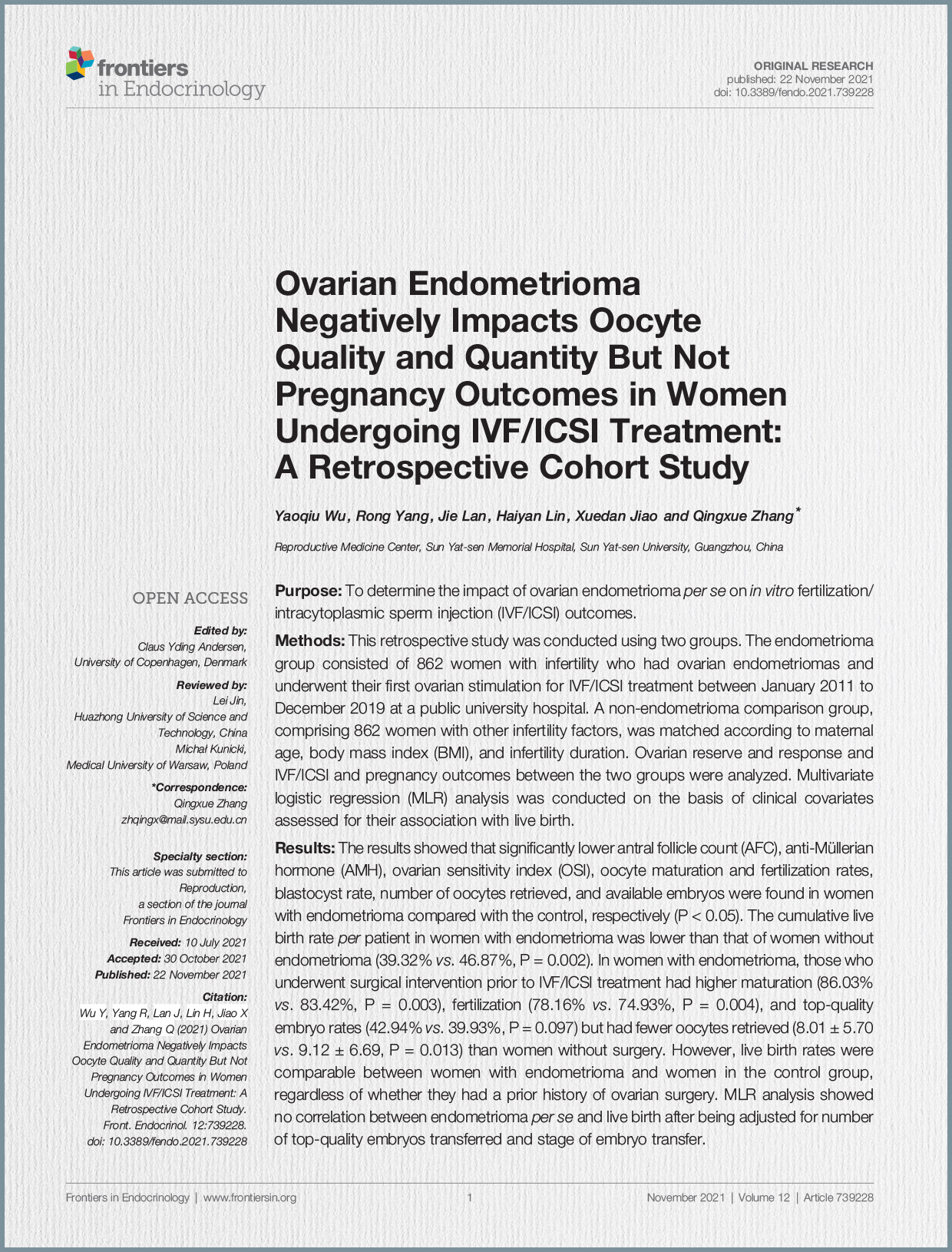
Similarly, for women with intact endometriomas, while the number of retrieved oocytes may be reduced, clinical pregnancy and live birth rates are comparable to controls without endometriomas. Hamdan et al. reported similar clinical pregnancy and live birth rates in women with intact endometriomas compared to controls. However, most published meta-analyses on ART outcomes in women with endometrioma are retrospective, which may affect the reliability of the estimates.
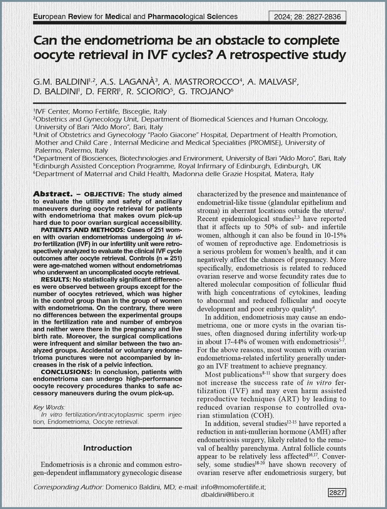
Risks of Conservative Management During ART Conservative management during ART carries potential risks, including abnormal oocyte competence, technical difficulties during oocyte retrieval, endometrioma rupture, injury to adjacent organs, infection, and follicular fluid contamination. The most expected risk without surgery is infection, with an incidence of 0.6%, suggesting routine antibiotic administration should be considered during oocyte retrieval. Importantly, accidental or voluntary endometrioma punctures were not associated with increased pelvic infection risk in one study, especially when punctures were followed by total aspiration and washing of the cysts.
Disease Progression and Ovarian Cancer Risk Low-quality evidence suggests deep infiltrating endometriosis might progress with controlled ovarian stimulation, but moderate evidence indicates IVF-ET does not worsen pain or increase recurrence risk. The likelihood of developing ovarian cancer within an endometrioma is rare at reproductive age. While general lifetime ovarian cancer risk is 1.31%, it rises to 1.80% in women with endometriosis (relative risk 1.42%). However, a Dutch study found higher incidence of clear-cell and endometrioid ovarian cancer in women with histologically proven endometriosis, with many diagnoses occurring synchronously after average menopausal age, suggesting a need for long-term follow-up.
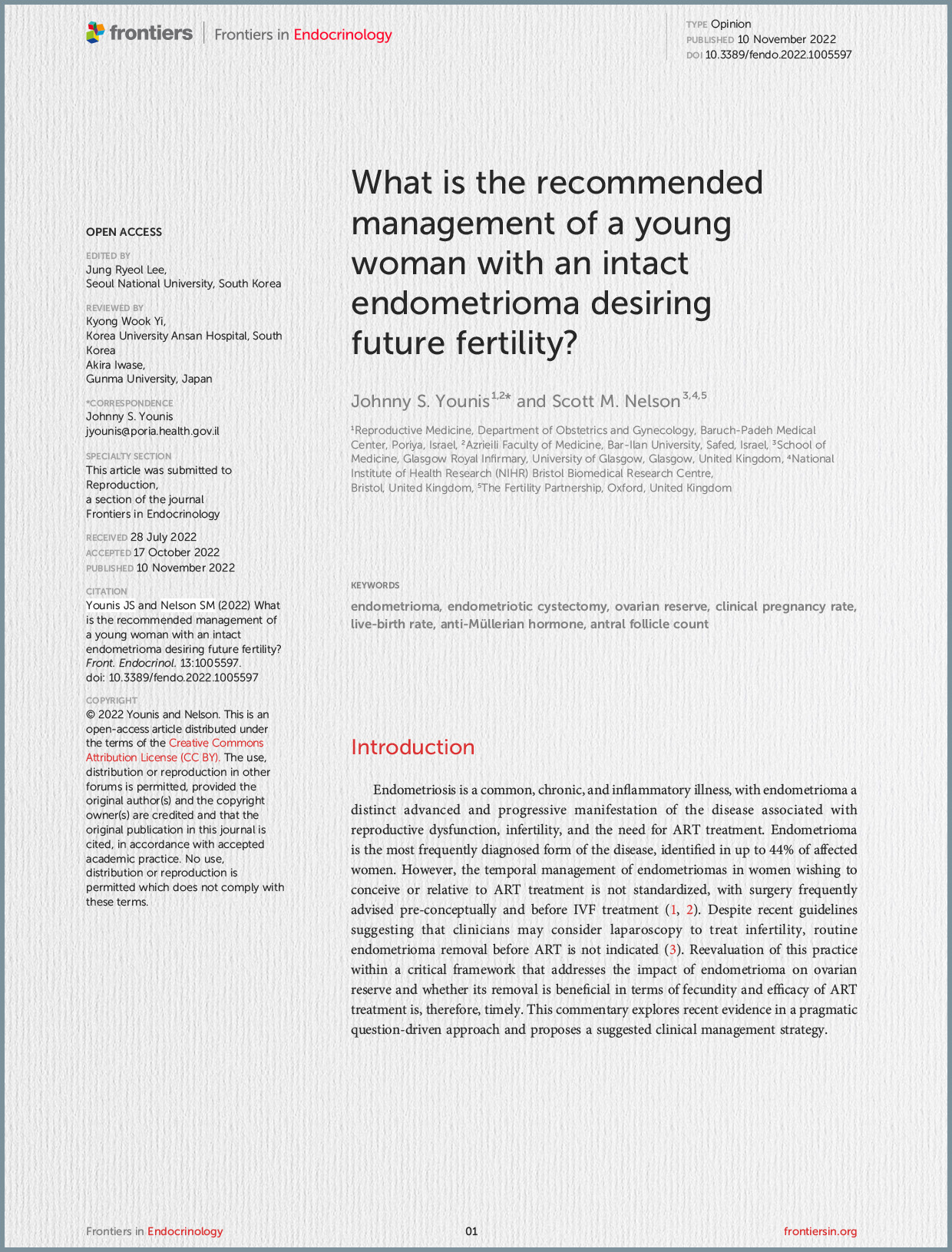
Conclusion and Clinical Management Strategy The authors conclude that current data support conservative management of endometriomas until reproductive aspirations are realized, advocating against routine endometriotic cystectomy due to its deleterious and sustained effects on ovarian reserve and its limited benefit for pregnancy probability. Conservative management is encouraged even with ART, unless there’s significant risk of endometrioma complications.
Surgical intervention should be considered in specific scenarios:
- Women with endometriosis-associated pelvic pain unresponsive to medical therapy.
- Cases where developing follicles are inaccessible during oocyte retrieval due to the endometrioma’s presence.
- In patients with endometrioma showing manifestations of endometriosis-associated ovarian cancer, despite its rarity at reproductive age, surgery and histological evaluation are necessary for diagnosis and treatment.
The authors acknowledge the retrospective nature of many studies and call for future prospective studies to refine risk estimates. They also suggest exploring other advanced non-conservative modalities like ultrasound-guided sclerotherapy or laser vaporization. Finally, fertility preservation should be discussed when surgery is unavoidable or ovarian reserve is a priori impaired.


