Bibliographic and Educational Resources in Cytogenomics

This platform is designed to serve as a comprehensive educational and bibliographic resource for healthcare professionals involved in cytogenomics. Covering a wide range of up-to-date topics within the field, it offers structured access to recent scientific literature and a variety of pedagogical tools tailored to clinicians, educators, and trainees.

Each topic is grounded in a curated selection of recent publications, accompanied by in-depth summaries that go far beyond traditional abstracts—offering clear, clinically relevant insights without the time burden of reading full articles. These summaries act as gateways to the original literature, helping users identify which articles warrant deeper exploration.

In addition to these detailed reviews, users will find a rich library of supplementary materials: topic overviews, FAQs, glossaries, synthesis sheets, thematic podcasts, fully structured course outlines adaptable for teaching, and ready-to-use PowerPoint slide decks. All resources are open access and formatted for easy integration into academic or clinical training programs.

By providing practical, well-structured content, the platform enables members of the cytogenomics community to efficiently update their knowledge on selected topics. It also offers educational materials that are easily adaptable for instructional use.

Low-Pass Whole-Genome Sequencing

Overview

Low-Pass Whole-Genome Sequencing (LP-WGS) is an emerging diagnostic and prognostic tool in genetics, particularly for detecting copy number variants (CNVs) and analyzing circulating tumor DNA (ctDNA) or circulating tumor cells (CTCs). It offers a cost-effective and efficient alternative to traditional sequencing methods and other genomic analyses.

LP-WGS is characterized by significantly lower sequencing coverage compared to traditional high read-depth Whole-Genome Sequencing (WGS), which typically has around 30-fold coverage and detects single-nucleotide variants (SNVs), small insertions and deletions (indels), CNVs, and structural variations (SVs) simultaneously. In contrast, LP-WGS usually operates under 1-fold coverage, focusing primarily on CNV analysis, often at a resolution of 50–100 kb. Ultra-low-pass whole-genome sequencing (ULP-WGS) is an even lower coverage variant, typically at approximately 0.1x to 1x, or specifically defined as ≤0.5x coverage for plasma cell-free DNA (cfDNA) applications. This lower depth provides a broader, though less detailed, view of the genome.

LP-WGS and ULP-WGS have diverse applications in both clinical diagnostics and cancer prognosis:

  • Clinical Genetic Diagnosis:
    • In China, LP-WGS is emerging as an alternative diagnostic test for CNV analysis.
    • It is routinely applied for four primary indications: high-risk pregnancies, spontaneous abortions, couples with adverse pregnancy history, and children with congenital birth defects.
    • In 2019, a joint Chinese expert group consensus recommended LP-WGS as a first-tier diagnostic test for pregnant women referred for or electing prenatal diagnosis, couples with balanced chromosomal translocations, and/or couples with pregnancy loss.
    • Spontaneous abortions accounted for the largest proportion of cases referred for LP-WGS-based CNV analysis in a survey of Chinese tertiary hospitals.
  • Cancer Diagnosis and Prognosis (Liquid Biopsy):
    • LP-WGS and ULP-WGS of circulating tumor DNA (ctDNA) or circulating tumor cells (CTCs) from liquid biopsies (e.g., plasma, cerebrospinal fluid (CSF), ascites) provide a minimally invasive method to identify key molecular alterations.
    • It can aid in diagnosis, determine prognosis, identify therapeutic targets, and detect early relapse.
    • Prognostic Value in Solid Tumors: The presence of positive ctDNA (tumor fraction ≥10%) detected by ULP-WGS is associated with a significantly higher risk of death and disease progression in patients with various solid tumors, including metastatic non-small cell lung cancer, localized osteosarcoma, metastatic castration-resistant prostate cancer, and advanced hepatocellular carcinoma.
    • Hepatocellular Carcinoma (HCC): ULP-WGS of ctDNA has demonstrated prognostic implications, with losses of 5q and 16q identified as independent prognostic factors for poor overall survival in HCC patients receiving systemic treatment.
    • Central Nervous System (CNS) Tumors: LP-WGS can detect genome-wide copy number aberrations (CNAs) in cell-free DNA from CSF in pediatric patients, demonstrating its feasibility in upfront diagnostic and monitoring settings. It has been used for measurable residual disease (MRD) detection in medulloblastoma.
    • Malignant Ascites (MA): A single-cell LP-WGS-based protocol (scMet-Seq) accurately detects CTCs for diagnosing MA across over 10 cancer types, showing high diagnostic sensitivity (79%) and nearly 100% diagnostic specificity and positive predictive value (PPV), outperforming clinical cytology.
    • Metastatic Small-Cell Lung Cancer (SCLC): ScMet-Seq enables blood-based diagnosis of metastatic SCLC with 90% sensitivity and 100% specificity and PPV, indicating that elevated CTC counts are associated with metastatic stage.

LP-WGS offers several advantages that make it a compelling choice for specific clinical and research applications:

  • Cost-Effectiveness: It requires much lower costs and computational resources compared to traditional WGS, making it a more affordable and accessible approach.
  • High Throughput and Automation: It provides higher throughput and is feasible for automation, suitable for laboratories with increasing demands.
  • Reduced DNA Requirement and Lower Failure Rates: Requires a smaller amount of DNA and results in lower experimental failure rates.
  • Faster Turnaround Time: Offers a shorter turnaround time.
  • Genome-Wide Coverage: Provides better genome-wide coverage and higher resolution of CNV detection compared to chromosomal microarray analysis (CMA).
  • Sensitivity for Mosaicism: Offers higher sensitivity in detecting low-level mosaicism.
  • Non-Invasive: Liquid biopsy approaches using LP-WGS are minimally invasive, allowing for repeated sampling and dynamic monitoring of disease progression and treatment response.
  • Reflecting Tumor Heterogeneity: CtDNA analysis via LP-WGS can provide a more precise depiction of the comprehensive mutational profile and heterogeneity across various lesions within a patient.
  • Accurate CTC Detection: Enables accurate detection of CTCs by analyzing copy number alterations, which are nearly ubiquitous in solid tumors and sporadically found in benign tissues.

Despite its advantages, LP-WGS faces several challenges:

  • Lack of Standardization: There are no detailed international or national recommendations for wet-lab and dry-lab procedures or variant nomenclature guidelines, leading to difficulties in achieving consensus and standard practice.
  • Quality Control (QC): Challenges remain in establishing standard criteria for LP-WGS-based CNV analysis and data reporting formats.
  • Variability in Practices: Survey results indicate variability in sequencing methods (single-end vs. paired-end), parameters (read counts, read length), and reported detection limits among different hospitals.
  • Sensitivity for Low Tumor Burden: LP-WGS and ULP-WGS have lower sensitivity and may struggle to detect ctDNA in patients with low tumor burden or in earlier stages of cancer.
  • Resolution for Detailed Variants: The low resolution limits the detection of detailed genetic variants.
  • Nomenclature Compliance: Compliance with internationally recognized criteria like ISCN 2016 and HGVS for CNV reporting nomenclature remains a challenge, with common errors including lack of prefix for reference sequences, absence of sequencing technology, variant type, or genome build.
  • Retrospective Studies and Sample Size: Many studies utilizing ULP-WGS have been retrospective and included limited sample sizes, necessitating larger prospective studies for validation.
  • Inadequate cfDNA Yield: In some cases, especially in pediatric patients, the cfDNA yield from CSF can be too low for analysis.
  • Tumors Without Detectable CNAs: A small fraction of tumors may not have detectable CNAs, potentially leading to false negatives in CNA-based diagnostic methods.
  • Inability to Trace Tissue Origin: CNA profiling may not be able to trace the tissue origin of CTCs or identify the specific cancer type.

Successful implementation of LP-WGS involves careful consideration of technical aspects:

  • Sequencing Platforms: Various platforms are used, including those from MGI Tech Co., Ltd., Illumina, and Thermo Fisher Scientific.
  • Sequencing Modes: Both single-end and paired-end sequencing are utilized, with single-end being more common due to cost and time efficiency, though paired-end provides more reliable information on CNV boundaries.
  • Read Counts and Length: Mean read count for LP-WGS in Chinese hospitals was 13.75 million reads/case, with a median read-length of 65 bp. Higher read depth typically yields higher resolution.
  • DNA Input: For CSF liquid biopsy in pediatric CNS tumors, 5 ng of DNA input is required.
  • Workflow: LP-WGS testing involves three major processes: wet-lab assays (DNA extraction, library preparation, sequencing), bioinformatic analysis (alignment, normalization, variant calling, annotation), and reporting (manual variant review, interpretation).
  • Internal vs. Outsourced Processes: Hospitals may conduct all steps internally (Pattern I), outsource wet-lab work (Pattern II), outsource data analysis and interpretation (Pattern III), or outsource the entire procedure (Pattern IV).
  • Cell Fixation and Amplification: For single-cell LP-WGS of CTCs, methods like click chemistry-based cell fixation and Tn5 transposome-based protocols are developed to improve sequencing quality, success rates, and reduce cost and processing time.
  • CNA Burden: A threshold for CNA burden (e.g., >0.02) is used to define cells with detectable CNAs, and a positive diagnosis often requires multiple cells to exhibit concordant CNA profiles to minimize false positives.

FAQ

LP-WGS is a sequencing method that focuses on analyzing Copy Number Variants (CNVs) with a relatively low sequencing read depth, typically under 1-fold. It is a variant of whole-genome sequencing (WGS) that provides a broader, though less detailed, view of the genomic landscape compared to traditional high read-depth WGS (usually ~30-fold). Ultra-low-pass whole-genome sequencing (ULP-WGS) is an even lower coverage variant, typically at ≤0.5x coverage.

Compared to high read-depth WGS, LP-WGS focuses primarily on CNV analysis (typically 50–100 kb resolution) rather than simultaneously detecting single-nucleotide variants (SNVs), small insertions/deletions (indels), and structural variations (SVs). Compared to routine CMA, LP-WGS offers better genome-wide coverage with reads more evenly aligned across most genomic regions, leading to higher resolution of genome-wide CNV detection and higher sensitivity for detecting low-level mosaicism. LP-WGS also requires less DNA, has higher throughput, and results in lower experimental failure rates.

CNVs are defined as DNA segments of at least 50 base-pairs (bp) that exhibit a variable copy number compared to a reference genome. They contribute to human genome diversity and are associated with a wide array of human disorders, such as DiGeorge syndrome and Charcot–Marie–Tooth disease, type 1A. In cancer, CNAs (copy number alterations) refer to gains or losses of parts of chromosomes and play a crucial role in tumor development and progression, often leading to overexpression of oncogenes or loss of tumor suppressor genes.

LP-WGS offers several advantages:

    • Cost-effectiveness: It is a significantly more affordable option than traditional WGS or high-depth targeted NGS panels, especially for large-scale screening and detecting major genomic alterations.
    • High throughput and shorter turnaround time: LP-WGS requires less manpower and is suitable for laboratories with high demands.
    • Genome-wide coverage: It provides a broad overview of the genome, which is advantageous for assessing large structural variations and inferring CNAs.
    • Lower DNA input requirement: It needs a smaller amount of DNA compared to traditional methods.
    • Feasibility for automation: The process is more amenable to automation.

Despite its advantages, LP-WGS has limitations:

    • Lower resolution and sensitivity: It provides less granular information than high-depth WGS and can be challenged by low tumor burden, potentially missing detailed genetic variants or subtle changes.
    • Interpretation difficulties: There can be difficulties interpreting noise-related data.
    • Lack of standardization: In China, there are no detailed recommendations or standard criteria for wet-lab and dry-lab procedures or data reporting formats, leading to variations in practice.
    • Inability to detect specific somatic variants: LP-WGS is not designed to detect specific SNVs or small indels, which are covered by high-depth targeted NGS panels.

LP-WGS is increasingly used to analyze circulating tumor DNA (ctDNA) or circulating tumor cells (CTCs) from liquid biopsy samples like plasma or cerebrospinal fluid (CSF). It helps detect CNAs in ctDNA/CTCs, which can serve as prognostic biomarkers and aid in non-invasive tumor genetic profiling. It can also be used for measurable residual disease (MRD) detection and monitoring tumor evolution.

A meta-analysis revealed that patients with positive ctDNA status at baseline (tumor fraction ≥10%) detected by ULP-WGS (≤0.5x coverage) had a significantly higher risk of death (HR = 2.60) and disease progression (HR = 2.28) compared to those with negative ctDNA. This indicates that detectable ctDNA can serve as a noninvasive biomarker for worse prognosis in patients with advanced-stage cancers.

The surveyed Chinese hospitals utilized sequencing platforms from three main manufacturers: MGI Tech Co., Ltd. (MGI), Illumina, and Thermo Fisher Scientific. Specific platforms mentioned include MGISEQ-2000, BGISEQ-500, NovaSeq 6000, NextSeq 500, Annoroad NextSeq 550AR, Berry Genomics NextSeq CN500, MiSeq/MiSeqDx, HiSeq 2000, and Ion Proton.

The most frequently reported detection limit for CNV size (resolution) in Chinese tertiary hospitals using LP-WGS was 100 kb. However, the detection limits varied, ranging from 50 kb to 4 Mb for spontaneous abortion cases, and 50 kb to 1 Mb for other applications.

Yes, studies demonstrate the feasibility of LP-WGS for detecting CNAs in cell-free DNA (cfDNA) from CSF in pediatric patients with CNS tumors. It has shown high concordance with primary tumor copy number profiles. This approach can aid in diagnosis and monitoring response to therapy and relapse.

Frequent gains reported include those in 3q, 8q, 1q, 7q, and 5p, while losses frequently encompass 13q, 8p, 4q, 16q, and 5q. These CNAs often involve well-known oncogenes (e.g., PIK3CA, SOX2, MYC, MCL1, MET, TERT) or tumor suppressor genes (e.g., CSMD1, ITM2B, GTF2H2, NAIP, OCLN, E-cadherin, ING2, RB1).

A single-cell low-pass WGS-based protocol (scMet-Seq) has been developed to detect circulating tumor cells (CTCs) in ascites for diagnosing MA. By identifying suspicious CTCs (sCTCs) and then using single-cell low-pass WGS to profile their CNAs, genuine CTCs are determined by requiring at least two sCTCs to exhibit concordant CNA profiles. This method has shown high diagnostic sensitivity (79% across >10 cancer types) and ~100% diagnostic specificity and positive predictive value (PPV) for MA diagnosis, superior to clinical cytology.

Yes, scMet-Seq, which utilizes single-cell low-pass WGS, has been investigated for blood-based diagnosis of metastatic SCLC. It achieved a diagnostic sensitivity of 90% and 100% diagnostic specificity and PPV in a preliminary study. Elevated CTC counts, identified by scMet-Seq, are associated with the metastatic stage of SCLC.

A click chemistry-based cell fixation method has been developed to improve sequencing quality by minimizing DNA degradation during sample processing and immunostaining, leading to approximately a 30% increase in sequencing coverage compared to traditional methods. Additionally, a Tn5 transposome-based single-cell WGS protocol combines whole genome amplification (WGA) and sequencing library construction into a single step, significantly shortening processing time (from 8.5h to 3h) and achieving a 97% cost reduction compared to the MALBAC protocol.

Instead of relying on epithelial markers, the scMet-Seq method uses a CNA-based CTC definition to identify genuine CTCs. This definition states that if at least two suspicious CTCs (sCTCs) exhibit concordant CNA profiles, these cells are identified as genuine CTCs, and a positive scMet-Seq test is generated for establishing deterministic cancer diagnosis. This approach leverages the fact that CNAs are nearly ubiquitous in solid tumors but occur sporadically and randomly in benign tissues.

The current variability in sequencing methods, parameters (read counts, read length, depth of coverage), and reported detection limits among different hospitals highlights the need for establishing standard criteria for LP-WGS-based CNV analysis. Challenges also remain in data reporting formats and consistent compliance with international nomenclature guidelines. Standardization, potentially aided by in vitro diagnostic (IVD) medical device manufacturers, is crucial for cross-laboratory referencing and quality assessment programs.

Bibliography

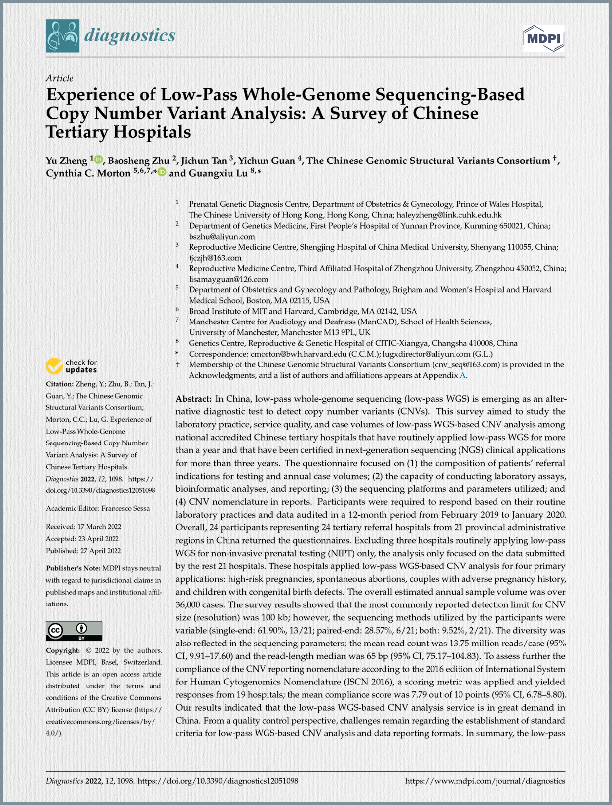
Zheng, Y., Zhu, B., Tan, J., Guan, Y., The Chinese Genomic Structural Variants Consortium, Morton, C. C., & Lu, G. (2022).
Experience of Low-Pass Whole-Genome Sequencing-Based Copy Number Variant Analysis: A Survey of Chinese Tertiary Hospitals. Diagnostics, 12(5), 1098. https://doi.org/10.3390/diagnostics12051098

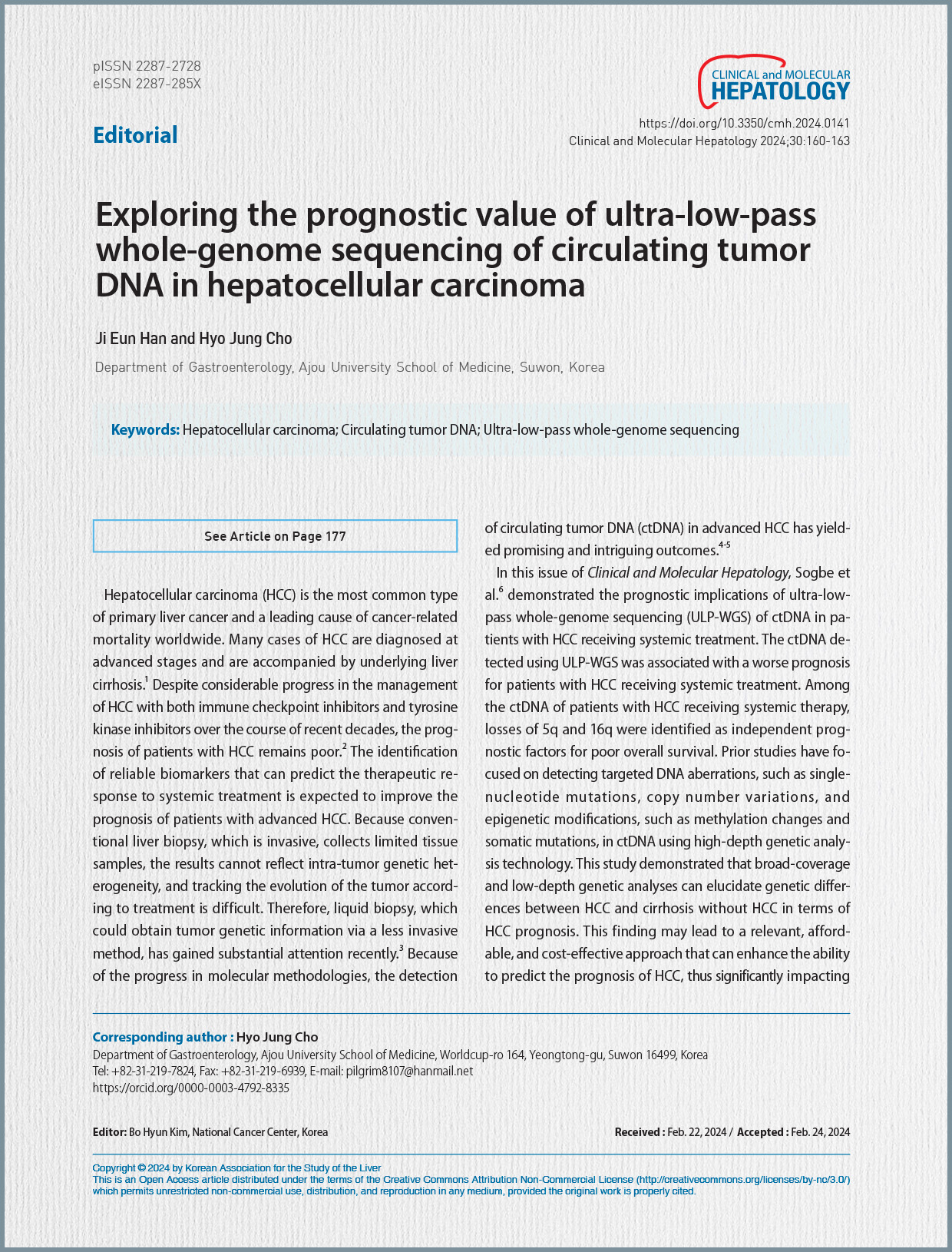
Han, J. E., & Cho, H. J. (2024).
Exploring the prognostic value of ultra-low-pass whole-genome sequencing of circulating tumor DNA in hepatocellular carcinoma. Clinical and Molecular Hepatology, 30, 160–163. https://doi.org/10.3350/cmh.2024.0141

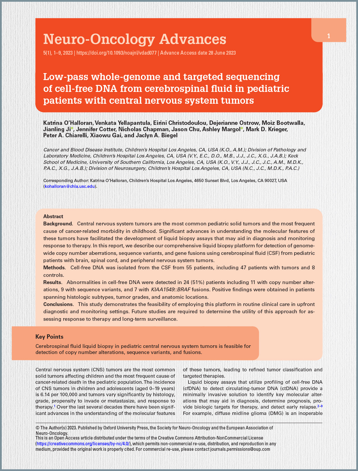
O’Halloran, K., Yellapantula, V., Christodoulou, E., Ostrow, D., Bootwalla, M., Ji, J., Cotter, J., Chapman, N., Chu, J., Margol, A., Krieger, M. D., Chiarelli, P. A., Gai, X., & Biegel, J. A. (2023).
Low-pass whole-genome and targeted sequencing of cell-free DNA from cerebrospinal fluid in pediatric patients with central nervous system tumors. Neuro-Oncology Advances, 5(1), 1–9. https://doi.org/10.1093/noajnl/vdad077

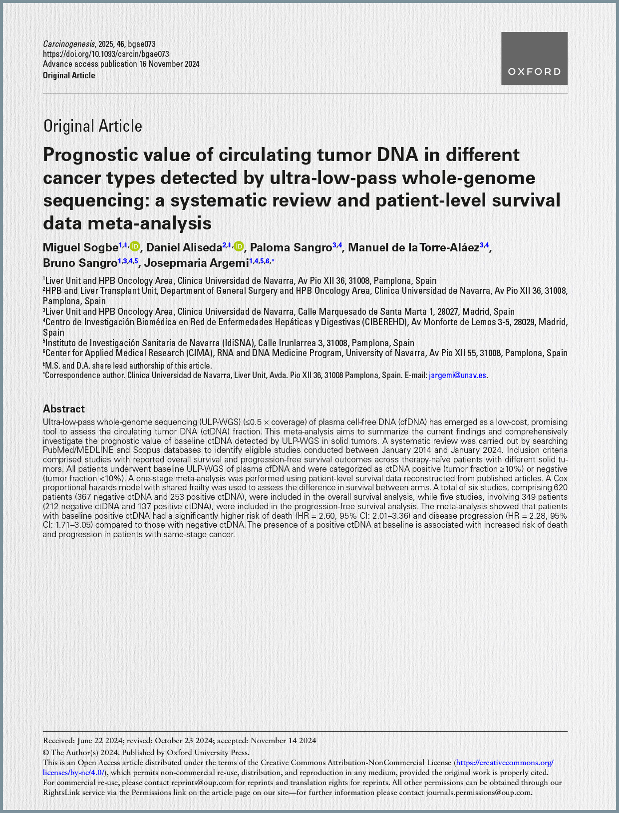
Sogbe, M., Aliseda, D., Sangro, P., de la Torre-Aláez, M., Sangro, B., & Argemi, J. (2025).
Prognostic value of circulating tumor DNA in different cancer types detected by ultra-low-pass whole-genome sequencing: a systematic review and patient-level survival data meta-analysis. Carcinogenesis, 46(Advance access publication 16 November 2024), bgae073. https://doi.org/10.1093/carcin/bgae073

Shen, X., Dai, J., Guo, L., Liu, Z., Yang, L., Gu, D., Xie, Y., Wang, Z., Li, Z., Xu, H., & Shi, Q. (2024).
Single-cell low-pass whole genome sequencing accurately detects circulating tumor cells for liquid biopsy-based multi-cancer diagnosis. npj Precision Oncology, 8(1), 30. https://doi.org/10.1038/s41698-024-00520-1

CNVs are defined as DNA segments of at least 50 base-pair (bp) in size that exhibit a variable copy number compared to a reference genome. These genetic variations contribute to human genome diversity and are associated with numerous human disorders, such as DiGeorge syndrome and Charcot–Marie–Tooth disease, type 1A. Traditionally, chromosomal microarray analysis (CMA) has been the first-tier test for identifying clinically significant CNVs. However, CMA has limitations including relatively low throughput and uneven probe coverage, which can lead to missed clinically significant CNVs.

Low-pass WGS offers several advantages over CMA, such as better genome-wide coverage, leading to higher resolution and increased sensitivity for detecting low-level mosaicism. Additionally, it provides higher throughput, is more amenable to automation, requires smaller amounts of DNA, and results in lower experimental failure rates. Unlike high read-depth WGS (typically ~30-fold), which detects various genetic alterations simultaneously (single-nucleotide variants, small insertions/deletions, CNVs, and structural variations), low-pass WGS (usually under 1-fold) primarily focuses on CNV analysis, typically detecting variants in the 50–100 kb range. This method requires less manpower for both laboratory and data-analysis procedures, has higher throughput, and a shorter turnaround time, making it particularly suitable for laboratories facing increasing demands for prenatal diagnosis. In 2019, a joint Chinese expert group consensus recommended low-pass WGS as the first-tier diagnostic test for specific patient groups, including pregnant women referred for or electing prenatal diagnosis, couples with balanced chromosomal translocations, and/or couples experiencing pregnancy loss. Despite its increasing adoption in Chinese laboratories, there was a noted absence of detailed recommendations for laboratory and bioinformatic procedures, as well as variant nomenclature guidelines, leading to a lack of standardization.

Purpose and Methodology of the Survey: This study aimed to investigate the current laboratory practices, service quality, and case volumes of low-pass WGS-based CNV analysis among nationally accredited Chinese tertiary hospitals. The survey targeted hospitals that had been routinely using low-pass WGS for over a year and were certified in next-generation sequencing (NGS) clinical applications for more than three years. An online questionnaire was designed and distributed in March 2020, covering several key areas: (1) the composition of patients’ referral indications and annual case volumes; (2) the capacity for conducting wet laboratory assays, bioinformatic analyses, and reporting internally; (3) the sequencing platforms and parameters utilized; and (4) CNV nomenclature in reports. Participants were instructed to base their responses on their routine laboratory practices and audited data from a 12-month period between February 2019 and January 2020. Of the 24 tertiary referral hospitals that returned questionnaires, three were excluded because they exclusively applied low-pass WGS for non-invasive prenatal testing (NIPT). The analysis therefore focused on data from the remaining 21 hospitals. Data collection was further confirmed through phone interviews and email correspondence.

Key Findings of the Survey:

  1. Patient Referrals and Annual Sample Volumes: The survey revealed a significant demand for low-pass WGS-based CNV analysis in China, with an estimated annual sample volume of over 36,000 cases across the 21 analyzed hospitals. The four primary applications for which these hospitals used low-pass WGS-based CNV analysis were:
  • High-risk pregnancies: This was the most frequent indication, accounting for 41.87% (15,254 cases) of the annual sample volume and utilized by 61.9% (13/21) of hospitals.
  • Spontaneous abortions: Comprised 33.82% (12,322 cases) of the volume and was applied by 90.5% (19/21) of hospitals. The high prevalence of chromosomal abnormalities in miscarriages likely contributes to this demand.
  • Couples with adverse pregnancy history: Represented 19.66% (7,162 cases) of the volume and was used by 76.2% (16/21) of hospitals. This demand is expected to increase due to national policies.
  • Children with congenital birth defects: Accounted for the smallest portion, 4.65% (1,694 cases), and was used by 52.4% (11/21) of hospitals. Despite the high overall demand, the study noted that low-pass WGS-based CNV analysis might be underutilized in prenatal diagnosis for high-risk pregnancies and particularly in pediatric cases.
  1. Hospital Capacity and Outsourcing: The survey identified diverse operational models among the hospitals:
  • Wet lab work: 57.1% (12/21) of hospitals performed wet lab work in-house, while 38.1% (8/21) outsourced it. One hospital employed a hybrid model, conducting prenatal wet lab internally but outsourcing postnatal samples.
  • Bioinformatics analysis: A higher proportion, 71.4% (15/21) of hospitals, analyzed sequencing data internally, with 28.57% (6/21) outsourcing this task. Notably, hospitals that handled wet lab work internally also performed bioinformatics internally, but only 25% of those outsourcing wet lab also performed bioinformatics internally. These varied patterns (e.g., fully internal, outsourcing wet-lab, outsourcing both) present significant challenges for quality assessment programs.
  1. Sequencing Platforms and Parameters: Participating hospitals used sequencing platforms from three main manufacturers: MGI Tech Co., Ltd., Illumina, and Thermo Fisher Scientific.
  • The most commonly reported detection limit (resolution) for CNV size was 100 kb. However, detection limits varied, ranging from 50 kb to 4 Mb for spontaneous abortion cases, and 50 kb to 1 Mb for other applications.
  • Regarding sequencing modes, 61.90% (13/21) of hospitals used single-end sequencing, 28.57% (6/21) used paired-end, and 9.52% (2/21) used both. Single-end sequencing was more common, likely due to its lower cost and time requirements.
  • The mean read count was 13.75 million reads/case (95% CI, 9.91–17.60), and the read-length median was 65 bp (95% CI, 75.17–104.83). The average depth of coverage varied considerably even for the same reported detection limit, highlighting the lack of standardized parameters.
  1. Compliance with CNV Reporting Nomenclature: Of the 19 participants who submitted reporting nomenclatures for representative pathogenic CNVs (DiGeorge Syndrome and Charcot-Marie-Tooth Disease type 1A), 12 different types of nomenclature were received. A scoring metric based on the ISCN 2016 (International System for Human Cytogenomics Nomenclature) and HGVS (Human Genome Variation Society) recommendations was applied.
  • The mean compliance score was 7.79 out of 10 points (95% CI, 6.78–8.80).
  • Only Hospital 16 achieved a perfect score of 10, fully complying with both ISCN 2016 and HGVS.
  • In contrast, Hospital 3 scored 0, providing only a basic description without following the established nomenclature formats.
  • The most frequent error observed was the absence of the prefix for reference sequences “g.” (52.63% of hospitals). Other common errors included not mentioning the sequencing-based technology symbol (10.52%), not using “del” or “dup” to describe the variant type (10.52%), and various minor inconsistencies (89.47%). While all hospitals correctly listed the affected chromosome, arm, and band, many used “hg19” instead of the recommended “GRCh37” for the genome build. The authors suggested that shared errors among hospitals using the same platforms might indicate reliance on inappropriate software-generated nomenclatures. The study also noted the publication of the updated ISCN 2020 after the survey, which further necessitates adoption of new guidelines.

Conclusion: The study concludes that low-pass WGS-based CNV analysis is a common and highly demanded diagnostic approach in China, transforming genetic diagnoses for patients. However, it is still in its early stages of development and lacks comprehensive standardization. Key challenges include establishing standard criteria for laboratory procedures (e.g., detection limits, sequencing parameters, depth of coverage) and ensuring consistent compliance with internationally recognized data reporting formats, particularly CNV nomenclature. The observed diversity in practices highlights the urgent need for standardized guidelines and external quality assessment programs to enhance the overall quality and reliability of this important genetic testing service in China.

Understanding Hepatocellular Carcinoma and the Need for Better Diagnostics Hepatocellular carcinoma (HCC) stands as the most common form of primary liver cancer and is a leading cause of cancer-related mortality globally. A significant number of HCC cases are diagnosed at advanced stages, frequently accompanied by underlying liver cirrhosis, contributing to a generally poor prognosis despite recent advancements in systemic treatments, including immune checkpoint inhibitors and tyrosine kinase inhibitors. The persistent challenge in managing HCC underscores a critical need for the identification of reliable biomarkers that can accurately predict a patient’s therapeutic response to systemic treatments, thereby potentially improving their prognosis.

Conventional methods for obtaining tumor genetic information, such as liver biopsy, are invasive and offer limited tissue samples. These limitations mean that such biopsies often fail to fully capture the intra-tumor genetic heterogeneity, and it becomes challenging to track the evolution of the tumor in response to ongoing treatment. In light of these challenges, liquid biopsy has emerged as a promising alternative, attracting substantial attention due to its ability to yield tumor genetic information through a less invasive approach. Specifically, the detection of circulating tumor DNA (ctDNA) in advanced HCC patients has shown intriguing and promising results, offering a non-invasive avenue for diagnosis, prognosis, and monitoring treatment response. CtDNA fragments, derived from cancer cells, are present in the bloodstream and can quantitatively and qualitatively reflect the genetic and molecular characteristics of the tumor.

Introduction to Ultra-Low-Pass Whole-Genome Sequencing (ULP-WGS) In the evolving landscape of genomic research, ultra-low-pass whole-genome sequencing (ULP-WGS) has emerged as a notable alternative to traditional whole-genome sequencing (WGS). While traditional WGS provides a comprehensive and detailed overview of the genome with high sequencing coverage (typically more than 30x), it is associated with substantial costs and demanding data processing requirements. In contrast, ULP-WGS operates at a significantly lower coverage, ranging approximately from 0.1x to 1x, offering a broader, albeit less detailed, view of the genomic landscape. This method is particularly advantageous for large-scale screening and the identification of major genomic alterations, such as copy number variations (CNVs), due to its significantly lower costs and reduced computational demands.

ULP-WGS’s cost-effectiveness and efficiency in detecting large-scale genomic changes make it a compelling choice for specific research and clinical applications that do not necessitate a highly detailed genetic map. The prognostic implications of ctDNA evaluated by ULP-WGS have been reported across various cancers, including metastatic squamous non-small cell lung cancer, Ewing sarcoma, osteosarcoma, metastatic castration-resistant prostate cancer, cervix cancer, and metastatic triple-negative breast cancer.

Key Findings from the Sogbe et al. Study as Discussed in the Editorial The editorial emphasizes the significance of the study by Sogbe et al., which delved into the prognostic implications of ULP-WGS of ctDNA in patients with HCC receiving systemic treatment. The key findings highlighted are:

  • The ctDNA detected using ULP-WGS was associated with a worse prognosis for HCC patients undergoing systemic treatment.
  • Among the ctDNA from these patients, losses of 5q and 16q were identified as independent prognostic factors for poor overall survival.
  • The study demonstrated that broad-coverage and low-depth genetic analyses can elucidate genetic differences between HCC and cirrhosis without HCC, specifically in terms of HCC prognosis. This approach may lead to a relevant, affordable, and cost-effective method to enhance the prediction of HCC prognosis, significantly impacting both clinical research and practice.
  • The authors of the Sogbe et al. study investigated the copy number alterations of ctDNA across various chromosomal loci using ULP-WGS, highlighting the loci where losses (8p, 4p, 13q, 16q, and 5q) and gains (1q, 8q, 7q, and 5p) were most prevalent.

Implications of ULP-WGS for Copy Number Alterations (CNAs) The detection of ctDNA and the exploration of copy number alterations (CNAs) using ULP-WGS carry significant implications. CNAs, defined as gains or losses of parts of chromosomes, are crucial in the development and progression of tumors. By analyzing the copy number alterations of ctDNA with ULP-WGS, researchers and clinicians can non-invasively gain insights into the genetic landscape of tumors at a relatively low cost. The identification of specific losses like 5q and 16q as independent biomarkers for poor survival contributes significantly to improving treatment strategies and enabling personalized approaches for HCC patients.

Limitations of ULP-WGS for Clinical Applications Despite its economic viability and potential for cost-effective genomic feature identification, the editorial acknowledges several limitations of ULP-WGS in clinical applications:

  • Low sensitivity: This method poses a challenge when detecting ctDNA in patients with a low tumor burden.
  • Low resolution: ULP-WGS limits the detection of detailed genetic variants.
  • Interpretation difficulties: Challenges are encountered when interpreting noise-related data, which further complicates its application.
  • Study design: The majority of studies that have utilized ULP-WGS, including the Sogbe et al. study, were retrospective and included limited sample sizes.
  • Treatment bias: The predominance of sorafenib treatment among patients in the Sogbe et al. study introduced a potential bias.

Future Directions To overcome these limitations and validate the clinical effectiveness of ULP-WGS, the editorial stresses the necessity of further prospective studies with larger sample sizes. Furthermore, with the current shift towards immunotherapy-based first-line treatments for HCC, such as atezolizumab plus bevacizumab or durvalumab plus tremelimumab, future research on the role of ctDNA analyzed using ULP-WGS in predicting progression-free survival or tumor response to immunotherapy is expected to have significant implications.

In conclusion, the editorial posits that the study by Sogbe et al. makes a significant contribution to understanding the role of ctDNA in HCC prognosis through ULP-WGS. While ULP-WGS presents itself as a non-invasive and cost-effective approach for tumor genetic profiling, it is associated with challenges like low sensitivity and resolution that must be addressed. The continued investigation of ctDNA using ULP-WGS is anticipated to more clearly define its potential as a prognostic biomarker for patients with advanced HCC.

Background and Significance of Pediatric CNS Tumors Central nervous system (CNS) tumors are the most common solid tumors in children and represent the leading cause of cancer-related mortality in the pediatric population. These tumors exhibit significant variations in histology, grade, metastatic potential, and response to treatment. Over recent decades, substantial advancements in understanding the molecular features of these tumors have led to refined classification and the development of targeted therapies.

Traditional methods for obtaining molecular information, such as direct brainstem biopsy for tumors like diffuse midline glioma (DMG), can be highly invasive and associated with potential morbidity. Furthermore, tissue biopsies may not fully capture intra-tumoral heterogeneity and can be challenging for repeated sampling to monitor disease evolution. In this context, liquid biopsy, which analyzes cell-free DNA (cfDNA) to detect circulating tumor DNA (ctDNA), offers a minimally invasive solution for identifying crucial molecular alterations. This approach can aid in diagnosis, prognosis, identification of therapeutic targets, and early detection of relapse. Serial liquid biopsies also hold the potential to facilitate monitoring of treatment response and provide insights into tumor evolution.

Advantages of CSF for Liquid Biopsy in CNS Tumors Cerebrospinal fluid (CSF) has been demonstrated to yield the highest fraction of ctDNA in patients with brain tumors compared to plasma. Initial liquid biopsy assays in pediatric neuro-oncology, such as digital droplet PCR (ddPCR), showed high sensitivity even with low tumor DNA input. For example, ddPCR successfully detected the H3K27M mutation (present in over 70% of pediatric DMG patients and correlated with poor outcomes) in 87% of CSF samples from DMG patients. The variant allele frequency (VAF) of this mutation in CSF-derived ctDNA also appeared to be prognostic, with a decrease in VAF correlating with prolonged survival in patients treated with ONC201.

More advanced liquid biopsy studies have utilized hybridization capture or amplicon-based next-generation sequencing (NGS) for detecting recurrent somatic mutations and structural variants. Studies using panels like MSK-IMPACT detected alterations in 46% of pediatric brain tumor patients, with higher positivity rates in those with leptomeningeal disease. A larger-scale study confirmed CSF’s enrichment for ctDNA compared to other biofluids, evaluating copy number alterations (CNAs) by Low-pass whole-genome sequencing (LP-WGS) and point mutations by hybrid gene capture. Notably, CNAs or mutations were primarily detected in high-grade tumors. LP-WGS has also been employed for measurable residual disease (MRD) detection in medulloblastoma, showing high positivity rates at diagnosis (54% for localized, 85% for metastatic disease) and strong correlation between MRD positivity at the end of therapy and relapse rate.

Integrated Liquid Biopsy Platform: LP-WGS and Targeted Sequencing While previous studies often focused on either LP-WGS for CNAs or targeted panels for mutations, this study aimed to develop an integrated platform for comprehensive detection of genome-wide CNAs, mutations, and gene fusions using CSF liquid biopsy in pediatric nervous system tumors. This integrated approach includes LP-WGS to detect CNAs and a custom capture hybridization-based panel to detect somatic single nucleotide variants (SNVs), indels, and gene fusions.

Methods Overview The study included 55 patients (47 with tumors, 8 controls) aged 2 months to 21 years. CSF samples (3–5mL) were collected, centrifuged to separate supernatant and pellet, and immediately frozen. DNA was extracted from the CSF supernatant using a specific kit. Libraries were prepared using the xGen Prism DNA Library Prep Kit, incorporating unique molecular identifiers (UMIs), with 5 ng of DNA input. The library aliquot was split:

  • One aliquot for LP-WGS sequenced to an average depth of coverage of 4x. (Note: UMI-based read-collapsing was not performed for LP-WGS due to low targeted coverage).
  • A second aliquot for targeted hybrid gene capture using a custom panel designed to cover specific genomic regions frequently mutated in gliomas, including BRAF exons and introns 8-11, sequenced on an Illumina NextSeq 500. The coverage depth for the panel averaged 331x. Bioinformatic analysis involved processing data using Illumina’s Dragen Tool, aligning reads to the human reference genome (build hs37d5), and using the ichorCNA algorithm for LP-WGS to obtain segmented calls and estimate tumor fraction (positivity defined as tumor fraction >10%). Illumina Manta was used for structural variant detection, and Integrative Genomics Viewer (IGV) for verifying mutations and fusions.

Key Results

  • Patient Characteristics: Of 79 consented patients, 55 had sufficient cfDNA for analysis (47 tumor cases, 8 controls). Tumor cases generally yielded more cfDNA than controls (mean 18.9 ng/mL vs. 0.5 ng/mL), with high-grade tumors yielding more than low-grade lesions (33.6 ng/mL vs. 4.2 ng/mL). The median age of evaluable patients was 8 years.
  • LP-WGS for CNAs:
    • 11 of 22 patients (50%) with abnormal copy number profiles in their primary tumor (assessed by CMA) had CNAs detected by LP-WGS in cfDNA, consistent with ctDNA presence.
    • Overall, 24.4% of evaluated tumor cases had CNAs detected in cfDNA.
    • CSF and primary tumor copy number analyses showed high concordance.
    • CNAs detected included partial and whole chromosome gains and losses, as well as focal amplifications.
    • Examples of CNAs consistent with specific tumor types were observed, such as PDGFRA amplification in diffuse midline glioma, 1q gain in ependymoma (a known poor prognostic marker), and monosomy 6 in Wnt subgroup medulloblastoma.
  • Targeted Gene Capture for Sequence Variants:
    • 9 of 15 (60%) evaluable patients with known pathogenic or likely pathogenic variants in their primary tumors had identical variants detected in cfDNA from CSF.
    • Variants in genes like H3-3A (H3K27M), BRAF, FGFR1, PIK3CA, PIK3R1, ATRX, and TP53 were detected in both low- and high-grade tumors.
    • VAF for somatic variants ranged widely from 1.5% to 91.4%.
  • Targeted Gene Capture for Fusion Genes:
    • 7 of 10 (70%) patients with primary tumors harboring KIAA1549::BRAF fusions (common in pilocytic/pilomyxoid astrocytomas) also had a detectable fusion in cfDNA from CSF.
    • All seven patients with positive CSF KIAA1549::BRAF fusions had nonmetastatic disease. The study detected fusions using a custom panel tiling BRAF exons and introns 8-11 and bioinformatics methods capable of detecting fusions from small fractions of tumor-derived cfDNA.

Clinical Utility Demonstrated The study highlights the utility of LP-WGS for CNA detection in CSF for detecting residual/recurrent disease. A specific case of an 11-year-old male with medulloblastoma demonstrated this: despite negative CSF cytology at the end of therapy, LP-WGS of cfDNA showed persistent CNAs consistent with residual disease, influencing the clinical decision for further therapy and monitoring.

Discussion and Limitations The developed platform provides a robust clinical liquid biopsy protocol for pediatric CNS tumors, requiring only 5 ng of DNA and applicable when surgical biopsy is not feasible. It allows for comprehensive analysis of CNAs, sequence variants, and gene fusions from a single library. The study’s results, showing detection rates of 60% for mutations and 70% for fusions in CSF compared to primary tumor assays, support the platform’s utility. While LP-WGS detected significant CNAs (e.g., 22q deletion in AT/RT, monosomy 6 in WNT medulloblastoma), some differences between primary tumor and CSF CNAs were observed, likely due to tumor heterogeneity. The platform successfully detected ctDNA in various low-grade lesions, including pilocytic and pilomyxoid astrocytomas, and identified KIAA1549::BRAF fusions, which is significant given the historical technical challenges of detecting low-grade glioma DNA in liquid biopsies. The ability to monitor ctDNA burden longitudinally could provide insights into therapy response, risk stratification, and patient selection for adjuvant therapy.

However, the study has limitations:

  • Small cohort size: This prevented stratification of results for individual histopathologic diagnostic categories.
  • Low cfDNA yield: In some patients, the cfDNA yield from CSF was too low for analysis, which may be challenging to overcome in infants or unstable patients.
  • Need for prospective trials: The authors emphasize that future prospective clinical trials with larger sample sizes and dedicated liquid biopsy aims are necessary to validate the clinical effectiveness and define optimal clinical use, particularly within specific disease categories.

Conclusion This pilot study affirms the feasibility and potential clinical utility of employing this CSF liquid biopsy platform for the detection of copy number alterations, sequence variants, and fusions in pediatric CNS tumors for both upfront diagnostic and monitoring settings. It lays a path forward for future implementation of liquid biopsy evaluation into clinical trial design and routine clinical care.

Background and Rationale for Liquid Biopsy Precision medicine endeavors to assess the unique characteristics of each patient’s disease, often relying on next-generation sequencing (NGS) of tumor tissue obtained through conventional biopsy procedures. However, traditional tumor tissue sampling presents significant challenges, particularly for patients with metastatic cancer, as repeated biopsies are often impractical and may fail to capture the dynamic evolution or intra-tumor genetic heterogeneity of the disease over time. This highlights a clear need for novel, non-invasive biomarkers that can help predict prognosis and monitor treatment response, thereby guiding more personalized therapeutic strategies.

Liquid biopsy, specifically the genomic analysis of cell-free DNA (cfDNA) extracted from plasma, has emerged as a promising non-invasive method for both prognostication and evaluating treatment response. cfDNA consists of short DNA fragments originating from both normal and tumor cells, including ctDNA (also referred to as tumor fraction). Unlike a single tumor tissue biopsy, ctDNA provides a more accurate representation of the comprehensive mutational profile and heterogeneity across various lesions within an individual patient. The ability to repeatedly analyze ctDNA over time allows for dynamic monitoring of disease progression and treatment response, offering valuable insights into prognostic differences even among patients at the same tumor stage.

While short-read sequencing NGS panel assays are commonly used for detecting single nucleotide variations (SNVs) or small insertions/deletions (indels) with high sequencing depth (5000X to 12000X), whole-genome sequencing (WGS), with its lower depth but broader genomic coverage, is preferred for assessing large structural variations, inferring Copy Number Alterations (CNAs), or calculating the ctDNA fraction. CNAs, defined as amplifications or deletions of chromosomal regions, are a crucial subset of somatic mutations that contribute to carcinogenesis by causing overexpression of oncogenes or loss of tumor suppressor genes (TSGs). Despite the potential of WGS, high sequencing expenses associated with depths like 5x have limited its routine clinical application. Even low-pass WGS (LP-WGS) with 1.5x coverage has remained cost-prohibitive.

To overcome these cost barriers, ultra-low-pass whole-genome sequencing (ULP-WGS), with coverage typically ≤0.5x, has emerged as a more cost-effective and promising alternative for estimating ctDNA amount and detecting CNAs. This approach is significantly more affordable for routine clinical practice compared to higher coverage WGS on platforms like Nextseq, Novaseq, and MGI-400. However, detecting ctDNA using ULP-WGS can be challenging in patients with minimal tumor burden due to often-low tumor fractions against a high background of non-tumoral cfDNA.

Methods of the Study This systematic review and meta-analysis involved a comprehensive literature search in PubMed/MEDLINE and Scopus for English-language studies published between January 2014 and January 2024, supplemented by a manual review of reference lists. Included studies were prospective or retrospective, focusing on patients with solid tumors, comparing overall survival (OS) and/or progression-free survival (PFS) outcomes between groups with positive and negative ctDNA. A positive ctDNA status was defined as a tumor fraction ≥10%. This threshold was based on ichorCNA’s validated performance, which demonstrated 91% sensitivity for tumor detection and 100% specificity for confirming tumor absence at this threshold. Only studies using ULP-WGS of plasma cfDNA with an average genome-wide fold coverage of ≤0.5x were eligible.

Data extracted included study characteristics, cfDNA input, sequencing parameters (median depth, ctDNA detection rate), prevalent CNAs, and survival outcomes (P-values and Hazard Ratios). The risk of bias was assessed using the Newcastle-Ottawa Scale (NOS). For the meta-analysis, patient-level survival data were reconstructed from published Kaplan-Meier plots. A Cox proportional hazards model with shared frailty was used for the primary analysis to account for between-study heterogeneity. Statistical heterogeneity was evaluated using the Cochrane Q test and the I2 statistic.

Key Results The meta-analysis included six studies for overall survival (OS), encompassing 620 patients (367 negative ctDNA, 253 positive ctDNA), and five studies for progression-free survival (PFS), involving 349 patients (212 negative ctDNA, 137 positive ctDNA). The solid tumors studied included metastatic non-small cell lung cancer, localized osteosarcoma, metastatic castration-resistant prostate cancer, and advanced hepatocellular carcinoma.

  • Feasibility of ULP-WGS: ULP-WGS was performed on plasma samples with a median coverage of 0.3X (range: 0.2–0.5X) and cfDNA inputs ranging from 1 to 50 ng. The detection rate of ctDNA varied from 28% to 61% across the included studies.
  • Copy Number Alteration Analysis: ULP-WGS of plasma cfDNA effectively facilitated the analysis of CNAs.
    • Most frequently reported gains: 3q, 8q, 1q, 7q, and 5p.
    • Most frequently reported losses: 13q, 8p, 4q, 13q, 16q, and 5q.
    • Specific CNAs showed prognostic associations: in advanced hepatocellular carcinoma, loss of 5q and 16q was linked to shorter OS. In metastatic castration-resistant prostate cancer, loss of 8p and gain of 9q were associated with worse OS. For localized osteosarcoma, a gain in 8q showed a trend towards worse OS.
  • Survival Analysis:
    • Overall Survival (OS): The analysis revealed that having a positive ctDNA status was significantly associated with an increased risk of death (Hazard Ratio [HR] = 2.60; 95% CI, 2.01–3.36; P < 0.0001) compared to having a negative ctDNA. The positive ctDNA group also demonstrated a significant decrease of 6.3 months in restricted median survival time (RMST) at 3 years (P < 0.001), which translates to a 22.9% relative decrease in life expectancy.
    • Progression-Free Survival (PFS): A positive ctDNA status was significantly associated with an increased risk of progression (HR = 2.28; 95% CI, 1.71–3.05; P < 0.0001). The positive ctDNA group had a 3.7 months lower RMST at 3 years (P < 0.019), corresponding to a 25.2% relative decrease in progression-free expectancy.
    • Sensitivity Analysis: When studies focusing on early-stage patients were excluded, the presence of ctDNA in advanced stages showed an even more pronounced correlation with an elevated hazard of death and progression, along with a wider gap in survival rates.

Discussion and Limitations This study is reported as the first meta-analysis to comprehensively investigate the prognostic role of ctDNA detected by ULP-WGS in patients with various solid tumors. The results consistently demonstrate that patients with a positive ctDNA status at baseline have a significantly worse prognosis in terms of both PFS and OS. This suggests that detectable ctDNA could serve as a non-invasive biomarker of worse prognosis in patients with advanced stages of cancer, enabling more accurate risk stratification, treatment planning, and follow-up.

The advantages of ULP-WGS include its easy processing, low cost, and rapid readout. Its primary utility lies not in detecting specific somatic variants (which are covered by high-depth targeted NGS panels) but in identifying copy number gains or losses through genomic inference and precisely calculating the tumor fraction. This approach offers a unique capacity to gather comprehensive somatic information from a circulating analyte, which can be repeatedly measured throughout a patient’s treatment journey, and may also capture tumor heterogeneity.

However, the study acknowledges several limitations of ULP-WGS. It has lower sensitivity and necessitates a relatively high tumor burden for effective ctDNA and CNA detection, often showing poor performance in patients with minimal tumor burden, such as those with localized prostate cancer or early hepatocellular carcinoma. This lower sensitivity may be attributed to reduced necrosis and vascularization in localized small tumors. More sensitive methods, like droplet digital PCR or deep-targeted sequencing for SNVs, might be required for earlier stages or minimal residual disease detection. Other limitations include disparities in pre-analytical conditions and technical sequencing aspects across included studies, which could affect comparability. The diverse efficacy of treatments received in different clinical scenarios also poses a limitation. Furthermore, not all studies included survival analyses incorporating CNAs, and the need for sequential analysis for treatment response evaluation remains important.

Conclusion In summary, this meta-analysis indicates that the detection of ctDNA using ULP-WGS of plasma cfDNA warrants further exploration as a prognostic marker for advanced-stage patients with various cancer types. Despite certain limitations, the findings underscore its potential as a valuable tool for future clinical applications in cancer management.


Background and Challenges in Cancer Diagnosis Traditional cancer diagnosis primarily relies on biopsy, which involves collecting tissue samples for microscopic examination. While considered the gold standard, biopsy is an invasive procedure with inherent concerns regarding feasibility, safety, and a significant requirement for specialized expertise. Furthermore, conventional biopsies may not fully capture the dynamic evolution or intra-tumor genetic heterogeneity of cancer.

Liquid biopsy has emerged as a less invasive and safer alternative, involving the analysis of tumor-derived components present in body fluids, such as circulating tumor DNA (ctDNA) or circulating tumor cells (CTCs). CtDNA analysis, while promising, faces challenges due to its low concentration against a high background of non-tumor cell-free DNA (cfDNA), which can lead to false negative results. Additionally, clonal hematopoiesis can generate false positives, compromising ctDNA analysis accuracy. In contrast, CTCs are intact tumor cells that can be isolated individually, offering 100% tumor content, equivalent to tumor tissues, for genomic analysis. However, prior single-cell DNA sequencing methods for CTCs have been limited by low throughput, accuracy, and high costs. Existing CTC detection methods, often based on epithelial markers (e.g., EpCAM), may yield false positives in blood and are unsuitable for non-blood body fluids containing numerous benign epithelial cells.

Principle and Methodology of scMet-Seq To address these limitations, the study developed scMet-Seq, a method that accurately detects CTCs for deterministic cancer diagnosis across many cancer types and body fluids. The protocol involves two main steps:

  1. Detection of Suspicious CTCs (sCTCs): scMet-Seq rapidly identifies sCTCs in body fluids using immunostaining with Hexokinase 2 (HK2), a metabolic function-associated marker. HK2 activity is increased in a wide range of cancers due to aberrant energy metabolism, making it a general marker for sCTC detection. This immunostaining is combined with cytokeratin (CK, an epithelial marker), CD45 (a leukocyte marker), and DAPI (for nucleus identification) to screen sCTCs from millions of cells.
  2. Determination of Genuine CTCs: Once sCTCs are identified, single-cell low-pass whole-genome sequencing (WGS) (mean depth: 0.2x) is performed to profile copy number alternations (CNAs). CNAs are chosen because they are nearly ubiquitous in solid tumors but occur sporadically in benign tissues. The source indicates that mean CNA burdens in various cancers range from 23 to 90 times higher than in normal controls, and CNAs detected in non-tumor cells are mostly random and not recurrent.

A positive scMet-Seq test is defined by two criteria: the number of sCTCs is higher than a predetermined threshold, and at least two sCTCs exhibit concordant CNA profiles with a CNA burden greater than 0.02. This CNA-based definition is crucial for eliminating false positives and establishing a deterministic cancer diagnosis, as opposed to solely relying on epithelial markers.

Technical Advancements in scMet-Seq The study addressed key technical bottlenecks to ensure high accuracy and cost-effectiveness for single-cell WGS of CTCs.

  • Improved Cell Fixation: A click chemistry-based cell fixation method using dithiobis[succinimidylpropionate] (DSP) and N-succinimidyl 3-(2-pyridyldithio) propionate (SPDP) was developed. This method forms crosslinks among biomolecules, protecting nucleic acids from degradation and maintaining protein immunoreactivity. Compared to traditional paraformaldehyde (PFA)-based fixation, this new method showed a significant improvement (approximately 30% increase) in sequencing coverage and quality for single tumor cells after immunostaining and storage.
  • Low-Cost Tn5-based Single-Cell WGS: A Tn5 transposome-based protocol was developed, which combines whole genome amplification (WGA) and sequencing library construction into a single step. This significantly reduces processing time (from 8.5 hours to 3 hours compared to MALBAC protocol) and cost (97% reduction compared to MALBAC). This protocol demonstrates comparable genome-wide CNA profiling quality to the MALBAC protocol but with much higher efficiency and affordability.

Clinical Applications and Diagnostic Performance

The scMet-Seq protocol was investigated in two clinical contexts:

  • Malignant Ascites (MA) Diagnosis: MA is an end-stage manifestation of cancer metastases, often presenting as the initial sign of malignancy. Current cytology methods for MA diagnosis have limited sensitivity (50-60%).
    • A training cohort of 20 MA and 20 benign ascites (BA) patients determined an sCTC count threshold of ≥2.0 sCTCs/ml for further single-cell WGS.
    • In a validation cohort of 109 patients, scMet-Seq demonstrated a diagnostic sensitivity of 79%, a diagnostic specificity of 100%, and a positive predictive value (PPV) of 100% for MA diagnosis across more than 10 cancer types. This performance was superior to cytology alone (52% sensitivity).
    • The study showed that CTC-derived CNA profiles were concordant with those of tumor tissues, providing compelling evidence of their malignant origin.
  • Metastatic Small-Cell Lung Cancer (SCLC) Diagnosis: SCLC is characterized by rapid growth and early metastasis, with distant metastases significantly impacting prognosis.
    • A training cohort of 26 SCLC patients (20 extensive disease, 6 limited disease) and 20 high-risk controls identified an sCTC count threshold of ≥3.0 sCTCs/ml for discriminating metastatic SCLC patients.
    • In a preliminary validation cohort of 18 SCLC patients, scMet-Seq achieved a diagnostic sensitivity of 90% for metastatic SCLC, with 100% specificity and 100% PPV.
    • CTCs exhibited identical CNA patterns compared to bulk tumor tissues, confirming their tumor origin and suggesting that peripheral blood analysis can be equivalent to tumor biopsies.

Advantages and Limitations of scMet-Seq

Key Advantages:

  • Deterministic Cancer Diagnosis: ScMet-Seq provides a highly accurate cancer diagnosis with nearly 100% diagnostic specificity and PPV. This is achieved by requiring consistent CNA profiles among multiple sCTCs, which biologically characterizes malignant cells and eliminates false positives.
  • Multi-Cancer Diagnostic Method: Due to the ubiquitous nature of somatic CNAs and increased HK2 activity in most solid tumors, scMet-Seq has high sensitivity across different body fluids and numerous cancer types.
  • Diagnosis of Cancer Metastasis: The method can diagnose cancer metastasis by detecting CTCs in body fluids distant from primary tumors, a capability that ctDNA analysis lacks in discriminating metastatic vs. localized diseases.

Limitations:

  • Lower Sensitivity in Some Tumors: A small fraction of tumors may be absent of detectable CNAs, which can reduce the diagnostic sensitivity of scMet-Seq.
  • Preliminary Clinical Utility: The current cohort sizes are small, necessitating larger-scale prospective clinical trials for validation.
  • No Tissue Origin Tracing: CNA profiling in scMet-Seq cannot trace the tissue origin of CTCs or identify the specific cancer type. Clinical imaging techniques typically provide clues for tissue origin.
  • Threshold Determination: The sCTC threshold needs to be determined individually for each specific body fluid and cancer type based on a training cohort to achieve optimal diagnostic accuracy.
  • Metabolic Activity Dependence: HK2, as a metabolic marker, may be less sensitive for tumors with low glycolytic activity or CTCs with low HK2 levels due to cell death, quiescence, or diverse metabolic dependencies.

Conclusion The study concludes that scMet-Seq represents an innovative and low-cost multi-cancer diagnostic method for liquid biopsy that complements traditional biopsy-based cancer diagnosis. By accurately detecting CTCs through a CNA-based definition and utilizing advanced cell fixation and sequencing protocols, scMet-Seq offers significant improvements over current diagnostic methods, particularly in its ability to provide deterministic cancer diagnosis and identify metastatic disease.

Summary sheet

Introduction
Low-Pass Whole-Genome Sequencing (LP-WGS)
is an emerging whole-genome sequencing (WGS) method characterized by low sequencing coverage, typically under 1x. Unlike traditional WGS, which aims for high read-depth (around 30x) to detect single-nucleotide variants (SNVs), small insertions/deletions (indels), CNVs, and structural variations (SVs) simultaneously, LP-WGS primarily focuses on Copy Number Variant (CNV) detection. This method offers a resolution for CNVs generally ranging from 50 to 100 kb.

Ultra-Low-Pass Whole-Genome Sequencing (ULP-WGS) is a variant of LP-WGS, operating at even lower coverage, typically ≤0.5x. Both LP-WGS and ULP-WGS are considered cost-effective and rapid alternatives to high-depth sequencing and chromosomal microarray analysis (CMA).

Key Applications
LP-WGS has a wide range of clinical and research applications, particularly in settings requiring genome-wide CNV analysis:

  • Prenatal Diagnosis: A common diagnostic tool for high-risk pregnancies, spontaneous abortions, and couples with adverse pregnancy history.
  • Pediatrics: Applied for children with congenital birth defects and suspected genetic disorders.
  • Oncology (Liquid Biopsy):
    • Circulating Tumor DNA (ctDNA) Analysis: ULP-WGS of plasma cell-free DNA (cfDNA) is a promising, low-cost tool to estimate the ctDNA fraction and detect Copy Number Alterations (CNAs) in solid tumors. CtDNA analysis offers a non-invasive way to gain insights into tumor genetic landscape and heterogeneity.
    • Circulating Tumor Cell (CTC) Detection: Protocols like scMet-Seq utilize single-cell low-pass WGS to accurately detect intact CTCs in various body fluids (e.g., ascites, blood) for multi-cancer diagnosis and metastasis detection. This method defines positive detection by identifying multiple suspicious CTCs exhibiting concordant CNA profiles.
    • Measurable Residual Disease (MRD): LP-WGS has been used for MRD detection, for example, in medulloblastoma, where positivity correlates with relapse rates.

Technical Aspects & Parameters

  • Coverage & Resolution: LP-WGS balances low coverage with sufficient resolution (50-100 kb) for CNV detection across the genome. Higher resolutions typically require more sequencing reads.
  • Sequencing Modes: Both single-end (more common, 61.9%) and paired-end sequencing (28.57%) are used. Paired-end sequencing and longer read lengths provide more reliable information for CNV boundary detection.
  • Processes: Involves wet-lab assays (DNA extraction, library preparation, sequencing), bioinformatic analysis (alignment, variant calling), and reporting. These processes may be conducted in-house or outsourced.
  • CtDNA Threshold: For ULP-WGS of cfDNA, a tumor fraction of ≥10% is often used to define a positive ctDNA status for accurate genomic profiling.
  • CTC Detection (scMet-Seq): Incorporates a metabolic marker (e.g., Hexokinase 2 (HK2)) for sensitive identification of suspicious CTCs (sCTCs), followed by single-cell low-pass WGS to confirm genuine CTCs via CNA profiling. A Tn5 transposome-based protocol significantly reduces processing time and cost for single-cell WGS.

Advantages & Limitations

Advantages:

  • Cost-effectiveness and speed compared to high-depth WGS.
  • Enhanced CNV detection: Better genome-wide coverage and higher resolution than CMA, plus increased sensitivity for low-level mosaicism.
  • Non-invasive liquid biopsy: Enables genetic insights from body fluids without invasive tissue biopsies.
  • High diagnostic specificity/PPV for CTCs: ScMet-Seq achieves nearly 100% diagnostic specificity and positive predictive value.
  • Multi-cancer applicability: CNAs and HK2 activity are common across many solid tumors, making LP-WGS based methods versatile.

Limitations:

  • Standardization: Lack of detailed guidelines for wet-lab, dry-lab, and reporting parameters.
  • Sensitivity: Can have low sensitivity for detecting ctDNA in patients with very low tumor burden.
  • Resolution: Lower resolution than high-depth WGS means it cannot detect detailed genetic variants like SNVs or small indels.
  • Interpretation Challenges: Potential difficulties with noise-related data.
  • Limited Tumor Origin Information: CNA profiling alone may not trace the exact tissue origin of CTCs or identify the specific cancer type.

Reporting Standards (Nomenclature)

Clinical reports for LP-WGS based CNV analysis should follow internationally recognized criteria:

  • ISCN (International System for Human Cytogenomic Nomenclature): For describing numerical and/or structural chromosomal changes (e.g., ISCN 2016 or the newer ISCN 2020).
  • HGVS (Human Genome Variation Society): For describing single nucleotide-level changes.
  • Common reporting errors include missing the sequencing technology symbol, genome build, “g.” prefix for reference sequences, or precise variant type (e.g., “del” or “dup”). Manual checking of automatically generated nomenclature is crucial.

Podcast

Course Outline:
Low-Pass Whole Genome Sequencing (LP-WGS): A Comprehensive Overview

  • Definition and Core Concept:
    • LP-WGS is an emerging whole-genome sequencing (WGS) method characterized by low sequencing coverage, typically under 1x.
    • It is specifically designed for Copy Number Variant (CNV) detection.
    • Ultra-Low-Pass Whole-Genome Sequencing (ULP-WGS) is a variant with even lower coverage, typically ≤0.5x.
  • Distinction from High-Depth WGS:
    • Unlike traditional high read-depth WGS (usually around 30-fold), which detects single-nucleotide variants (SNVs), small insertions/deletions (indels), CNVs, and structural variations (SVs) simultaneously, LP-WGS primarily focuses on CNV analysis.
  • Cost-Effectiveness and Speed:
    • LP-WGS offers a cost-effective and rapid alternative to high-depth sequencing and chromosomal microarray analysis (CMA). It requires less manpower for both wet-lab and data-analysis procedures, leading to higher throughput and shorter turnaround times.
  • Coverage and Resolution:
    • LP-WGS balances low coverage with sufficient resolution for CNV detection across the genome.
    • The most commonly reported detection limit (resolution) for CNV size is 100 kb, though it can be as precise as 50 kb.
    • Higher resolutions typically require larger amounts of sequencing reads.
    • Observed average depths of coverage for 100 kb resolution ranged from 0.03x to 1.88x.
  • Sequencing Modes:
    • Both single-end (61.9%) and paired-end sequencing (28.57%) are utilized.
    • Paired-end sequencing and longer read lengths generally provide more reliable information for CNV boundary detection.
  • DNA Input:
    • LP-WGS can analyze low DNA input, such as 5 ng of cfDNA from CSF, or 1 to 50 ng of plasma cfDNA.
  • Bioinformatic Analysis:
    • Involves alignment, normalization, variant calling, and variant annotation.
    • Algorithms like ichorCNA are used to obtain segmented calls and estimate tumor fraction, with a tumor fraction of ≥10% often defining positive ctDNA status for accurate genomic profiling.
  • Sequencing Platforms:
    • Platforms from manufacturers such as MGI Tech Co., Ltd., Illumina, and Thermo Fisher Scientific are commonly used.
  • A. Clinical Diagnostics (Prenatal & Pediatrics)
    • LP-WGS is emerging as a diagnostic test for detecting CNVs.
    • Prenatal Diagnosis: Frequently used for high-risk pregnancies, spontaneous abortions, and couples with adverse pregnancy history.
    • Pediatrics: Applied for children with congenital birth defects and suspected genetic disorders.
    • Advantages over CMA: LP-WGS provides better genome-wide coverage (reads are more evenly aligned), resulting in higher resolution of genome-wide CNV detection and higher sensitivity in the detection of low-level mosaicism compared to chromosomal microarray analysis (CMA). It also offers higher throughput and feasibility of automation, requires a smaller amount of DNA, and results in lower experimental failure rates.
  • B. Oncology (Liquid Biopsy)
    • Circulating Tumor DNA (ctDNA) Analysis:
      • Non-invasive approach to obtain tumor genetic information.
      • ULP-WGS of plasma cell-free DNA (cfDNA) is a promising, low-cost tool to estimate the ctDNA fraction and detect Copy Number Alterations (CNAs) in solid tumors.
      • Prognostic Value: The presence of positive ctDNA at baseline is associated with a significantly higher risk of death and disease progression in patients with various solid tumors, including hepatocellular carcinoma (HCC), metastatic non-small cell lung cancer, localized osteosarcoma, and metastatic castration-resistant prostate cancer.
      • Provides insights into tumor heterogeneity and dynamic monitoring of disease progression and treatment response.
    • Cerebrospinal Fluid (CSF) Analysis in CNS Tumors:
      • Studies demonstrate the feasibility of LP-WGS for detecting genome-wide CNAs, alongside sequence variants and gene fusions, in CSF from pediatric patients with central nervous system (CNS) tumors.
      • CSF has been shown to yield the highest fraction of ctDNA in patients with brain tumors compared to plasma.
      • Used for Measurable Residual Disease (MRD) detection (e.g., in medulloblastoma), where positivity correlates with relapse rates.
      • Can provide insights into tumor evolution and help monitor response to therapy.
    • Circulating Tumor Cell (CTC) Detection (scMet-Seq):
      • scMet-Seq is a single-cell low-pass WGS-based protocol that accurately detects intact CTCs in blood and non-blood body fluids (e.g., ascites).
      • It uses single-cell low-pass WGS (mean depth 0.2x) to profile CNAs in individual CTCs.
      • A CNA-based CTC definition is employed: a positive test is defined by at least two suspicious CTCs (sCTCs) exhibiting concordant CNA profiles. This provides a “deterministic cancer diagnosis”.
      • Achieves nearly 100% diagnostic specificity and positive predictive value (PPV), often superior to clinical cytology.
      • Applications include malignant ascites (MA) diagnosis across multiple cancer types and blood-based diagnosis of metastatic small-cell lung cancer (SCLC).
      • Technical advancements include a click chemistry-based cell fixation method to improve sequencing quality and a Tn5 transposome-based protocol that significantly reduces processing time and cost for single-cell WGS.
  • Advantages:
    • Cost-Effectiveness and Speed: LP-WGS/ULP-WGS are significantly more affordable and faster than high-depth WGS.
    • Enhanced CNV Detection: Offers better genome-wide coverage and higher resolution for CNVs than CMA, and increased sensitivity for low-level mosaicism.
    • Non-Invasive Liquid Biopsy: Enables genetic insights from easily accessible body fluids like plasma, CSF, and ascites, reducing the need for invasive tissue biopsies.
    • High Diagnostic Specificity/PPV for CTCs: When combined with CNA profiling of multiple cells (e.g., scMet-Seq), it yields very high specificity and positive predictive value.
    • Multi-Cancer Applicability: CNAs are common across many solid tumors, making LP-WGS and related methods versatile for various cancer types and body fluids.
    • Capture of Tumor Heterogeneity: The broader genomic coverage of WGS (even low-pass) can better capture tumor heterogeneity compared to targeted panels.
  • Limitations:
    • Resolution for Detailed Variants: LP-WGS’s lower resolution means it cannot reliably detect detailed genetic variants like SNVs or small indels. High-depth targeted NGS panels are still required for these.
    • Sensitivity for Low Tumor Burden: Can have low sensitivity for detecting ctDNA or CNAs in patients with very low tumor burden or in early disease stages. This is due to the low tumor fraction in a high background of non-tumoral cfDNA.
    • Standardization Challenges: A current lack of detailed guidelines for wet-lab, dry-lab procedures, and reporting parameters hinders widespread standardization.
    • Interpretation Challenges: Potential difficulties with noise-related data.
    • Limited Tumor Origin Information: CNA profiling alone may not precisely trace the exact tissue origin of CTCs or identify the specific cancer type.
    • Study Design Limitations: Many current studies are retrospective and involve limited sample sizes, necessitating larger prospective validation.
  • Processes and QC: LP-WGS testing involves three major processes: wet-lab assays, bioinformatic analysis, and reporting. Each step requires quality control (QC) measures to ensure reliability.
  • Reporting Nomenclature: Clinical reports for LP-WGS-based CNV analysis should follow internationally recognized criteria, such as:
    • ISCN (International System for Human Cytogenomic Nomenclature): For describing numerical and/or structural chromosomal changes (e.g., ISCN 2016 or the newer ISCN 2020, recommended since April 2020).
    • HGVS (Human Genome Variation Society): For describing single nucleotide-level changes.
  • Common Reporting Errors: Survey data from China showed common errors, including missing the sequencing technology symbol, genome build (e.g., hg19 vs. GRCh37/38), the “g.” prefix for reference sequences, or precise variant type (“del” or “dup”). Manual checking of automatically generated nomenclature is crucial.
  • External Quality Assessment (EQA): Currently, there are no widespread EQA programs for LP-WGS-based CNV analysis testing, posing challenges for quality assessment due to variations in laboratory practices and outsourcing models.
  • Summary: LP-WGS is rapidly gaining traction as a common and powerful diagnostic approach, particularly in China, transforming possibilities for genetic diagnoses. Its ability to non-invasively detect CNVs/CNAs in various liquid biopsy contexts (ctDNA, CTCs) is revolutionizing cancer diagnosis, prognosis, and monitoring.
  • Future Directions: Despite its advantages, challenges remain, especially regarding standardization of procedures and reporting formats, and the need for larger prospective studies to fully define its clinical utility. Continued development and integration into clinical trials will further refine its applications.
  • Open for Questions: This segment allows students to clarify concepts and delve deeper into specific areas of interest.

Powerpoint Slides

Slide 1: Introduction to Low-Pass Whole Genome Sequencing (LP-WGS)

  • LP-WGS is an emerging whole-genome sequencing (WGS) method characterized by low sequencing coverage, typically under 1x.
  • It is specifically designed for Copy Number Variant (CNV) detection.
  • Ultra-Low-Pass Whole-Genome Sequencing (ULP-WGS) is a variant with even lower coverage, typically ≤0.5x, for assessing circulating tumor DNA (ctDNA) fraction and detecting CNAs.
  • LP-WGS offers a cost-effective and rapid alternative to high-depth sequencing and chromosomal microarray analysis (CMA).

Slide 2: Core Principles of LP-WGS

  • Unlike traditional high read-depth WGS (~30-fold), which detects SNVs, small indels, CNVs, and SVs simultaneously, LP-WGS (under 1-fold) primarily focuses on CNV analysis.
  • It requires less manpower for both wet-lab and data-analysis procedures.
  • This leads to higher throughput and shorter turnaround times, making it suitable for laboratories with increasing demands.
  • The approach provides a broader, albeit less detailed, view of the genomic landscape, proving advantageous for large-scale screening and major genomic alterations.

Slide 3: LP-WGS vs. Chromosomal Microarray Analysis (CMA)

  • CMA, while adopted as a first-tier test for clinically significant CNVs, is limited by its relatively low throughput and uneven probe coverage.
  • LP-WGS provides better genome-wide coverage (reads are more evenly aligned) compared to CMA.
  • This results in higher resolution of genome-wide CNV detection and higher sensitivity in detecting low-level mosaicism.
  • LP-WGS also offers higher throughput, feasibility of automation, requires a smaller amount of DNA, and leads to lower experimental failure rates.
  • It shares the same sequencing platform with other NGS-based genetic tests, like non-invasive prenatal testing (NIPT).

Slide 4: Technical Parameters: Coverage and Resolution

  • LP-WGS balances low coverage with sufficient resolution for CNV detection across the genome.
  • The most commonly reported detection limit (resolution) for CNV size is 100 kb, though it can be as precise as 50 kb.
  • Higher resolutions typically require larger amounts of sequencing reads.
  • Observed average depths of coverage for 100 kb resolution ranged from 0.03x to 1.88x.

Slide 5: Technical Parameters: Sequencing Modes and DNA Input

  • Both single-end (61.9%) and paired-end (28.57%) sequencing are utilized in LP-WGS.
  • Paired-end sequencing and longer read lengths generally provide more reliable information for CNV boundary detection, improving CNV calling.
  • LP-WGS can analyze low DNA input, such as 5 ng of cfDNA from CSF for pediatric CNS tumor analysis.
  • For plasma cfDNA, inputs ranging from 1 to 50 ng are common for ULP-WGS.

Slide 6: Bioinformatic Analysis of LP-WGS

  • The process involves alignment, normalization, variant calling, and variant annotation.
  • Algorithms like ichorCNA are utilized to obtain segmented calls and estimate tumor fraction from sequencing data.
  • A tumor fraction of ≥10% is often used as a threshold to define positive ctDNA status for accurate genomic profiling in ULP-WGS studies.
  • In LP-WGS, UMI (unique molecular identifier) based read-collapsing is typically not performed due to the low targeted coverage.

Slide 7: LP-WGS Sequencing Platforms

  • Sequencing platforms from three major manufacturers are commonly used: MGI Tech Co., Ltd. (e.g., MGISEQ-2000, BGISEQ-500).
  • Illumina (e.g., NovaSeq 6000, NextSeq 500, MiSeq/MiSeqDx, HiSeq 2000) platforms are also widely utilized.
  • Thermo Fisher Scientific (e.g., Ion Proton) is another key platform provider.
  • The majority of hospitals typically use one platform, but some may flexibly use multiple platforms for different clinical indications requiring varied detection scopes or read depths.

Slide 8: Diverse Applications of LP-WGS – Overview

  • LP-WGS is emerging as a significant diagnostic test for detecting CNVs in clinical settings.
  • It is widely applied in prenatal diagnosis and pediatrics, particularly in China, for specific high-risk patient populations.
  • LP-WGS plays a crucial role in oncology through liquid biopsy by analyzing circulating tumor DNA (ctDNA) and circulating tumor cells (CTCs).
  • This approach offers cost-effective and non-invasive means to gain genetic insights into various diseases.

Slide 9: LP-WGS in Prenatal and Pediatric Diagnosis

  • In Chinese tertiary hospitals, LP-WGS is used for four primary applications: high-risk pregnancies (41.87% of cases), spontaneous abortions (33.82%), couples with adverse pregnancy history (19.66%), and children with congenital birth defects (4.65%).
  • A 2019 Chinese expert consensus recommended LP-WGS as a first-tier diagnostic test for pregnant women referred for prenatal diagnosis, couples with balanced chromosomal translocations, and/or couples with pregnancy loss.
  • It is increasingly utilized in Chinese laboratories due to lower costs, reduced manpower requirements, and high throughput with shortened turnaround times.
  • LP-WGS provides higher sensitivity in the detection of low-level mosaicism compared to CMA, which is important in prenatal settings.
  • Despite its potential, LP-WGS may be underutilized in pediatric cases in some regions, with suggestions for further training and studies on diagnostic strategies.

Slide 10: LP-WGS for ctDNA Analysis in Oncology

  • LP-WGS, particularly ULP-WGS of plasma cell-free DNA (cfDNA), is a non-invasive approach to obtain tumor genetic information.
  • It is a promising, low-cost tool to estimate the ctDNA fraction and detect Copy Number Alterations (CNAs) in solid tumors.
  • The presence of positive ctDNA at baseline is associated with a significantly higher risk of death and disease progression in patients with various solid tumors.
  • ctDNA analysis provides a more precise depiction of mutational profiles and heterogeneity across lesions, allowing for dynamic monitoring of disease progression and treatment response over time.

Slide 11: LP-WGS in Hepatocellular Carcinoma (HCC): Prognostic Value

  • ULP-WGS of ctDNA in patients with HCC receiving systemic treatment has shown significant prognostic implications, with ctDNA detection associated with a worse prognosis.
  • Specific chromosomal alterations, such as losses of 5q and 16q, have been identified as independent prognostic factors for poor overall survival in HCC patients.
  • This broad-coverage, low-depth genetic analysis offers a relevant, affordable, and cost-effective approach to enhance the ability to predict HCC prognosis.
  • ULP-WGS contributes to understanding genetic differences between HCC and cirrhosis without HCC in terms of prognosis.

Slide 12: LP-WGS in CNS Tumors via Cerebrospinal Fluid (CSF)

  • Studies demonstrate the feasibility of LP-WGS for detecting genome-wide CNAs, alongside sequence variants and gene fusions, in CSF from pediatric patients with central nervous system (CNS) tumors.
  • CSF has been shown to yield the highest fraction of ctDNA in patients with brain tumors compared to plasma.
  • LP-WGS is used for Measurable Residual Disease (MRD) detection (e.g., in medulloblastoma), where positivity correlates with relapse rates.
  • This approach can provide insights into tumor evolution and help monitor response to therapy in CNS tumors.

Slide 13: scMet-Seq: LP-WGS for Circulating Tumor Cell Detection

  • scMet-Seq (single-cell metabolic assay and sequencing) is a single-cell low-pass WGS-based protocol that accurately detects intact circulating tumor cells (CTCs) in blood and non-blood body fluids.
  • It combines immunostaining of a metabolic function-associated marker (Hexokinase 2, HK2) with a Tn5 transposome-based WGS method and an improved cell fixation strategy.
  • A CNA-based CTC definition is employed: a positive test is defined by at least two suspicious CTCs (sCTCs) exhibiting concordant CNA profiles, providing a “deterministic cancer diagnosis”.
  • This method achieves nearly 100% diagnostic specificity and positive predictive value (PPV), often superior to clinical cytology.

Slide 14: scMet-Seq: Technical Innovations

  • A click chemistry-based cell fixation method has been developed to improve sequencing quality and DNA integrity, showing approximately 30% increase of sequencing coverage compared to traditional paraformaldehyde (PFA) fixation.
  • A Tn5 transposome-based protocol for single-cell WGS significantly reduces processing time (from 8.5 hours to 3 hours) and cost (up to 97% reduction) by combining whole genome amplification and library construction into a single step.
  • These technical advancements address major limitations in single-cell WGS, such as low throughput, low accuracy, and high cost, making cell-based genomic profiling feasible.
  • The protocol maintains genome integrity and minimizes sequence-dependent bias, crucial for accurate CNA detection from minimal DNA input (~6 pg).

Slide 15: scMet-Seq in Malignant Ascites (MA) Diagnosis

  • Malignant ascites (MA) refers to fluid containing tumor cells in the abdomen, manifesting as an end-stage event with poor prognosis and typically originating from cancer metastases.
  • scMet-Seq has been investigated as a multi-cancer diagnostic method for MA diagnosis across more than 10 cancer types.
  • It demonstrated a diagnostic sensitivity of 79%, with 100% diagnostic specificity and positive predictive value (PPV) in a validation cohort.
  • This performance is superior to clinical cytology, which exhibits a diagnostic sensitivity of only 52% in MA diagnosis.
  • CTC-derived CNA profiles are found concordant with those of tumor tissues, providing compelling evidence of tumor origin and enabling a deterministic cancer diagnosis.

Slide 16: scMet-Seq in Metastatic Small-Cell Lung Cancer (SCLC)

  • Small-Cell Lung Cancer (SCLC) is characterized by rapid growth, early metastasis, and poor prognosis, with a majority of patients having metastatic disease at diagnosis.
  • scMet-Seq enables a blood-based diagnosis of metastatic SCLC, which has major implications for management and prognosis.
  • It showed a diagnostic sensitivity of 90%, and specificity and PPV of 100% in diagnosing metastatic SCLC.
  • Elevated CTC counts detected by scMet-Seq are associated with the metastatic stage of SCLC.
  • CTCs exhibited identical CNA patterns compared with bulk tumor tissues, indicating tumor origin and that peripheral blood liquid biopsy is equivalent to tumor biopsies for genomic information.

Slide 17: Key Advantages of Low-Pass Whole Genome Sequencing

  • Cost-Effectiveness and Speed: LP-WGS/ULP-WGS are significantly more affordable and faster than high-depth WGS and CMA, requiring less manpower and offering high throughput.
  • Enhanced CNV Detection: Provides better genome-wide coverage and higher resolution for CNVs than CMA, with increased sensitivity for low-level mosaicism.
  • Non-Invasive Liquid Biopsy: Enables genetic insights from easily accessible body fluids like plasma, CSF, and ascites, reducing the need for invasive tissue biopsies.
  • High Diagnostic Specificity/PPV for CTCs: When combined with CNA profiling of multiple cells (e.g., scMet-Seq), it yields very high specificity and positive predictive value.
  • Multi-Cancer Applicability: CNAs are common across many solid tumors, making LP-WGS and related methods versatile for various cancer types and body fluids.
  • Capture of Tumor Heterogeneity: The broader genomic coverage of WGS (even low-pass) can better capture tumor heterogeneity compared to targeted panels.

Slide 18: Limitations of Low-Pass Whole Genome Sequencing

  • Resolution for Detailed Variants: LP-WGS’s lower resolution means it cannot reliably detect detailed genetic variants like SNVs or small indels, requiring high-depth targeted NGS panels for these.
  • Sensitivity for Low Tumor Burden: Can have low sensitivity for detecting ctDNA or CNAs in patients with very low tumor burden or in early disease stages due to low tumor fraction.
  • Lack of Standardization: A current lack of detailed guidelines for wet-lab, dry-lab procedures, and reporting parameters hinders widespread standardization.
  • Interpretation Challenges: Potential difficulties with noise-related data and the need for careful interpretation.
  • Limited Tumor Origin Information: CNA profiling alone may not precisely trace the exact tissue origin of CTCs or identify the specific cancer type.
  • Study Design Limitations: Many current studies are retrospective and involve limited sample sizes, necessitating larger prospective validation.

Slide 19: Quality Control and Reporting Standards

  • LP-WGS testing involves three major processes—wet-lab assays, bioinformatic analysis, and reporting—each requiring quality control (QC) measures to ensure reliability.
  • Clinical reports for LP-WGS-based CNV analysis should follow internationally recognized criteria such as ISCN (International System for Human Cytogenomic Nomenclature) for chromosomal changes (e.g., ISCN 2016 or the newer ISCN 2020).
  • They should also follow HGVS (Human Genome Variation Society) for describing single nucleotide-level changes.
  • Common reporting errors include missing the sequencing technology symbol, genome build (e.g., hg19 vs. GRCh37/38), the “g.” prefix for reference sequences, or precise variant type (“del” or “dup”).
  • Manual checking of automatically generated nomenclature is crucial to prevent errors that could lead to incorrect variant interpretation or misleading clinical guidance.

Slide 20: Conclusion and Future Outlook

  • LP-WGS is rapidly gaining traction as a common and powerful diagnostic approach, particularly in China, transforming possibilities for genetic diagnoses.
  • Its ability to non-invasively detect CNVs/CNAs in various liquid biopsy contexts (ctDNA, CTCs) is revolutionizing cancer diagnosis, prognosis, and monitoring.
  • Despite its advantages, challenges remain, especially regarding standardization of procedures and reporting formats.
  • Continued development and integration into clinical trials are necessary to fully define its clinical utility and further refine its applications.