


Demo

Demo
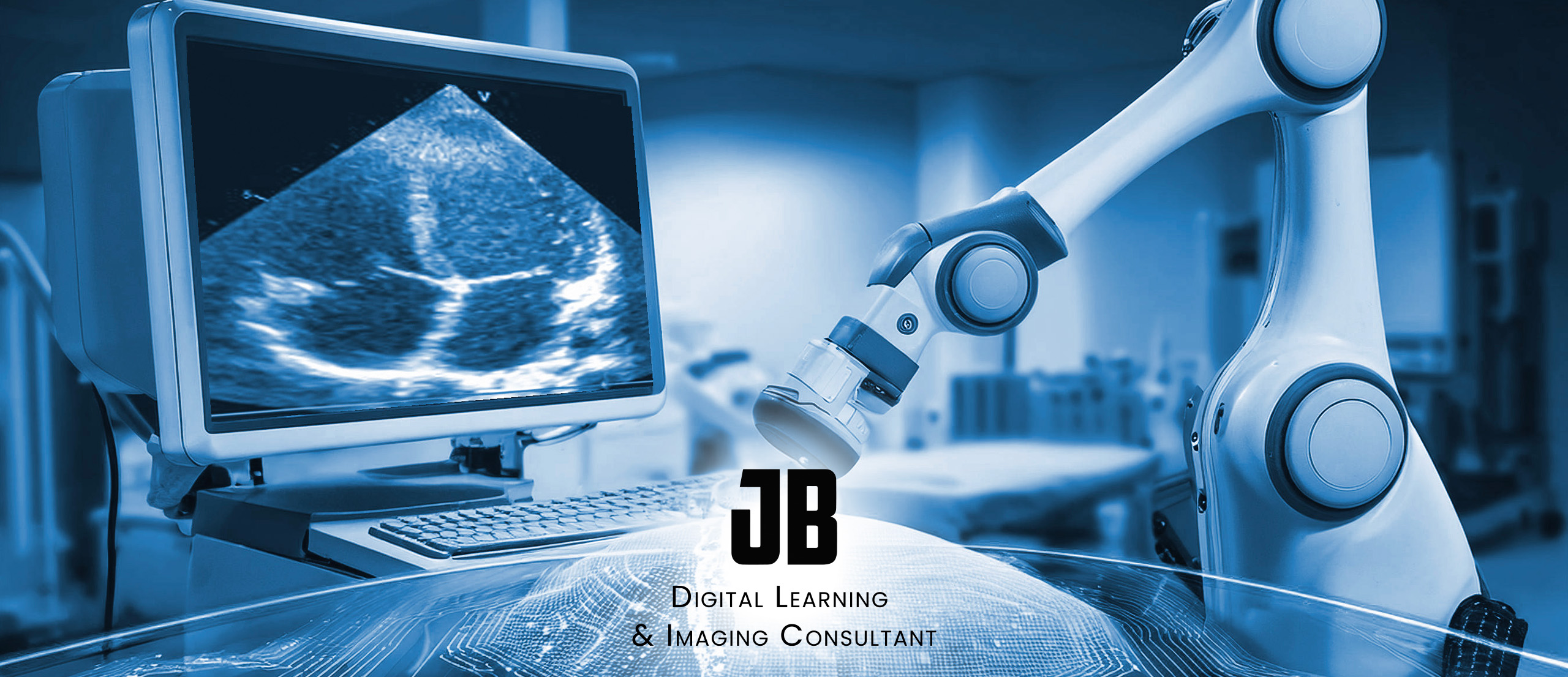
Artificial Intelligence in Fetal Cardiology: Advances in Prenatal Screening and Diagnosis
Congenital heart disease (CHD) remains the most common congenital anomaly worldwide, significantly affecting neonatal outcomes. Early and accurate prenatal detection is essential for timely intervention and improved prognosis. With the advent of artificial intelligence (AI), novel machine-learning approaches, including deep learning and neural networks, are revolutionizing fetal echocardiography and electrocardiography. This course explores the intersection of AI and fetal cardiology, examining the latest advancements in automated cardiac imaging, AI-assisted biometrics, and deep-learning-based screening methods for detecting CHD. Through a review of current research, case studies, and AI applications, participants will gain a comprehensive understanding of how AI is enhancing fetal heart diagnostics.
course outline
Time | Topic |
0:00 – 05:00 | Introduction to Fetal Cardiology and CHD – Epidemiology, significance of early detection, challenges in traditional fetal echocardiography. |
05:00 – 15:00 | Fundamentals of AI in Medical Imaging – Introduction to deep learning, machine learning, and their applications in medical imaging. |
15:00 – 30:00 | AI-Assisted Fetal Echocardiography – Review of AI-based detection systems, automation of cardiac imaging, standardization, and accuracy improvements. |
30:00 – 40:00 | Fetal Electrocardiography and AI Integration – Role of AI in analyzing fetal ECG, detection of critical CHDs, and overcoming current limitations. |
40:00 – 50:00 | Case Studies and Clinical Applications – Review of AI-driven models in detecting atrioventricular septal defects, coarctation of the aorta, and other CHDs. |
50:00 – 55:00 | Challenges and Ethical Considerations – Bias in AI models, operator-AI interaction, limitations in current AI systems. |
55:00 – 60:00 | Future Directions and Conclusion – The potential of AI for real-time ultrasound guidance, enhanced diagnostic accuracy, and integration into routine prenatal care. |
1.1 Epidemiology of Congenital Heart Disease (CHD)
- Incidence rates and global statistics.
- Most common types of CHD (e.g., ventricular septal defect, atrioventricular septal defect, hypoplastic left heart syndrome).
- Long-term outcomes and survival rates with/without prenatal diagnosis.
1.2 Importance of Early Detection
- Role of prenatal screening in improving neonatal outcomes.
- Impact of CHD detection on perinatal management and postnatal interventions.
- Limitations of traditional fetal echocardiography in identifying complex CHDs.
1.3 Challenges in Conventional Fetal Cardiology
- Operator dependency and variability in diagnostic accuracy.
- Technical difficulties: fetal movement, maternal obesity, poor acoustic windows.
- Need for standardized assessment methods.
2.1 Introduction to Artificial Intelligence (AI)
- Definition and key concepts (machine learning, deep learning, neural networks).
- Role of AI in medical imaging.
- Supervised vs. unsupervised learning in fetal cardiology applications.
2.2 How AI Processes Medical Images
- Image acquisition and preprocessing.
- Feature extraction using convolutional neural networks (CNNs).
- Role of segmentation models in cardiac structure identification.
2.3 AI Training and Validation in Fetal Cardiology
- Data collection: importance of large annotated datasets.
- Training neural networks: challenges in fetal ultrasound image variability.
- Bias and generalization issues in AI-based diagnostics.
3.1 The Role of AI in Fetal Echocardiography
- AI-driven standardization of fetal heart image acquisition.
- Automatic identification of key cardiac planes:
- Four-chamber view.
- Three-vessel and trachea view.
- Outflow tracts and aortic arch.
- Real-time AI-assisted quality control in ultrasound acquisition.
3.2 AI for CHD Detection and Classification
- AI models for detecting specific CHDs (e.g., atrioventricular septal defects, transposition of the great arteries).
- Comparison of AI performance vs. human sonographers in CHD detection.
- Integration of AI into existing echocardiographic workflows.
3.3 Clinical Validation and Real-World Application
- Sensitivity and specificity of AI-based echocardiography systems.
- Studies validating AI-assisted echocardiography in CHD detection.
- Challenges in implementing AI tools in clinical settings.
4.1 The Basics of Fetal Electrocardiography (fECG)
- What is fECG? How is it obtained?
- Differences between fECG and traditional fetal heart rate monitoring.
- Current challenges in using fECG for CHD detection.
4.2 AI Applications in fECG Analysis
- How AI enhances signal processing and noise reduction in fECG.
- AI models for detecting arrhythmias and conduction defects.
- Early detection of CHD through AI-assisted ECG waveform analysis.
4.3 fECG vs. Echocardiography: Complementary Approaches?
- Strengths and weaknesses of each modality.
- AI models combining echocardiography and fECG for enhanced CHD screening.
- Future potential of AI-driven multimodal diagnostics.
5.1 AI in Diagnosing Atrioventricular Septal Defects (AVSD)
- How AI models detect AVSD from four-chamber ultrasound images.
- Accuracy rates compared to human experts.
- Role of AI confidence scoring and clinician-AI collaboration.
5.2 AI in Screening for Coarctation of the Aorta (CoA)
- Automated cardiac biometric measurements.
- Key echocardiographic findings AI models use for CoA detection.
- AI-based risk stratification models.
5.3 AI for Rare and Hard-to-Detect CHDs
- Deep learning for detecting transposition of the great arteries (TGA).
- AI-assisted identification of hypoplastic left heart syndrome (HLHS).
- Future possibilities for expanding AI-based CHD screening.
6.1 AI Model Bias and Performance Variability
- Dataset limitations leading to biased AI models.
- Ensuring AI generalizability across diverse populations.
- Addressing false positives and false negatives in AI predictions.
6.2 Operator-AI Interaction and Clinical Trust
- How clinicians perceive AI assistance in fetal echocardiography.
- Risks of automation bias: over-reliance on AI outputs.
- The need for explainable AI in clinical decision-making.
6.3 Regulatory and Ethical Considerations
- AI approval pathways in medical imaging.
- Liability issues: who is responsible for AI-driven misdiagnosis?
- Ethical concerns regarding AI replacing human expertise.
7.1 The Future of AI in Fetal Cardiology
- Real-time AI integration into ultrasound machines.
- AI-assisted telemedicine and remote fetal cardiac screening.
- Personalized risk assessment using AI-driven predictive models.
7.2 Research Gaps and Unsolved Challenges
- Need for larger, more diverse training datasets.
- Improving AI interpretability for clinician adoption.
- Addressing legal and ethical challenges in AI-driven diagnostics.
7.3 Key Takeaways and Final Thoughts
- Summary of AI’s impact on fetal cardiology.
- Clinical implications and practical considerations for implementation.
- The evolving role of AI in improving congenital heart disease detection.
course text
1.1 Epidemiology of Congenital Heart Disease (CHD)
Congenital heart disease (CHD) is the most common congenital anomaly worldwide, affecting approximately 8 per 1,000 live births. Among these cases, nearly one-third require intervention within the first year of life. CHD encompasses a broad spectrum of structural abnormalities, including ventricular septal defects (VSDs), atrial septal defects (ASDs), tetralogy of Fallot (TOF), and hypoplastic left heart syndrome (HLHS).
Despite advancements in imaging technology, the prenatal detection rate remains suboptimal, with detection rates varying significantly between 30% and 80%, depending on the type of CHD and the expertise of the sonographer. This variability highlights the need for improved diagnostic tools such as artificial intelligence (AI)-assisted echocardiography.
1.2 Importance of Early Detection
Prenatal diagnosis of CHD has profound implications for neonatal outcomes. Early identification allows for:
- Parental counseling and decision-making, including potential interventions.
- Optimized perinatal care, such as planned delivery at tertiary centers with cardiac surgical facilities.
- Early postnatal management, reducing morbidity and mortality.
Studies indicate that neonates diagnosed prenatally with critical CHD have better neurological outcomes and lower mortality rates than those diagnosed postnatally.
1.3 Challenges in Conventional Fetal Cardiology
Despite its importance, fetal echocardiography remains a highly operator-dependent technique, requiring substantial training and expertise. Several challenges persist, including:
- Maternal factors: Obesity, previous abdominal surgeries, and poor acoustic windows.
- Fetal factors: Movement, positioning, and small heart size in the first trimester.
- Technical limitations: Variability in machine settings and inconsistency in obtaining standard views.
The low reproducibility and inter-operator variability of echocardiographic assessments necessitate standardized, AI-driven solutions that can enhance accuracy and reproducibility.
2.1 Introduction to Artificial Intelligence (AI)
What is Artificial Intelligence?
Artificial Intelligence (AI) is a branch of computer science that enables machines to perform tasks that typically require human intelligence. These tasks include pattern recognition, decision-making, problem-solving, and learning from experience. In medical imaging, AI is revolutionizing how images are analyzed, interpreted, and used for diagnosis.
AI encompasses several subfields, including:
- Machine Learning (ML) – AI models that improve their performance by learning from large datasets.
- Deep Learning (DL) – A specialized form of ML that uses artificial neural networks to mimic the human brain’s ability to recognize patterns.
- Natural Language Processing (NLP) – AI that understands and processes human language, used in clinical documentation and decision support.
Why is AI Important in Medical Imaging?
Medical imaging plays a crucial role in disease diagnosis, treatment planning, and patient monitoring. However, traditional imaging analysis is often time-consuming, operator-dependent, and subject to interpretation variability. AI offers the potential to:
- Improve diagnostic accuracy by detecting features that may be missed by the human eye.
- Automate repetitive tasks, reducing workload for radiologists and sonographers.
- Enhance image quality through noise reduction and standardization.
- Speed up analysis and reporting, allowing for quicker medical decision-making.
2.2 How AI Processes Medical Images
The Role of AI in Image Analysis
AI is particularly well-suited for pattern recognition, making it an excellent tool for medical imaging applications, including fetal cardiology. AI can help detect subtle anatomical abnormalities in fetal ultrasound images that may indicate congenital heart disease (CHD).
Three Key Steps in AI-Based Image Processing
AI follows a structured approach to analyzing medical images, typically involving three main steps:
1. Preprocessing – Optimizing Image Quality
Before AI can analyze an image, it must be preprocessed to ensure it is clear, standardized, and free of artifacts. This includes:
- Noise reduction – Eliminating background interference from ultrasound images.
- Image enhancement – Adjusting brightness, contrast, and resolution to improve visibility.
- Normalization – Standardizing images across different ultrasound machines for consistency.
2. Feature Extraction – Identifying Key Anatomical Structures
Once an image has been optimized, AI extracts relevant features that are essential for diagnosis. In fetal cardiology, these features include:
- Fetal heart chambers – Identifying atria and ventricles.
- Major blood vessels – Detecting abnormalities in the aorta and pulmonary artery.
- Heart motion patterns – Analyzing how the fetal heart contracts and relaxes.
3. Classification and Segmentation – Detecting Abnormalities
In the final step, AI classifies the image as normal or abnormal based on extracted features. AI can also segment the image, which means labeling different parts of the heart to highlight specific anomalies.
For example, AI models trained on thousands of fetal echocardiograms can distinguish between a normal four-chamber heart view and one with an atrioventricular septal defect (AVSD). This allows for early detection and intervention.
2.3 AI Training and Validation in Fetal Cardiology
How AI Learns from Data
For AI to perform accurately in medical imaging, it must be trained on a large dataset of labeled images. The training process involves:
- Feeding thousands of fetal ultrasound images into the AI model.
- Teaching the model to recognize key anatomical patterns and anomalies.
- Using statistical algorithms to refine its accuracy.
- Validating the AI model on new, unseen images to ensure reliability.
AI training requires high-quality, well-annotated datasets, typically labeled by expert fetal cardiologists. These datasets must be diverse and representative of different populations to ensure the AI model can generalize well across various patient groups.
Key Factors Affecting AI Performance in Fetal Cardiology
Several factors influence how well AI models perform in fetal cardiology:
1. Dataset Quality and Size
- AI models require large volumes of high-resolution ultrasound images to learn effectively.
- If the dataset is too small or not diverse, the AI may struggle with real-world cases.
2. Data Labeling and Annotation
- Training data must be accurately labeled by experts to ensure AI models learn correctly.
- Incorrect or inconsistent labeling can lead to misdiagnosis and model bias.
3. Variability in Ultrasound Machines
- Different hospitals use different ultrasound equipment, leading to variations in image quality.
- AI models must be trained to handle differences in imaging protocols and resolutions.
4. Real-World Testing and Validation
- AI models are first tested in controlled environments using pre-existing datasets.
- They must then be validated in clinical settings to confirm their effectiveness in real-time diagnosis.
Key Takeaways from Chapter 2
- AI plays a transformative role in medical imaging, particularly in fetal cardiology.
- AI image analysis involves three main steps: preprocessing, feature extraction, and classification.
- Training AI requires large, diverse datasets and rigorous validation.
- Challenges such as data variability, labeling accuracy, and clinical validation must be addressed.
- AI has the potential to improve diagnostic accuracy, efficiency, and consistency in CHD detection.
3.1 The Role of AI in Fetal Echocardiography
Why is AI Important in Fetal Echocardiography?
Fetal echocardiography is the primary imaging method used to assess fetal heart development and detect congenital heart disease (CHD) before birth. However, this process is highly operator-dependent, meaning that the accuracy of the diagnosis can vary based on the experience of the clinician performing the scan.
AI has emerged as a transformative tool in fetal echocardiography, offering enhanced accuracy, automation, and real-time decision support. The key roles of AI in fetal echocardiography include:
Standardizing Image Acquisition – AI can help ensure correct probe positioning and angle selection.
Automating Image Analysis – AI can identify key cardiac structures and measure heart dimensions.
Detecting CHD Earlier and More Accurately – AI models can recognize subtle anomalies that may be missed by human observers.
Reducing Variability Between Operators – AI improves diagnostic consistency across different medical professionals and settings.
By integrating AI into fetal echocardiography, clinicians can enhance the early detection of CHD, optimize workflow efficiency, and reduce diagnostic errors.
3.2 AI for CHD Detection and Classification
How AI Detects CHD from Fetal Echocardiography
AI models are trained to recognize normal and abnormal fetal heart structures by analyzing thousands of ultrasound images. AI can process echocardiographic data using deep learning algorithms that learn from expert-labeled images to detect CHD with high accuracy.
Key AI Capabilities in CHD Detection
Automatic Identification of Cardiac Planes – AI ensures that correct echocardiographic views (e.g., four-chamber view, three-vessel view) are captured.
Image Segmentation and Annotation – AI highlights heart structures and measures key dimensions.
Classification of Normal vs. Abnormal Hearts – AI assigns probability scores indicating the likelihood of CHD.
Real-Time Decision Support – AI can provide clinicians with immediate feedback on image quality and potential abnormalities.
Which CHDs Can AI Detect?
AI has been trained to identify a range of CHDs with high sensitivity and specificity, including:
1. Atrioventricular Septal Defect (AVSD)
AI analyzes the four-chamber view to detect missing or misaligned septa.
Identifies abnormal blood flow patterns associated with AVSD.
2. Coarctation of the Aorta (CoA)
AI measures ventricular size ratios and great vessel dimensions to assess risk.
Improves early diagnosis by detecting subtle narrowing of the aortic arch.
3. Tetralogy of Fallot (TOF)
AI identifies right ventricular hypertrophy and overriding aorta.
Detects pulmonary stenosis by analyzing outflow tract views.
4. Transposition of the Great Arteries (TGA)
AI detects misalignment of the great vessels in the three-vessel view.
Helps in differentiating between TGA and other outflow tract abnormalities.
How Accurate is AI in CHD Detection?
Studies show that AI-assisted echocardiography can achieve:
Sensitivity >90% for detecting certain CHDs.
High specificity, reducing false positives and unnecessary follow-ups.
Improved diagnostic consistency compared to traditional ultrasound interpretation.
AI’s role in CHD detection continues to grow as more high-quality datasets become available and models are validated across diverse populations.
3.3 Clinical Validation and Real-World Application
How AI is Tested Before Clinical Use
Before AI models can be used in clinical practice, they undergo rigorous validation and testing to ensure accuracy, safety, and reliability. The validation process includes:
Training Phase – AI learns from thousands of labeled ultrasound images.
Validation Phase – AI is tested on unseen datasets to evaluate performance.
Clinical Trials – AI is assessed in real-world settings to compare its accuracy against expert fetal cardiologists.
Regulatory Approval – AI tools must meet medical safety standards (e.g., FDA, CE Mark) before deployment.
Real-World Applications of AI in Fetal Echocardiography
AI is already being used in leading hospitals and research centers to enhance fetal echocardiography. Examples of real-world applications include:
AI-Guided Ultrasound Systems – AI helps sonographers capture optimal images by providing real-time feedback on probe positioning.
Automated Fetal Heart Screening Programs – AI assists in large-scale CHD screening, improving detection rates in routine obstetric care.
AI-Enabled Telemedicine – Remote AI analysis allows CHD specialists to review echocardiograms from distant locations, improving access to expert care.
Key Challenges in AI Adoption
Despite its potential, several challenges must be addressed before AI can be fully integrated into fetal echocardiography:
Data Quality and Variability – AI must be trained on diverse datasets to handle different imaging conditions.
Trust and Acceptance by Clinicians – AI should act as a decision-support tool, not a replacement for human expertise.
Regulatory and Ethical Considerations – AI systems must comply with patient safety and data privacy laws.
Cost and Infrastructure Requirements – Implementing AI in clinical practice requires investment in software, training, and workflow integration.
By addressing these challenges, AI can become a powerful tool in improving fetal CHD detection and patient outcomes.
Key Takeaways from Chapter 3
AI enhances fetal echocardiography by improving image acquisition, analysis, and CHD detection.
AI can accurately classify normal vs. abnormal fetal hearts with high sensitivity and specificity.
Key CHDs detected by AI include AVSD, CoA, TOF, and TGA.
Clinical validation is essential to ensure AI models perform reliably in real-world settings.
AI is already being integrated into ultrasound systems, large-scale CHD screening, and telemedicine services.
Challenges such as data variability, clinician acceptance, and regulatory compliance must be addressed for widespread adoption.
4.1 The Basics of Fetal Electrocardiography (fECG)
What is Fetal Electrocardiography (fECG)?
Fetal electrocardiography (fECG) is a non-invasive method used to record the fetal heart’s electrical activity. Unlike fetal echocardiography, which focuses on the structural aspects of the heart, fECG provides valuable information about heart rhythm, conduction, and arrhythmias.
The fetal heart generates electrical signals, which can be recorded using electrodes placed on the maternal abdomen. However, these signals are weak and often contaminated by maternal ECG activity, muscle movements, and background noise. AI helps filter, enhance, and analyze these signals, making fECG a more reliable tool for early fetal cardiac assessment.
Key Differences Between fECG and Echocardiography
Feature | Fetal Echocardiography | Fetal ECG |
Focus | Structural assessment | Functional and electrical assessment |
Detection | CHDs (e.g., AVSD, CoA) | Arrhythmias and conduction disorders |
Technology | Ultrasound imaging | Surface electrode recording |
Best Used For | Diagnosing congenital defects | Detecting abnormal heart rhythms |
By combining echocardiography and fECG, clinicians can get a more complete picture of fetal heart health, identifying both structural and electrical abnormalities.
4.2 AI Applications in fECG Analysis
How AI Enhances fECG Analysis
AI plays a crucial role in improving the accuracy and usability of fECG by:
Noise Reduction – AI algorithms filter out maternal ECG, muscle artifacts, and environmental noise.
Signal Enhancement – AI amplifies weak fetal heart signals to improve analysis.
Arrhythmia Detection – AI identifies abnormal rhythms such as atrioventricular block and premature beats.
Automated Diagnosis – AI classifies fECG patterns, helping to distinguish between normal and abnormal cases.
AI in fECG Signal Processing
fECG signals are often distorted by interference, making it difficult to extract clear heart rhythms. AI models use advanced signal processing techniques, such as:
Convolutional Neural Networks (CNNs) – Extract relevant features from fECG waveforms.
Recurrent Neural Networks (RNNs) – Detect patterns over time to classify arrhythmias.
Bayesian Filtering – Eliminates background noise while preserving fetal heart signals.
AI in Early Detection of Fetal Arrhythmias
Fetal arrhythmias occur in 1-2% of pregnancies and can lead to fetal heart failure or intrauterine demise if not detected early. AI can help identify:
Atrioventricular Block (AV Block) – A conduction disorder where electrical signals are delayed or blocked.
Supraventricular Tachycardia (SVT) – A fast heart rate that can lead to fetal hydrops.
Long QT Syndrome – A condition that increases the risk of dangerous ventricular arrhythmias.
AI-enhanced fECG analysis allows for earlier detection and better management of these conditions, improving fetal outcomes.
4.3 fECG vs. Echocardiography: Complementary Approaches?
Why Use Both fECG and Echocardiography?
While echocardiography is the gold standard for detecting structural defects, it is limited in its ability to assess electrical function. fECG, on the other hand, provides detailed insights into cardiac conduction and rhythm but cannot visualize heart anatomy.
By combining AI-driven fECG with fetal echocardiography, clinicians can:
Detect both structural and electrical abnormalities.
Assess overall fetal heart function more comprehensively.
Improve prenatal screening for conditions requiring early intervention.
Real-World Applications of AI in Multimodal Fetal Cardiology
AI-Assisted CHD Screening – Combining AI-driven echocardiography with fECG to improve CHD detection.
AI-Based Risk Stratification – Using AI to assess fetal heart risk based on combined structural and electrical analysis.
AI for Remote Monitoring – Allowing pregnant women to monitor fetal heart function from home.
AI’s ability to synchronize and analyze both modalities is paving the way for more advanced fetal cardiac care.
4.4 Clinical Validation of AI in fECG and Multimodal Screening
How AI is Tested and Validated in fECG
Before AI-based fECG models can be used in clinical practice, they must undergo rigorous validation. This process includes:
Training on Large Datasets – AI models are trained on fECG recordings from thousands of pregnancies.
Testing on Independent Datasets – AI is evaluated on new, unseen data to ensure accuracy.
Clinical Trials and Real-World Testing – AI tools are tested in hospital settings for reliability.
Regulatory Approval – AI models must meet medical safety standards (FDA, CE) before deployment.
Current Research and AI Performance Metrics
Recent studies have demonstrated that AI-enhanced fECG analysis can achieve:
90-95% sensitivity for detecting fetal arrhythmias.
High specificity, reducing false positives and unnecessary interventions.
Improved signal clarity, leading to more reliable diagnoses.
AI-driven fECG has the potential to become a standard tool in prenatal cardiology, complementing traditional ultrasound screening.
4.5 Challenges and Future Directions in AI-Based fECG
Challenges in AI-Enhanced fECG
Despite its promise, AI-based fECG faces several challenges:
Variability in Signal Quality – Differences in maternal body composition, fetal position, and recording conditions can affect fECG accuracy.
Limited Datasets – AI models need more diverse and representative fECG datasets to improve performance.
Regulatory and Ethical Concerns – AI in fECG must be thoroughly tested to ensure patient safety and reliability.
Integration with Clinical Workflows – AI tools must be user-friendly and seamlessly integrated into existing prenatal care.
Future Directions for AI in fECG
Looking ahead, AI is expected to play an even greater role in fetal cardiac care, with innovations including:
Real-Time AI-Enhanced fECG Monitoring – AI models that provide continuous fetal heart monitoring at home.
Cloud-Based AI Diagnostics – Remote AI interpretation of fECG recordings for improved telemedicine applications.
AI-Guided Treatment Planning – AI predicting the best management strategies for fetal arrhythmias.
Multimodal AI Integration – Combining AI analysis of fECG, echocardiography, and genetic data to create personalized fetal heart risk assessments.
By addressing current challenges and continuing research, AI will further revolutionize fetal cardiac screening and monitoring, ensuring better prenatal outcomes.
Key Takeaways from Chapter 4
fECG is a valuable tool for detecting fetal arrhythmias and assessing electrical heart function.
AI improves fECG by enhancing signal quality, detecting arrhythmias, and automating diagnosis.
Combining AI-driven echocardiography and fECG provides a more complete assessment of fetal heart health.
Clinical validation is crucial to ensure AI-based fECG models are accurate and reliable.
Future advancements in AI-enhanced fECG will expand its role in remote monitoring and personalized fetal heart risk prediction.
5.1 AI in Diagnosing Atrioventricular Septal Defects (AVSD)
What is Atrioventricular Septal Defect (AVSD)?
Atrioventricular septal defect (AVSD) is a congenital heart defect characterized by a malformation of the atrial and ventricular septa along with abnormal development of the atrioventricular valves. It is commonly associated with chromosomal abnormalities, especially Down syndrome (trisomy 21).
Challenges in AVSD Detection Using Traditional Echocardiography
AVSD is primarily diagnosed using the four-chamber view in fetal echocardiography. However, detection can be challenging due to:
Variability in image quality, making subtle septal defects harder to visualize.
Fetal movement and poor acoustic windows affecting heart visualization.
Operator experience, as detecting AVSD requires expertise in recognizing abnormal valve morphology and ventricular symmetry.
How AI Improves AVSD Detection
AI models trained on large datasets of fetal echocardiograms can significantly enhance AVSD detection rates by:
Automatically identifying the four-chamber view and ensuring image quality.
Segmenting and highlighting septal defects to assist clinicians in identifying abnormalities.
Comparing normal and abnormal heart structures to detect abnormal atrioventricular valve attachment.
Providing probability scores indicating the likelihood of AVSD presence.
Clinical Validation of AI in AVSD Diagnosis
AI has shown higher sensitivity and specificity compared to traditional human interpretation.
Studies report up to 90% accuracy in AI-based AVSD detection when using deep learning models.
AI-assisted echocardiography can serve as a decision-support tool, flagging high-risk cases for expert review.
By improving AVSD detection, AI contributes to earlier interventions, better prenatal counseling, and improved neonatal outcomes.
5.2 AI in Screening for Coarctation of the Aorta (CoA)
What is Coarctation of the Aorta (CoA)?
Coarctation of the aorta (CoA) is a narrowing of the aortic arch, restricting blood flow and leading to hypertension, heart failure, or circulatory collapse after birth if untreated. CoA accounts for 5-8% of all congenital heart defects but is one of the most commonly missed diagnoses in prenatal screening.
Why is CoA Difficult to Diagnose Prenatally?
The aortic narrowing is often subtle, making it difficult to detect with standard echocardiography.
Normal fetal circulation (via ductus arteriosus) can mask CoA, making the defect appear minor before birth.
Traditional detection relies on indirect signs, such as ventricular size asymmetry, which can be highly variable.
How AI Enhances CoA Detection
AI improves CoA detection by:
Measuring key fetal cardiac biometrics such as right vs. left ventricular size ratios.
Analyzing the three-vessel and trachea (3VT) view to identify narrowing in the aortic arch.
Using deep learning models to compare CoA cases with normal fetal hearts to recognize patterns.
Reducing false positives and false negatives by combining multiple imaging markers.
Clinical Impact of AI in CoA Detection
AI-assisted echocardiography has been shown to increase CoA detection rates by up to 50% compared to standard methods.
Earlier detection allows for specialized perinatal management, ensuring that affected newborns receive early intervention.
AI can help identify borderline cases, providing risk stratification for postnatal follow-up.
By improving CoA detection rates, AI plays a critical role in preventing life-threatening complications in affected neonates.
5.3 AI for Rare and Hard-to-Detect CHDs
Challenges in Diagnosing Rare CHDs
While common congenital heart defects like AVSD and CoA receive significant research attention, rare and complex CHDs remain difficult to detect due to:
Limited training datasets, as rare CHDs occur less frequently.
Atypical anatomical presentations, making automated detection more challenging.
Variability in fetal growth, affecting heart morphology and diagnostic criteria.
How AI is Helping with Rare CHD Detection
AI models can improve the detection of rare congenital heart defects such as:
1. Hypoplastic Left Heart Syndrome (HLHS)
AI can analyze fetal cardiac structure asymmetry to detect underdeveloped left heart structures.
Studies have shown that AI improves HLHS detection sensitivity, particularly in early gestation.
2. Transposition of the Great Arteries (TGA)
AI detects abnormal great artery alignment in the three-vessel view.
Machine learning models have shown improved specificity in differentiating TGA from normal cardiac outflow patterns.
3. Ebstein’s Anomaly
AI identifies displacement of the tricuspid valve, an essential marker of Ebstein’s anomaly.
AI-guided Doppler analysis can help detect abnormal blood flow patterns.
Benefits of AI in Rare CHD Screening
Automated analysis of multiple cardiac planes, improving detection rates.
Standardized screening protocols, reducing variability between clinicians.
Earlier referrals for fetal cardiology specialists, ensuring timely interventions.
AI is expanding the boundaries of CHD detection, offering hope for improved diagnosis of rare and complex congenital heart anomalies.
5.4 AI in Real-World Clinical Applications
How AI is Currently Used in Hospitals and Clinics
AI is already being integrated into fetal cardiology programs worldwide, with real-world applications including:
AI-Guided Fetal Echocardiography
AI helps sonographers acquire optimal heart views by providing real-time feedback.
Ensures standardized image acquisition, reducing operator dependency.
Automated CHD Screening in Obstetric Clinics
AI-enhanced ultrasound machines can pre-screen for CHD, flagging cases for expert review.
Improves CHD detection in routine obstetric scans, especially in low-resource settings.
AI-Assisted Telemedicine for Remote Diagnosis
Cloud-based AI models allow specialists to analyze fetal heart scans remotely.
Expands access to expert fetal cardiology services in rural areas.
AI in Risk Stratification and Predictive Analytics
AI models predict high-risk pregnancies based on fetal heart biometrics.
Enables personalized prenatal care strategies, optimizing fetal outcomes.
Challenges in Clinical AI Adoption
Despite AI’s promise, several challenges remain:
Ensuring AI reliability and accuracy across diverse populations.
Training clinicians to interpret AI outputs effectively.
Integrating AI into existing hospital workflows without disrupting care.
By addressing these challenges, AI can be fully leveraged as a tool to improve prenatal cardiac care worldwide.
Key Takeaways from Chapter 5
AI significantly enhances the detection of CHDs like AVSD, CoA, and HLHS.
Machine learning models improve accuracy, reducing missed diagnoses.
AI-driven echocardiography is already being implemented in hospitals for CHD screening.
Telemedicine applications enable remote CHD diagnosis, expanding access to expert care.
Challenges such as data variability and AI integration into clinical workflows must be addressed.
Conclusion
This chapter has provided real-world examples of AI applications in fetal cardiology, demonstrating how AI is improving the detection and management of CHD. AI is no longer just a research tool—it is actively transforming clinical workflows, improving diagnostic accuracy, and expanding access to fetal cardiac care.
6.1 AI Model Bias and Performance Variability
What is Bias in AI Models?
Bias in artificial intelligence (AI) refers to systematic errors that cause an AI model to produce inaccurate, unfair, or misleading results for certain populations. In fetal cardiology, AI bias can affect CHD detection rates, leading to false positives or false negatives that may disproportionately impact specific groups.
Bias can stem from:
Imbalanced training data, where certain patient demographics are underrepresented.
Variability in ultrasound imaging settings, leading to inconsistent AI performance.
Subjectivity in clinician-labeled datasets, which may introduce human error.
Differences in fetal anatomy across ethnic backgrounds, affecting model generalizability.
How Bias Affects CHD Detection
When AI models are trained primarily on datasets from high-resource hospitals, they may struggle when applied to:
Low-resource settings, where ultrasound equipment and imaging quality differ.
Diverse patient populations, leading to lower detection rates for certain ethnic groups.
Rural healthcare systems, where AI might not be fine-tuned for specific local conditions.
Addressing Bias in AI-Based Fetal Cardiology
To minimize bias and improve AI reliability, researchers must:
Use diverse, representative training datasets that include various ethnicities, geographic regions, and socioeconomic backgrounds.
Ensure multi-center validation of AI models across different healthcare settings.
Regularly audit AI performance metrics to identify and correct disparities in CHD detection.
Develop adaptive learning models, allowing AI to refine its performance over time in real-world clinical use.
By tackling these challenges, AI can become a more equitable and trustworthy tool in fetal cardiology.
6.2 Operator-AI Interaction and Clinical Trust
Why is Clinician Trust in AI Important?
For AI to be effectively integrated into fetal cardiology practice, clinicians must:
Understand how AI makes decisions to validate its recommendations.
Trust AI outputs while maintaining their own clinical judgment.
Use AI as a support tool rather than a replacement for expertise.
However, many healthcare providers remain skeptical about AI’s role in CHD diagnosis due to:
Lack of transparency in AI decision-making (AI as a “black box”).
Concerns over AI accuracy, especially in complex cases.
Fear of over-reliance on technology, leading to deskilling of clinicians.
The Role of Explainable AI (XAI) in Building Trust
Explainable AI (XAI) addresses these concerns by providing visual and logical explanations for AI decisions. Methods include:
Gradient-Weighted Class Activation Mapping (Grad-CAM) – Highlights which areas of an image the AI used to make its diagnosis.
Probability Scoring – Shows clinicians how confident the AI is in its prediction.
AI-Assisted Decision Trees – Provides step-by-step reasoning behind AI-based classifications.
When AI transparency improves, clinicians feel more confident using AI in their decision-making process.
How AI Should Support, Not Replace, Clinicians
AI is designed to function as a decision-support tool, helping clinicians:
Prioritize high-risk cases for further evaluation.
Reduce diagnostic variability across different healthcare providers.
Enhance workflow efficiency, allowing more time for patient care.
Clinician training programs should include AI literacy education, ensuring that fetal cardiologists understand how to interpret AI outputs correctly.
6.3 Regulatory and Ethical Considerations
Why is Regulation Essential for AI in Fetal Cardiology?
AI applications in fetal medicine must undergo rigorous validation before being used in clinical settings. Without proper regulation, AI models may:
Produce incorrect diagnoses, leading to mismanagement of pregnancies.
Lack accountability, making it unclear who is responsible for errors.
Violate patient privacy, if data security measures are inadequate.
Regulatory frameworks ensure that AI:
Meets safety and accuracy standards before clinical deployment.
Complies with patient data protection laws (e.g., HIPAA, GDPR).
Is continuously monitored for performance and fairness.
Current Regulatory Frameworks for AI in Healthcare
S. Food and Drug Administration (FDA) – Requires AI models to be tested for clinical accuracy and patient safety before approval.
European CE Mark Certification – Ensures AI compliance with medical device regulations in Europe.
International Standards (ISO 13485, IEC 62304) – Guidelines for AI development in medical imaging and diagnostics.
AI developers must work closely with regulatory agencies to ensure compliance and maintain patient safety.
Ethical Considerations in AI-Based CHD Screening
Ethical concerns surrounding AI in fetal cardiology include:
Informed Consent – Patients should be aware of AI’s role in their diagnosis.
Data Privacy – AI systems must comply with strict confidentiality regulations.
AI Accountability – Clear guidelines are needed to determine who is responsible for AI-based misdiagnoses.
Algorithmic Fairness – AI should not favor one demographic group over another.
Clinicians and AI developers must work together to ensure ethical AI deployment in fetal cardiology.
6.4 Challenges in AI Integration into Clinical Workflows
Obstacles to AI Adoption in Fetal Cardiology
Even with its advantages, AI adoption faces several challenges, including:
Integration with Existing Ultrasound Systems – Many hospitals lack AI-compatible imaging technology.
Training and Education for Clinicians – Healthcare providers need training on how to interpret and validate AI recommendations.
Resistance to Change – Some clinicians fear that AI will replace human expertise rather than enhance it.
Cost and Infrastructure Requirements – Implementing AI solutions requires investment in software, cloud storage, and computing power.
How to Overcome These Challenges
Improving AI Interpretability – Providing clinicians with clear explanations of AI outputs.
Regulatory Guidelines for AI Implementation – Establishing protocols for safe and effective AI usage.
Enhancing AI Usability in Clinical Settings – Ensuring seamless integration with ultrasound machines and electronic health records (EHRs).
Developing AI Training Programs for Clinicians – Offering hands-on education in AI literacy and its applications in fetal medicine.
By addressing these challenges, AI can be smoothly integrated into clinical workflows, leading to improved CHD detection rates and patient outcomes.
Key Takeaways from Chapter 6
AI models can introduce bias, affecting diagnostic accuracy across different populations.
Clinicians must trust and understand AI to use it effectively in CHD screening.
Regulatory frameworks (FDA, CE) are necessary to ensure AI safety and accuracy.
Ethical concerns, including informed consent and data privacy, must be addressed.
Challenges such as workflow integration, clinician education, and cost barriers need solutions for AI adoption.
AI’s Growing Role in Prenatal Cardiac Care
Artificial Intelligence (AI) is rapidly transforming the field of fetal cardiology, offering enhanced CHD detection, predictive analytics, and personalized risk assessment. While AI has already demonstrated its potential in automated echocardiography analysis and fetal ECG interpretation, its future applications will further expand its clinical impact.
Key Areas of Future AI Development in Fetal Cardiology
Real-Time AI-Guided Echocardiography – AI will assist clinicians by providing instant feedback during ultrasounds, ensuring that optimal images are captured for accurate diagnosis.
AI-Powered Telemedicine – Remote AI-assisted fetal heart screening will enable global access to specialized cardiology care, particularly in underserved regions.
Predictive Analytics for Fetal Health – AI will integrate biometric, genetic, and maternal health data to predict fetal cardiovascular risks before structural anomalies develop.
Personalized CHD Management Plans – AI will help customize prenatal care strategies based on individualized fetal health assessments, optimizing outcomes.
Multi-Modal AI Systems – Future AI models will analyze echocardiography, ECG, Doppler ultrasound, and genetic markers together for comprehensive fetal heart screening.
How AI Can Reduce Congenital Heart Disease-Related Mortality
AI-driven innovations will contribute to:
Earlier detection of life-threatening CHDs such as Hypoplastic Left Heart Syndrome (HLHS).
Reduced diagnostic variability, ensuring standardized assessments across healthcare settings.
Improved postnatal survival rates, allowing for timely interventions and optimized neonatal care.
The future of AI in fetal cardiology lies in refining its accuracy, expanding its clinical applications, and making its benefits accessible to all healthcare providers worldwide.
7.2 Research Gaps and Unsolved Challenges
What Are the Current Limitations of AI in Fetal Cardiology?
Despite its advancements, AI still faces significant challenges that must be addressed before widespread clinical adoption. These include:
1. Data Privacy and Ethical Concerns
Large-scale AI models require access to massive medical datasets, raising concerns about patient privacy and data security.
Ensuring compliance with HIPAA, GDPR, and other regulatory frameworks is crucial for AI in fetal cardiology.
2. Limited Representation in AI Training Data
AI models must be trained on diverse datasets to avoid bias and ensure equitable CHD detection across all populations.
More multi-center studies are needed to validate AI effectiveness in different ethnic and geographical groups.
3. Interpretability and Clinician Confidence in AI
Many AI models function as “black boxes”, meaning that their decision-making process is not easily explainable to clinicians.
Developing explainable AI (XAI) tools, such as visual heatmaps and probability scores, will help build trust and transparency in AI-based CHD diagnosis.
4. Integration with Existing Medical Infrastructure
Many healthcare institutions lack AI-ready ultrasound machines, slowing adoption.
Seamless integration of AI with electronic health records (EHRs) and telemedicine platforms is essential for streamlined patient care.
By addressing these research gaps, AI can unlock its full potential in fetal cardiology, making CHD detection more precise, and clinically impactful.
7.3 Key Takeaways and Final Thoughts
The Transformative Potential of AI in Fetal Cardiology
Over the past decade, AI has demonstrated its ability to revolutionize congenital heart disease screening through:
Automated image analysis, reducing human error and improving early CHD detection.
Real-time clinical decision support, enhancing fetal echocardiography accuracy.
AI-driven predictive models, providing insights into fetal cardiac risk factors.
Telemedicine applications, expanding access to expert-level CHD assessment worldwide.
What Needs to Happen Next?
To maximize AI’s benefits in fetal cardiology, we must focus on:
Advancing AI model accuracy and interpretability to increase clinician confidence.
Expanding research efforts to validate AI in diverse patient populations.
Developing AI-assisted telehealth solutions to improve CHD screening access in remote areas.
Integrating AI seamlessly into existing clinical workflows, ensuring its usability for all healthcare professionals.
Final Thoughts: AI as a Collaborative Tool in Fetal Medicine
AI is not meant to replace human expertise, but rather to enhance the capabilities of fetal cardiologists and sonographers. By embracing AI-powered technologies, healthcare providers can:
Improve prenatal CHD detection rates.
Optimize perinatal care for high-risk pregnancies.
Ensure better postnatal outcomes for infants born with congenital heart defects.
The future of AI in fetal cardiology is promising, but continued collaboration between AI researchers, clinicians, and regulatory bodies is essential to ensure that AI-driven innovations are safe, effective, and accessible to all patients.
Key Takeaways from Chapter 7
AI is transforming fetal cardiology by improving CHD detection, telemedicine, and predictive analytics.
Future AI applications will focus on real-time guidance, personalized CHD management, and multimodal screening approaches.
Research gaps such as AI bias, model interpretability, and data privacy must be addressed.
Seamless AI integration into clinical workflows will be essential for widespread adoption.
AI should serve as a decision-support tool, empowering rather than replacing human expertise.
Conclusion
This chapter has provided an in-depth look into the future of AI in fetal cardiology, outlining both exciting innovations and ongoing challenges. As AI technology continues to evolve, its integration into prenatal CHD screening will lead to earlier diagnoses, improved perinatal care, and better lifelong outcomes for affected infants.
FAQ
AI is used in fetal cardiology to enhance the detection and classification of congenital heart disease (CHD). It automates image acquisition, improves diagnostic accuracy, reduces human error, and assists in interpreting fetal echocardiography and electrocardiography (ECG) results.
AI uses deep learning algorithms, such as convolutional neural networks (CNNs), to analyze ultrasound images and identify abnormal cardiac structures. It can detect subtle features that may be missed by human sonographers, increasing the sensitivity and specificity of CHD diagnosis.
- Standardization: Reduces inter-operator variability.
- Increased Accuracy: AI models can detect abnormalities with high sensitivity and specificity.
- Automation: AI can assist in real-time analysis of fetal heart images.
- Time Efficiency: Reduces the time required for manual interpretation and diagnosis.
AI models have been trained to detect various congenital heart defects, including:
- Atrioventricular Septal Defect (AVSD)
- Coarctation of the Aorta (CoA)
- Tetralogy of Fallot (TOF)
- Hypoplastic Left Heart Syndrome (HLHS)
- Transposition of the Great Arteries (TGA)
- Ventricular Septal Defects (VSDs)
AI enhances fECG analysis by filtering noise, identifying waveforms, and detecting arrhythmias. It can also provide insights into fetal heart rate variability and conduction disorders, improving early CHD diagnosis.
- Data Dependency: AI performance depends on the quality and quantity of training datasets.
- Interpretability: Some AI models function as a “black box,” making it difficult to understand their reasoning.
- Variability in Ultrasound Machines: AI models may require retraining for different imaging devices.
- Regulatory and Ethical Concerns: AI must comply with medical standards before clinical deployment.
No. AI is intended to assist, not replace, human sonographers. While AI can detect patterns and anomalies with high accuracy, human expertise is required for clinical validation, interpretation, and decision-making.
Studies have shown that AI-assisted detection of CHD can achieve:
- Sensitivity: >90% for some defects (e.g., AVSD, CoA).
- Specificity: High accuracy in distinguishing normal from abnormal hearts.
However, performance varies based on data quality, fetal positioning, and ultrasound machine settings.
- Acceptance by Clinicians: Some clinicians may be skeptical about trusting AI decisions.
- Training Requirements: Clinicians need training to use AI-assisted diagnostic tools effectively.
- Data Privacy Issues: AI models must comply with healthcare data protection laws (e.g., GDPR, HIPAA).
- Regulatory Approval: AI models must undergo rigorous clinical validation before approval.
AI models are trained on large datasets of annotated fetal ultrasound images and fECG recordings. These datasets include:
- Normal and abnormal fetal heart images labeled by expert sonographers.
- Echocardiographic views (e.g., four-chamber view, three-vessel view).
- Retrospective and prospective clinical data from CHD-diagnosed cases.
To ensure fairness and reliability, AI models should be trained on diverse datasets that include:
- Different fetal positions and gestational ages.
- Multiple ultrasound machines and settings.
- Diverse patient populations (ethnic, geographical, and genetic diversity).
Bias reduction strategies, such as data augmentation and domain adaptation, help improve generalization.
AI automates the measurement of key fetal cardiac parameters, including:
- Ventricular size and symmetry.
- Blood flow velocities using Doppler ultrasound.
- Cardiothoracic ratio (CTR) and heart shape analysis.
Automated biometrics enhance early detection of growth abnormalities and CHD.
AI serves as a decision-support tool by:
- Flagging potential CHD cases for review.
- Providing real-time feedback during ultrasound scans.
- Highlighting key anatomical features using AI visualization techniques (e.g., Grad-CAM).
Ultimately, the final diagnosis is confirmed by a trained fetal cardiologist.
Yes. AI-powered ultrasound analysis enables remote CHD screening and diagnosis.
- Telemedicine applications include:
- Remote AI-assisted CHD screening in underserved regions.
- AI-guided fetal heart assessments in mobile clinics.
- Cloud-based AI tools for consultation and second opinions.
These innovations can expand access to specialized fetal cardiology care worldwide.
The future of AI in fetal cardiology includes:
- Real-time AI-guided echocardiography for automated CHD detection.
- Integration of multimodal AI systems, combining ultrasound, fECG, and genetic data.
- AI-powered fetal heart simulations for predicting disease progression.
- Personalized risk assessment models using machine learning-based fetal health predictions.
- Regulatory advancements ensuring safe, ethical, and reliable AI applications in perinatal medicine.
MCQs
1. Which machine learning model is most commonly used for image analysis in fetal echocardiography? (Difficulty: 2)
A) Decision Trees
B) Convolutional Neural Networks (CNNs)
C) Support Vector Machines (SVMs)
D) K-Nearest Neighbors (KNN)
E) Naive Bayes
✅ Correct Answer: B
2. Which factor most affects the accuracy of AI models in CHD detection? (Difficulty: 2)
A) The speed of the ultrasound machine
B) The size and diversity of the training dataset
C) The color of the ultrasound image
D) The presence of AI-generated noise
E) The weight of the fetus
✅ Correct Answer: B
What is a major challenge in training AI models for fetal cardiology? (Difficulty: 3)
A) Lack of sufficient labeled fetal cardiac images
B) AI models’ inability to learn from past data
C) AI replacing pediatric cardiologists completely
D) The need for physical AI hardware inside the ultrasound machine
E) AI being only useful for postnatal cardiac screening
✅ Correct Answer: A
What technique is used to ensure AI models provide interpretable results in fetal cardiology? (Difficulty: 3)
A) Randomization of training datasets
B) Gradient-Weighted Class Activation Mapping (Grad-CAM)
C) Use of larger ultrasound transducers
D) Increasing the brightness of ultrasound images
E) Running AI models only on high-end ultrasound machines
✅ Correct Answer: B
Which echocardiographic view is most commonly used for AI-based CHD screening? (Difficulty: 1)
A) Parasternal long-axis view
B) Four-chamber view (4CV)
C) Apical two-chamber view
D) Suprasternal notch view
E) Coronary artery view
✅ Correct Answer: B
Which CHD is AI most effective at detecting in fetal ultrasound images? (Difficulty: 2)
A) Atrial fibrillation
B) Atrioventricular Septal Defect (AVSD)
C) Coronary artery disease
D) Left ventricular hypertrophy
E) Hypertrophic cardiomyopathy
✅ Correct Answer: B
What is the main advantage of AI-assisted fetal echocardiography? (Difficulty: 1)
A) Reduces operator dependency and increases diagnostic consistency
B) Eliminates the need for fetal heart screening
C) Provides 100% accuracy in CHD diagnosis
D) Works independently without any human intervention
E) Predicts future cardiac conditions in adulthood
✅ Correct Answer: A
What is one way AI improves fetal echocardiography image quality? (Difficulty: 3)
A) Automatically optimizing probe positioning
B) Increasing ultrasound probe temperature
C) Eliminating the need for Doppler imaging
D) Converting ultrasound images into 3D MRI scans
E) Reducing fetal heart rate variability
✅ Correct Answer: A
AI has been shown to improve detection rates of which condition when compared to standard human interpretation? (Difficulty: 3)
A) Gestational diabetes
B) Coarctation of the aorta (CoA)
C) Anencephaly
D) Pre-eclampsia
E) Spina bifida
✅ Correct Answer: B
What is the primary purpose of AI in fetal ECG (fECG)? (Difficulty: 2)
A) Diagnosing heart conditions in adults
B) Enhancing signal quality and detecting arrhythmias
C) Replacing Doppler-based fetal heart rate monitoring
D) Measuring fetal heart size
E) Increasing maternal blood pressure
✅ Correct Answer: B
Which AI technique is used to reduce noise in fetal ECG recordings? (Difficulty: 3)
A) Histogram equalization
B) Bayesian filtering
C) Convolutional signal processing
D) Non-invasive neural mapping
E) Signal segmentation using machine learning
✅ Correct Answer: E
Which fetal heart condition is most associated with abnormal fetal ECG patterns? (Difficulty: 3)
A) Atrioventricular block
B) Patent foramen ovale
C) Ventricular fibrillation
D) Mitral valve prolapse
E) Coronary artery disease
✅ Correct Answer: A
What is a major limitation of AI in fetal ECG interpretation? (Difficulty: 3)
A) AI cannot process ECG signals in real-time
B) Variability in fetal positioning affecting signal quality
C) The inability of AI to learn from previous fECG cases
D) AI requires genetic testing before use
E) AI cannot distinguish between different CHDs
✅ Correct Answer: B
How can AI be used to combine fetal ECG and echocardiography? (Difficulty: 3)
A) Using multimodal deep learning to correlate structural and electrical abnormalities
B) Replacing ultrasound-based CHD diagnosis with ECG
C) Predicting fetal arrhythmias using MRI
D) Measuring fetal blood glucose levels
E) Identifying congenital heart conditions based on maternal health records
✅ Correct Answer: A
What is the biggest ethical concern related to AI in fetal cardiology? (Difficulty: 2)
A) The high cost of AI implementation
B) The risk of bias in AI models leading to misdiagnosis
C) The inability of AI to function without human supervision
D) AI causing an increase in fetal congenital heart defects
E) AI requiring excessive computational power
✅ Correct Answer: B
What is a major limitation of AI in fetal cardiac screening? (Difficulty: 3)
A) AI models cannot be integrated into ultrasound machines
B) AI models are biased due to lack of diverse training data
C) AI completely eliminates the need for human interpretation
D) AI cannot detect abnormalities in early gestation
E) AI makes fetal echocardiography more time-consuming
✅ Correct Answer: B
Why is regulatory approval critical for AI in fetal cardiology? (Difficulty: 2)
A) To ensure AI models are tested and validated for accuracy and safety
B) To make AI more expensive for clinical use
C) To prevent AI from being used in developing countries
D) To allow AI to function independently of clinicians
E) To ensure AI can detect all fetal abnormalities, including genetic disorders
✅ Correct Answer: A
How can AI bias be minimized in fetal cardiology applications? (Difficulty: 3)
A) Training AI models with diverse and balanced datasets
B) Ensuring AI models are used only in specialized clinics
C) Relying solely on high-end ultrasound machines
D) Limiting AI use to postnatal cardiac screening
E) Preventing AI from making any diagnostic suggestions
✅ Correct Answer: A
What is the primary challenge of AI deployment in low-resource settings? (Difficulty: 2)
A) Lack of ultrasound machines capable of integrating AI
B) Limited access to trained personnel who can interpret AI results
C) High costs associated with AI-powered fetal screening
D) Variability in internet connectivity and infrastructure
E) All of the above
✅ Correct Answer: E
What is the role of Explainable AI (XAI) in fetal cardiology? (Difficulty: 3)
A) It makes AI decisions transparent and interpretable for clinicians
B) It increases the complexity of AI algorithms
C) It replaces the need for human validation of AI outputs
D) It eliminates the risk of AI misdiagnosis
E) It allows AI to work without regulatory oversight
✅ Correct Answer: A
What is an example of an AI-based decision-support tool in fetal cardiology? (Difficulty: 2)
A) AI models that provide real-time feedback on ultrasound image quality
B) AI that predicts maternal risk for gestational diabetes
C) AI that detects neurological abnormalities in the fetus
D) AI that determines the exact time of delivery
E) AI that prevents preterm labor
✅ Correct Answer: A
Why is AI considered a valuable tool in prenatal CHD screening? (Difficulty: 1)
A) It enhances detection rates by reducing human error
B) It automates image interpretation, reducing inter-operator variability
C) It allows for faster and more efficient fetal heart assessments
D) It provides decision support for less experienced clinicians
E) All of the above
✅ Correct Answer: E
What factor affects the performance of AI models in CHD diagnosis? (Difficulty: 2)
A) Quality of training data used for model development
B) The experience level of the sonographer using AI
C) Variability in fetal positioning and movement
D) Differences in ultrasound machine settings
E) All of the above
✅ Correct Answer: E
What is one possible future application of AI in fetal cardiology? (Difficulty: 2)
A) AI-assisted real-time guidance during fetal echocardiography
B) AI-based genetic screening for congenital defects
C) AI predicting long-term cardiovascular outcomes postnatally
D) AI automatically correcting ultrasound probe positioning
E) All of the above
✅ Correct Answer: E
How can AI improve telemedicine in fetal cardiology? (Difficulty: 3)
A) Enabling remote fetal heart assessments in underserved regions
B) Allowing AI-powered second opinions from expert clinicians
C) Reducing the need for in-person specialist visits
D) Facilitating cloud-based AI diagnostics for prenatal care
E) All of the above
✅ Correct Answer: E
What is the significance of multimodal AI systems in fetal cardiology? (Difficulty: 3)
A) They integrate multiple sources of fetal data (e.g., ultrasound, ECG, genetic testing)
B) They eliminate the need for manual CHD screening
C) They make ultrasound imaging more expensive
D) They replace all existing fetal screening protocols
E) They function without human oversight
✅ Correct Answer: A
How can AI optimize CHD risk prediction models? (Difficulty: 3)
A) By analyzing large-scale prenatal health records
B) By integrating genetic and imaging data for precision diagnosis
C) By continuously learning from new clinical cases
D) By identifying high-risk pregnancies early in gestation
E) All of the above
✅ Correct Answer: E
How does AI enhance automated fetal heart rate monitoring? (Difficulty: 2)
A) By detecting irregular heart rhythms in real-time
B) By analyzing beat-to-beat variability to assess fetal well-being
C) By reducing false alarms associated with traditional monitoring
D) By combining Doppler ultrasound with AI analysis
E) All of the above
✅ Correct Answer: E
What is the long-term vision for AI in fetal cardiology? (Difficulty: 3)
A) AI performing fully autonomous fetal heart assessments
B) AI improving global accessibility to prenatal CHD screening
C) AI providing personalized risk assessments for each pregnancy
D) AI integrating with wearable maternal health monitoring devices
E) All of the above
✅ Correct Answer: E
Quiz
Short Answer Exam Questions
✅ Answer: AI improves CHD detection by using deep learning models, such as convolutional neural networks (CNNs), to analyze ultrasound images and detect subtle abnormalities. AI reduces operator dependency, enhances image segmentation, and increases diagnostic accuracy by learning from large datasets. Studies show AI can detect CHDs like AVSD and CoA with high sensitivity and specificity, outperforming standard human interpretation in some cases.
✅ Answer: Challenges include data variability, requiring AI models to be trained on diverse datasets; bias in training data, leading to incorrect predictions for underrepresented populations; regulatory and ethical concerns, ensuring AI models meet medical safety standards; clinician acceptance, as AI must be integrated without replacing human expertise; and technical limitations, such as differences in ultrasound machine settings and fetal positioning.
✅ Answer: AI enhances fECG analysis by removing noise, identifying waveform patterns, and detecting arrhythmias or conduction defects. Deep learning models can analyze beat-to-beat variability and assess fetal well-being. AI-based fECG systems can detect abnormalities like atrioventricular block earlier than traditional methods, improving prenatal monitoring and intervention planning.
✅ Answer: AI improves early detection rates, enhances image interpretation consistency, reduces human error, and provides real-time feedback during ultrasounds. AI can automate biometric measurements, improving efficiency and accuracy. Additionally, AI-driven telemedicine applications expand access to fetal cardiology expertise in remote or underserved areas.
✅ Answer: AI-based image segmentation automatically identifies and highlights fetal heart structures, ensuring accurate chamber measurement, vessel identification, and defect localization. This reduces variability between sonographers and improves CHD detection by providing consistent, high-quality images for analysis.
✅ Answer: AI models trained on limited or non-representative datasets may struggle to generalize across diverse populations, leading to false negatives or false positives in CHD detection. Bias can affect clinical decisions, disproportionately impacting certain ethnic groups or geographical regions. Addressing bias requires diverse datasets, rigorous validation, and transparency in AI decision-making.
✅ Answer: Explainable AI (XAI) ensures transparency and interpretability of AI models by highlighting which features contribute to a diagnosis. Techniques like Gradient-Weighted Class Activation Mapping (Grad-CAM) help clinicians understand why AI flagged a particular CHD, increasing trust and reliability in AI-assisted diagnosis.
✅ Answer: AI enables remote CHD screening by assisting in real-time ultrasound interpretation. It allows automated image analysis, guiding less-experienced clinicians in rural areas. Cloud-based AI platforms can flag abnormal cases for expert review, improving access to fetal cardiology expertise worldwide.
✅ Answer: AI models require large, well-labeled datasets for accuracy. They may struggle with image artifacts, fetal movement, or poor ultrasound quality. AI models are not 100% accurate and still require human validation. Additionally, regulatory approval and clinician training are necessary before widespread adoption.
✅ Answer: Multimodal AI integrates ultrasound, fECG, and genetic data for a comprehensive CHD risk assessment. This improves diagnostic accuracy, provides personalized risk stratification, and allows early intervention planning by combining structural and functional fetal heart assessments.
✅ Answer: AI analyzes biometric measurements such as right/left ventricular size ratios and main pulmonary artery vs. aortic dimensions to predict CoA risk. AI models improve detection rates by identifying subtle abnormalities often missed in standard screenings.
✅ Answer: AI detects suboptimal probe positioning, flags blurry or misaligned images, and ensures adherence to standard imaging protocols. Automated quality control reduces operator-dependent errors, leading to more consistent and reliable ultrasound examinations.
✅ Answer: AI models must meet medical safety and accuracy standards before clinical use. Regulatory bodies like FDA (USA) and CE (Europe) require rigorous validation studies proving AI effectiveness. Issues like data privacy, liability concerns, and ethical approval further complicate AI deployment.
✅ Answer: AI reduces false positives by improving image interpretation consistency and flagging only high-risk cases. It minimizes false negatives by detecting subtle cardiac abnormalities in early-stage CHD. However, human validation remains essential to ensure accurate final diagnoses.
✅ Answer: Future advancements include real-time AI-guided echocardiography, cloud-based AI diagnostics for global access, personalized fetal cardiac risk prediction models, and AI-integrated wearable fetal monitoring devices. Continued research and multicenter validation trials will improve AI’s reliability and adoption in prenatal care.
Podcast
PowerPoint Slides

- Introduction to the course topic.
- Overview of AI applications in fetal cardiology.
- Importance of early detection of congenital heart disease (CHD).
- Course structure and key learning objectives.
- CHD is the most common congenital anomaly.
- Affects ~8 per 1,000 live births.
- Includes structural heart defects such as AVSD, CoA, and HLHS.
- Early detection improves neonatal outcomes
- Allows for parental counseling and decision-making.
- Enables specialized perinatal care and surgical planning.
- Reduces neonatal morbidity and mortality.
- Improves long-term neurodevelopmental outcomes.
- Operator dependency affects diagnostic accuracy.
- Fetal movement and positioning can obscure imaging.
- Variability in ultrasound machine quality and settings.
- Some CHDs are subtle and easily missed.
- AI mimics human cognitive functions using algorithms.
- Machine learning (ML) enables AI to improve with data.
- Deep learning (DL) processes complex medical images.
- Convolutional Neural Networks (CNNs) are widely used in ultrasound analysis.
- Preprocessing: Enhancing image clarity, reducing noise.
- Feature Extraction: Identifying key anatomical structures.
- Classification: Distinguishing normal vs. abnormal hearts.
- Requires large, labeled datasets for training.
- Uses statistical models to improve accuracy.
- Needs validation on diverse populations.
- Ensures AI generalization across different imaging settings.
- Standardizes fetal heart image acquisition.
- Identifies key cardiac planes automatically.
- Detects structural abnormalities with high sensitivity.
- Reduces inter-operator variability.
- Atrioventricular Septal Defect (AVSD): AI analyzes four-chamber views.
- Coarctation of the Aorta (CoA): AI evaluates ventricular size ratios.
- Tetralogy of Fallot (TOF): AI detects overriding aorta and RV hypertrophy.
- Transposition of the Great Arteries (TGA): AI improves detection of vessel misalignment.
- Accuracy: Comparison with human expert diagnosis.
- Sensitivity & Specificity: AI’s ability to detect CHD reliably.
- ROC Curves: Evaluating AI prediction strength.
- Ongoing research and clinical trials.
- AI enhances signal clarity by filtering noise.
- Identifies waveform patterns linked to CHD.
- Detects fetal arrhythmias earlier than conventional methods.
- Improves real-time fetal heart rate monitoring.
- Echocardiography: Best for structural analysis.
- fECG: Best for detecting arrhythmias and conduction defects.
- AI Integration: Combining both for better diagnosis.
- Future applications in multimodal CHD screening.
- AI improves identification of septal defects.
- Analyzes four-chamber heart views more consistently.
- Reduces missed diagnoses in early pregnancy.
- AI analyzes right vs. left ventricular size.
- Measures pulmonary artery and aortic dimensions.
- Improves early diagnosis rates.
- AI assists in detecting rare cardiac anomalies.
- Provides automated decision-support for clinicians.
- Reduces false-negative rates in routine screening.
- Bias in AI training datasets.
- Trust and interpretability issues.
- Ensuring regulatory compliance and patient safety.
- AI as a diagnostic aid, not a replacement.
- Clinicians validate AI findings with experience.
- Improving user trust through explainable AI.
- FDA and CE mark requirements for AI tools.
- Ensuring AI meets clinical safety standards.
- Need for continuous monitoring and updates.
- Importance of diverse training datasets.
- Avoiding overfitting to specific populations.
- Continuous performance audits and recalibration.
- Cardiothoracic ratio (CTR).
- Ventricular size and symmetry.
- Blood flow velocities and Doppler analysis.
- Real-time AI-guided echocardiography.
- AI-powered global telemedicine solutions.
- AI-driven personalized fetal risk prediction.
- Remote AI-assisted diagnostics.
- Cloud-based fetal echocardiography analysis.
- Bridging the gap in underserved regions.
- AI predicts high-risk pregnancies.
- Integrating genetic and imaging data.
- Tailoring perinatal care strategies.
- Using Grad-CAM for visual explanations.
- Improving clinician trust in AI results.
- Ensuring interpretability in clinical practice.
- AI enhances CHD detection and reduces errors.
- AI supports but does not replace human expertise.
- Future developments will expand AI’s role in prenatal care.
Free pics